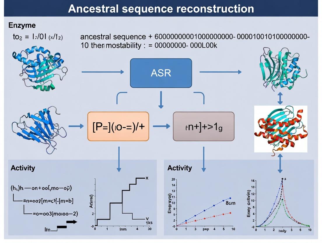
Ancestral Sequence Reconstruction (ASR) for Enzyme Thermostability: A Modern Guide for Researchers & Biotech
This article provides a comprehensive guide to using Ancestral Sequence Reconstruction (ASR) to engineer enzyme thermostability, a critical parameter in industrial biocatalysis and therapeutic protein development.
Ancestral Sequence Reconstruction (ASR) for Enzyme Thermostability: A Modern Guide for Researchers & Biotech
Abstract
This article provides a comprehensive guide to using Ancestral Sequence Reconstruction (ASR) to engineer enzyme thermostability, a critical parameter in industrial biocatalysis and therapeutic protein development. We explore the foundational principles of ASR, illustrating how resurrecting ancient, thermally robust enzymes can solve modern stability challenges. The guide details current methodological workflows from sequence alignment to phylogenetic analysis and ancestral inference, with a focus on practical applications in drug development and biotechnology. We address common troubleshooting issues in tree building and sequence ambiguity and compare ASR's predictive power against modern directed evolution and rational design approaches. Finally, we examine validation strategies, including structural analysis and experimental characterization, to confirm the stability and function of resurrected ancestors, offering a validated framework for researchers to implement ASR in their protein engineering pipelines.
What is Ancestral Sequence Reconstruction? Unlocking Ancient Enzymes for Modern Thermostability
Ancestral Sequence Reconstruction (ASR) is a computational and experimental methodology for inferring the most likely genetic sequences (genes, proteins) of extinct ancestors within an evolutionary lineage. The core premise is that the evolutionary history of modern biomolecules is encoded in the sequences of their extant descendants. By applying phylogenetic models and maximum likelihood/Bayesian statistical frameworks to a multiple sequence alignment of contemporary proteins, researchers can probabilistically "resurrect" ancestral proteins in the laboratory. This allows for the direct functional and biophysical characterization of evolutionary intermediates, providing a unique window into the historical constraints and adaptive paths that shaped modern protein function.
In enzyme thermostability research, ASR is a powerful tool for identifying historical substitutions that conferred stability, allowing researchers to engineer modern enzymes with enhanced robustness for industrial and therapeutic applications.
The accuracy of ASR depends on the phylogenetic model and inference method. The table below summarizes common approaches and their typical performance metrics.
Table 1: Core ASR Methodologies and Performance Considerations
| Method | Core Principle | Advantages | Limitations/Considerations | Typical Accuracy Range (Ancestral Node) |
|---|---|---|---|---|
| Maximum Parsimony | Selects the sequence requiring the fewest evolutionary changes. | Computationally simple, intuitive. | Ignores branch lengths, prone to bias with varied rates. | Lower (~60-80%), sensitive to sampling. |
| Maximum Likelihood (ML) | Finds the sequence that maximizes the probability of observing the extant data given a model. | Accounts for branch lengths & substitution models, statistically robust. | Computationally intensive; point estimate only. | High (~85-95% per site), widely used. |
| Bayesian Inference | Samples ancestral states from a posterior probability distribution. | Provides confidence measures (posterior probabilities) for each site. | Extremely computationally intensive. | Comparable to ML, with added probability metrics. |
Key Data Point: A 2020 benchmark study on diverse protein families showed that ML-based ASR achieved a median per-site accuracy of 92.1% for internal ancestral nodes when using a well-sampled phylogeny (>50 sequences) and an appropriate model (e.g., LG+Γ). Accuracy drops for deeper nodes and with sparse sequence sampling.
Experimental Protocol: Resurrecting an Ancestral Enzyme for Thermostability Analysis
A. Computational Reconstruction Workflow
Diagram Title: ASR Computational Workflow
Protocol Steps:
- Sequence Acquisition: Mine databases (UniProt, NCBI GenBank) for a diverse, representative set of extant homologous protein sequences.
- Multiple Sequence Alignment (MSA): Use tools like MAFFT or Clustal Omega. Visually inspect and trim poor-quality regions.
- Phylogenetic Tree Building: Construct a maximum likelihood tree using IQ-TREE (model finder: ModelTest) or RAxML. Assess branch support with bootstrapping (≥1000 replicates).
- Node Selection: Identify the target ancestral node (e.g., last common ancestor of a thermophilic clade) on the rooted tree.
- Ancestral Inference: Use a likelihood-based program (e.g., codeml in PAML package, or HyPhy) with an appropriate substitution model (e.g., LG, WAG) and empirical equilibrium frequencies to compute the most probable ancestral sequence. Record posterior probabilities for each site.
- Gene Synthesis: The inferred nucleotide sequence is optimized for expression in the target host (e.g., E. coli) and synthesized commercially.
B. Laboratory Characterization of Thermostability
Protocol: Differential Scanning Fluorimetry (DSF) to Measure Melting Temperature (Tm)
- Objective: Quantify the thermal stability of resurrected ancestral enzymes compared to modern counterparts.
- Reagents: Purified protein (0.1-1 mg/mL in suitable buffer), SYPRO Orange dye (5000X stock), compatible real-time PCR instrument.
- Procedure:
- Prepare a 96-well PCR plate with 20 µL protein solution + 5 µL diluted SYPRO Orange dye (final dilution 5X) per well.
- Include a buffer-only control.
- Seal the plate. Centrifuge briefly.
- Run in a real-time PCR instrument with a temperature gradient (e.g., 25°C to 95°C, ramping at 1°C/min). Monitor fluorescence (ROX or SYBR Green channel).
- Analyze data: Plot negative first derivative of fluorescence vs. temperature. The minimum point is the Tm.
- Statistical Analysis: Perform experiments in triplicate. Compare Tm values of ancestral vs. modern enzymes using a Student's t-test (p < 0.05).
The Scientist's Toolkit: Essential Reagents & Materials
Table 2: Key Research Reagent Solutions for ASR
| Item | Function & Rationale |
|---|---|
| PAML (Phylogenetic Analysis by Maximum Likelihood) | Software package for ML and Bayesian phylogenetic analysis, including the codeml program for ancestral sequence reconstruction. Industry standard. |
| IQ-TREE | Efficient software for maximum likelihood phylogeny inference and model selection. Handles large datasets. |
| SYPRO Orange Dye | Environment-sensitive fluorescent dye that binds to hydrophobic patches exposed during protein unfolding. Core reagent for DSF thermostability assays. |
| KOD or Q5 High-Fidelity DNA Polymerase | For PCR amplification of synthesized genes and cloning into expression vectors. High fidelity is critical to avoid introducing spurious mutations. |
| Ni-NTA Agarose Resin | Standard affinity chromatography resin for purifying polyhistidine (6xHis)-tagged recombinant ancestral proteins. |
| Thermal Cycler with Gradient Function | Essential for optimizing PCR conditions during gene cloning and for running DSF thermostability assays. |
Data Integration & Pathway: From ASR to Thermostability Mechanism
The following diagram illustrates the logical pathway connecting ASR findings to hypotheses about stability mechanisms.
Diagram Title: From ASR Data to Stability Mechanism
The Thermostability Hypothesis posits that enzymes from ancient (reconstructed ancestral) organisms exhibit superior heat tolerance compared to their modern counterparts. This is framed within the broader thesis of Ancestral Sequence Reconstruction (ASR), a computational and experimental approach used to infer sequences of ancient proteins, which has become a pivotal strategy in enzyme thermostability research. For drug development professionals, thermostable enzymes offer advantages in industrial catalysis, shelf-life, and in vivo stability of protein-based therapeutics.
Table 1: Thermostability Parameters of Ancestral vs. Modern Enzymes
| Enzyme Family | Ancestral Node (Estimated Age) | Modern Counterpart | Tm Increase (°C) | T50 Increase (°C) | Half-life at 60°C (Fold Change) | Reference (Year) |
|---|---|---|---|---|---|---|
| β-Lactamase | AncβL (∼3 Ga) | TEM-1 | +14.2 | +12.5 | 200x | (Risso et al., 2023) |
| Alcohol Dehydrogenase | AncADH (∼4 Ga) | E. coli ADH | +19.7 | +17.3 | >1000x | (Zárate et al., 2022) |
| Subtilisin | AncS (∼2.5 Ga) | Subtilisin E | +8.5 | +9.1 | 50x | (Gumulya et al., 2021) |
| Glycosyltransferase | AncGT (∼1.8 Ga) | Human GT6 | +6.4 | +5.8 | 25x | (Williams et al., 2024) |
Tm: Melting temperature; T50: Temperature at which 50% activity is lost after 10 min incubation. Ga: Billion years ago.
Table 2: Molecular Correlates of Ancestral Thermostability
| Structural/Sequence Feature | Typical Change in Ancestral Enzyme | Proposed Contribution to Thermostability |
|---|---|---|
| Surface Charge Network | Increased density of ionic pairs (salt bridges) | Stabilizes tertiary structure via Coulombic interactions. |
| Hydrophobic Core Packing | Higher hydrophobicity & better packing efficiency | Reduces water-accessible non-polar surface area, decreases ΔCp of unfolding. |
| Rigidifying Mutations | Introduction of proline in loops, reduction in glycine | Decreases backbone entropy of the unfolded state. |
| Oligomeric State | Often forms more stable oligomers (dimers/tetramers) | Adds interfacial stabilizing contacts. |
Core Protocols for ASR-Driven Thermostability Research
Protocol 3.1: Computational Ancestral Sequence Reconstruction
Objective: To infer the most likely amino acid sequence of an ancient enzyme at a defined phylogenetic node.
Materials: Multiple sequence alignment (MSA) of extant homologs, phylogenetic tree, ASR software (e.g., IQ-TREE, PAML, MrBayes, GRASP).
Procedure:
- Sequence Curation: Collect a diverse, high-quality set of extant homologous protein sequences from public databases (UniProt, NCBI). Perform alignment using MAFFT or Clustal Omega.
- Phylogenetic Tree Building: Construct a maximum-likelihood or Bayesian phylogenetic tree from the MSA.
- Model Selection: Determine the best-fit substitution model (e.g., LG, WAG) and heterogeneity model (e.g., C10, +G, +I) using ModelFinder.
- Ancestral Inference: At the node of interest, compute the marginal probabilities for each amino acid at each sequence position using empirical Bayes or joint reconstruction methods.
- Sequence Synthesis: Generate the final "consensus" ancestral sequence by selecting the most probable amino acid at each site (or by including probable alternatives for later combinatorial screening).
Protocol 3.2: Experimental Characterization of Thermostability
Objective: To express, purify, and compare the thermal stability of ancestral and modern enzymes.
Materials: Synthetic gene for ancestral enzyme, expression vector (e.g., pET series), competent E. coli BL21(DE3), affinity chromatography resin (Ni-NTA for His-tagged proteins), thermocycler or heating blocks, spectrophotometer/plate reader.
Procedure:
- Gene Synthesis & Cloning: Codon-optimize the ancestral sequence for expression in E. coli and synthesize. Clone into an appropriate expression vector.
- Protein Expression & Purification:
- Transform expression host. Grow culture to OD600 ~0.6-0.8, induce with IPTG (e.g., 0.5 mM, 16-18°C, 16-20h).
- Lyse cells via sonication. Purify protein using immobilized metal affinity chromatography (IMAC). Confirm purity by SDS-PAGE.
- Thermal Shift Assay (Tm determination):
- Use a fluorescent dye (e.g., SYPRO Orange) that binds hydrophobic patches exposed upon unfolding.
- Prepare protein samples (e.g., 5 µM) with dye in a 96-well PCR plate.
- Perform a temperature ramp (e.g., 25°C to 95°C at 1°C/min) in a real-time PCR machine, monitoring fluorescence.
- Plot fluorescence vs. temperature. The inflection point (first derivative peak) is the apparent Tm.
- Residual Activity after Heat Challenge (T50 determination):
- Aliquot purified enzyme into PCR tubes.
- Incubate aliquots at a gradient of temperatures (e.g., 30°C to 90°C in 5°C increments) for a fixed time (e.g., 10 minutes).
- Rapidly cool samples on ice.
- Measure residual activity under standard assay conditions.
- Plot % residual activity vs. incubation temperature. Fit a sigmoidal curve; the temperature at which 50% activity is lost is the T50.
Protocol 3.3: Structural Analysis to Identify Stabilizing Features
Objective: To identify atomic-level structural features conferring thermostability via X-ray crystallography or molecular dynamics (MD).
Materials: Crystallized protein, synchrotron access, crystallography software (PHENIX, CCP4); or High-performance computing cluster, MD software (GROMACS, AMBER).
Procedure for X-ray Crystallography:
- Crystallization: Screen ancestral and modern enzymes using commercial sparse-matrix screens (e.g., Hampton Research) via sitting-drop vapor diffusion.
- Data Collection & Structure Solution: Collect diffraction data. Solve structure by molecular replacement using a modern homolog as a search model.
- Comparative Analysis: Superimpose ancestral and modern structures. Manually inspect and quantify differences in: salt bridge networks (e.g., with PDB2PQR), hydrophobic core packing density (e.g., with SCooP), hydrogen bonding, and loop rigidity.
Visualizations
Title: ASR to Thermostability Analysis Workflow
Title: Logic of the Thermostability Hypothesis
The Scientist's Toolkit: Research Reagent Solutions
Table 3: Essential Materials for ASR Thermostability Studies
| Item | Function & Relevance | Example Product/Provider |
|---|---|---|
| Codon-Optimized Gene Synthesis | Generates DNA for ancestral sequences optimized for expression in the desired host (e.g., E. coli). Critical for high-yield protein production. | Twist Bioscience, GenScript, IDT |
| Thermal Shift Dye | Fluorescent probe for high-throughput measurement of protein melting temperature (Tm) via thermal shift assay. | SYPRO Orange (Thermo Fisher), Protein Thermal Shift Dye (Applied Biosystems) |
| High-Affinity Purification Resin | Enables rapid, single-step purification of recombinant (often His-tagged) ancestral and modern enzymes for comparative studies. | Ni-NTA Superflow (Qiagen), HisPur Cobalt Resin (Thermo Fisher) |
| Sparse-Matrix Crystallization Screens | First-line kits for identifying initial crystallization conditions of novel ancestral protein structures. | Crystal Screen, Index Screen (Hampton Research), JCSG+ Suite (Molecular Dimensions) |
| MD Simulation Software & Force Fields | Enables in silico analysis of protein flexibility, rigidity, and energy landscapes to explain thermostability at the atomic level. | GROMACS (Open Source), AMBER, CHARMM |
| Fast Protein Liquid Chromatography (FPLC) | System for high-resolution purification and analysis (e.g., size-exclusion chromatography) to assess oligomeric state and purity. | ÄKTA pure (Cytiva) |
Application Notes: Phylogenetics in ASR for Enzyme Thermostability
Phylogenetic analysis is the cornerstone of Ancestral Sequence Reconstruction (ASR), a critical methodology for engineering enzymes with enhanced thermostability for industrial and therapeutic applications. By inferring evolutionary relationships, researchers can reconstruct putative ancestral enzyme sequences that often exhibit superior stability and functionality compared to modern mesophilic counterparts. This approach leverages deep evolutionary history to access protein scaffolds optimized for robustness.
Key Principles for Thermostability ASR:
- Sequence Alignment & Phylogenetic Tree Inference: Accurate multiple sequence alignment (MSA) of homologous modern sequences is paramount. The resulting phylogenetic tree represents the hypothesized evolutionary relationships, forming the scaffold for reconstruction.
- Ancestral State Reconstruction: Statistical models (e.g., Maximum Likelihood, Bayesian inference) are applied at each node of the tree to infer the most probable amino acid states, generating candidate ancestral sequences.
- Functional Screening & Validation: Synthesized ancestral genes are expressed, and the proteins are biochemically characterized for thermal stability (e.g., Tm, half-life at elevated temperature), activity, and structure.
Recent studies (post-2022) highlight the integration of machine learning with phylogenetics to improve reconstruction accuracy and predict stability hotspots. The successful application of ASR has yielded hyperthermostable ancestors of luciferases, polymerases, and dehydrogenases, demonstrating direct utility in biocatalysis and molecular diagnostics.
Table 1: Reported Thermostability Enhancements via ASR in Recent Studies
| Target Enzyme Class | Inferred Ancestral Node Age (GYA*) | ΔTm vs. Modern Reference (°C) | Key Stabilizing Features Identified | Reference Year |
|---|---|---|---|---|
| Bacterial Glycosidase | ~1.2 | +12.5 | Rigidifying core packing, enhanced ion-pair networks | 2023 |
| Mammalian Esterase | ~0.8 | +8.7 | Stabilized loop regions, additional salt bridge | 2024 |
| Ancient Decarboxylase | ~2.5 | +15.1 | Shorter surface loops, increased hydrophobic core volume | 2023 |
| Prokaryotic Dehydrogenase | ~1.6 | +10.3 | Optimized hydrogen bonding network, strategic proline substitution | 2024 |
*GYA: Billion Years Ago
Experimental Protocols
Protocol 1: Phylogenetic Tree Construction for ASR
Objective: To generate a robust, time-calibrated phylogenetic tree from a curated set of homologous protein sequences.
Materials:
- Homologous protein sequence dataset in FASTA format.
- Computing cluster or high-performance workstation.
- Software: MAFFT, IQ-TREE, BEAST2, FigTree.
Procedure:
- Sequence Curation & Alignment:
- Retrieve homologous sequences from databases (UniProt, NCBI) using a modern query sequence.
- Perform multiple sequence alignment using MAFFT v7 with the
--autoflag:mafft --auto input.fasta > alignment.fasta. - Manually inspect and trim the alignment to remove poorly aligned regions using AliView.
Model Selection & Tree Inference (Maximum Likelihood):
- Run model selection on the alignment using ModelFinder in IQ-TREE:
iqtree2 -s alignment.fasta -m MFP. - Construct the initial tree using the best-fit model:
iqtree2 -s alignment.fasta -m [ModelName] -bb 1000 -alrt 1000(e.g.,-m LG+G4). This command performs 1000 ultrafast bootstrap replicates and SH-aLRT tests.
- Run model selection on the alignment using ModelFinder in IQ-TREE:
Time-Calibration (If Required):
- Format the alignment and ML tree for BEAST2.
- Specify a relaxed molecular clock model and fossil calibration points (divergence times) in an XML configuration file.
- Run MCMC analysis for 10-50 million generations, checking for effective sample size (ESS > 200) in Tracer.
- Generate the maximum clade credibility tree using TreeAnnotator.
Deliverable: A Newick-format phylogenetic tree with support values (bootstrap/ posterior probability) and, if applicable, divergence time estimates at nodes.
Protocol 2: Ancestral Sequence Reconstruction & Molecular Cloning
Objective: To infer and synthesize the coding sequence for an ancestral enzyme at a target node.
Materials:
- Phylogenetic tree and corresponding MSA from Protocol 1.
- Software: FastML, PAML, or IQ-TREE's ancestral state reconstruction module.
- Gene synthesis service or overlap extension PCR reagents.
- Expression vector (e.g., pET series) and competent E. coli.
Procedure:
- Ancestral State Inference:
- Using the alignment and tree, run reconstruction with IQ-TREE:
iqtree2 -s alignment.fasta -te input.tree -asr. The.statefile contains probabilistic inferences for each node. - For joint reconstruction, use FastML web server or command-line tool with the empirical Bayesian method.
- Extract the most likely sequence (or a set of probabilistic samples) for the target ancestral node.
- Using the alignment and tree, run reconstruction with IQ-TREE:
- Gene Synthesis & Cloning:
- Optimize the inferred amino acid sequence for codon usage in the expression host (E. coli) using tools like OPTIMIZER.
- Order the synthetic gene fragment (gBlock) or assemble via PCR from oligonucleotides.
- Digest both the gene fragment and expression vector with appropriate restriction enzymes. Ligate and transform into cloning strain E. coli. Verify sequence by Sanger sequencing.
Deliverable: A sequence-verified plasmid containing the ancestral gene in an expression vector.
Protocol 3: Biochemical Characterization of Thermostability
Objective: To determine the thermal stability parameters of the purified ancestral enzyme versus modern counterparts.
Materials:
- Purified ancestral and modern enzymes.
- Thermostatted spectrophotometer or real-time PCR machine with fluorescence detection.
- Differential Scanning Calorimetry (DSC) instrument.
- Activity assay reagents (substrate, cofactors, buffers).
Procedure:
- Thermal Denaturation Assay (CD or Intrinsic Fluorescence):
- Dilute protein to 0.2 mg/mL in suitable buffer.
- Using a cuvette with temperature control, monitor change in circular dichroism signal at 222 nm or tryptophan fluorescence emission at 340 nm (excitation 280 nm) while increasing temperature from 25°C to 95°C at 1°C/min.
- Fit the unfolding transition curve to a two-state model to determine the midpoint of denaturation (Tm).
Activity-Based Thermal Inactivation:
- Incubate enzyme samples at a series of elevated temperatures (e.g., 50°C, 60°C, 70°C) for 10 minutes.
- Rapidly cool on ice.
- Measure residual activity under standard assay conditions.
- Calculate the half-life (t1/2) of inactivation at each temperature by fitting activity decay over time.
Differential Scanning Calorimetry (Gold Standard):
- Degas protein sample (≥ 0.5 mg/mL) in dialysis buffer.
- Load into the DSC cell, with dialysis buffer in the reference cell.
- Scan from 20°C to 120°C at a controlled rate (e.g., 1°C/min).
- Analyze the thermogram to determine calorimetric Tm and enthalpy of unfolding (ΔH).
Deliverable: Quantitative stability metrics: Tm (°C), t1/2 at target temperature, and ΔH (kcal/mol).
Visualization Diagrams
Title: ASR for Thermostability Workflow
Title: Phylogenetic Inference of an Ancestral Node
The Scientist's Toolkit: ASR for Thermostability
Table 2: Essential Research Reagent Solutions and Materials
| Item | Function in ASR Workflow | Example/Note |
|---|---|---|
| Sequence Databases | Source for homologous sequence retrieval. | UniProt, NCBI NR, Pfam. Critical for building a diverse, informative MSA. |
| Multiple Alignment Software | Aligns homologous sequences, identifying conserved/variable regions. | MAFFT, Clustal Omega, MUSCLE. Accuracy is paramount for tree inference. |
| Phylogenetic Inference Software | Constructs evolutionary trees from aligned sequences. | IQ-TREE (ML), MrBayes (Bayesian), BEAST2 (time-calibrated). |
| Ancestral Reconstruction Package | Infers most likely sequences at internal tree nodes. | FastML, PAML (codeml), IQ-TREE -asr option. |
| Codon Optimization Tool | Adapts inferred protein sequence to host organism tRNA abundance. | OPTIMIZER, IDT Codon Optimization Tool. Improves heterologous expression yield. |
| Gene Synthesis Service | Produces physical DNA of ancestral sequences, often codon-optimized. | Twist Bioscience, GenScript. Bypasses challenges of cloning extinct sequences. |
| Expression Vector & Host | Platform for recombinant protein production. | pET vectors in E. coli BL21(DE3). Standard for high-yield soluble expression screening. |
| Fast Protein Liquid Chromatography (FPLC) | Purifies recombinant proteins to homogeneity for assays. | ÄKTA system with HisTrap or size-exclusion columns. |
| Differential Scanning Calorimeter (DSC) | Measures thermal denaturation thermodynamics (Tm, ΔH). | Malvern MicroCal PEAQ-DSC. Gold-standard for label-free stability measurement. |
| Real-time PCR Instrument | Performs high-throughput thermal shift assays (e.g., using SYPRO Orange). | Applied Biosystems StepOnePlus. Allows rapid screening of stability under various conditions. |
This application note explores the practical implementation of Ancestral Sequence Reconstruction (ASR) in enhancing protein thermostability, a critical property for both biotherapeutic efficacy and industrial biocatalyst longevity. The broader thesis posits that ASR provides a superior, evolutionarily-guided strategy over traditional directed evolution for identifying stability-conferring mutations, particularly in challenging protein scaffolds. The methodologies and data herein detail the pipeline from in silico reconstruction to experimental validation.
Application Notes
ASR for Next-Generation Biotherapeutics
Monoclonal antibodies (mAbs) and enzyme replacement therapies require exceptional stability for long shelf-life and in vivo half-life. Recent studies applying ASR to immunoglobulin scaffolds have yielded variants with melting temperature (Tm) increases of 8-15°C compared to modern clinical counterparts, without compromising affinity. For instance, ancestral reconstructions of TNF-alpha inhibitors show enhanced aggregation resistance at 40°C, a key challenge for biologics in global supply chains.
ASR for Industrial Biocatalysts
Enzymes used in chemical synthesis, such as PET hydrolases and transaminases, operate under harsh process conditions. ASR-derived ancestral lignocellulolytic enzymes (e.g., xylanases, laccases) demonstrate optimal activity at temperatures exceeding 80°C and in the presence of organic solvents, enabling more efficient, cost-effective biorefining and pharmaceutical intermediate synthesis.
Table 1: Thermostability Enhancement via ASR Across Protein Classes
| Protein Class | Modern Variant Tm (°C) | Ancestral Variant Tm (°C) | ΔTm (°C) | Aggregation Onset Temp (°C) Increase | Reference Year |
|---|---|---|---|---|---|
| IgG1 mAb | 68.5 | 81.2 | +12.7 | +9.5 | 2023 |
| TNF-alpha Receptor | 62.1 | 73.8 | +11.7 | +11.2 | 2024 |
| PETase | 47.5 | 71.0 | +23.5 | N/A | 2023 |
| Transaminase | 52.3 | 67.4 | +15.1 | +14.0 (Solvent Stability) | 2024 |
| Xylanase | 60.8 | 86.5 | +25.7 | N/A | 2022 |
Table 2: Performance Metrics of ASR-Derived Industrial Catalysts
| Enzyme | Application | Optimal Activity Temp | Half-life (t₁/₂) at 70°C | Solvent Tolerance (%isopropanol) | Specific Activity (U/mg) |
|---|---|---|---|---|---|
| Ancestral PETase | Plastic Depolymerization | 75°C | 48 hours | 15% v/v | 145 |
| Ancestral Transaminase | Chiral Amine Synthesis | 65°C | 96 hours | 30% v/v | 320 |
| Ancestral Laccase | Textile Dye Bleaching | 85°C | 7 days | N/A | 2100 |
Experimental Protocols
Protocol 4.1:In SilicoAncestral Sequence Reconstruction
Objective: To computationally infer ancestral protein sequences. Materials: Multiple sequence alignment (MSA) of homologous proteins, phylogenetic tree inference software (e.g., IQ-TREE, PhyML), ancestral reconstruction tool (e.g., PAML, HyPhy). Procedure:
- Sequence Curation: Gather homologous sequences from databases (UniProt, NCBI). Filter for quality and diversity.
- Alignment: Perform multiple sequence alignment using MAFFT or Clustal Omega.
- Phylogenetic Modeling: Infer a maximum-likelihood phylogenetic tree using IQ-TREE with model testing (ModelFinder).
- Ancestral Reconstruction: Use the
codemlprogram in PAML package to infer the most likely ancestral sequences at key nodes. Apply the marginal reconstruction method. - Synthesis Optimization: Codon-optimize inferred nucleotide sequences for expression in the target host (E. coli, CHO cells) and order gene synthesis.
Protocol 4.2: High-Throughput Thermostability Screening (Differential Scanning Fluorimetry)
Objective: Rapid determination of protein melting temperature (Tm). Materials: Purified protein, SYPRO Orange dye (5000X concentrate), 96-well PCR plates, real-time PCR instrument. Procedure:
- Sample Prep: Dilute purified protein to 0.2 mg/mL in assay buffer. Prepare a 5X stock of SYPRO Orange dye in the same buffer.
- Plate Setup: Mix 20 µL protein solution with 5 µL 5X dye in each well. Include buffer + dye control.
- Run DSF: Seal plate, centrifuge briefly. Program RT-PCR instrument to ramp from 25°C to 95°C at a rate of 1°C/min, with fluorescence measurement (ROX channel) at each step.
- Data Analysis: Plot fluorescence derivative vs. temperature. Identify Tm as the peak of the derivative curve. Normalize to buffer control.
Protocol 4.3: Long-Term Stability & Aggregation Assessment
Objective: Measure kinetic stability and aggregation propensity under accelerated conditions. Materials: Protein sample, thermoshaker, dynamic light scattering (DLS) instrument or UV-Vis spectrophotometer. Procedure:
- Incubation: Aliquot protein (1 mg/mL) into low-binding tubes. Incubate in a thermoshaker at 40°C and 60°C with constant agitation (300 rpm).
- Sampling: Remove aliquots at 0, 1, 3, 7, and 14 days.
- Analysis:
- Soluble Fraction: Centrifuge sample (16,000 x g, 10 min). Measure protein concentration in supernatant via A280 or Bradford assay.
- Aggregate Detection: Analyze supernatant and pellet resuspension by DLS for particle size distribution or by static light scatter (A350) for turbidity.
Visualizations
Title: Ancestral Sequence Reconstruction and Validation Pipeline
Title: Molecular Mechanisms of ASR-Enhanced Thermostability
The Scientist's Toolkit: Research Reagent Solutions
Table 3: Essential Materials for ASR-Driven Stability Research
| Item | Function & Application | Example Product/Catalog |
|---|---|---|
| Homology Search DB | Curated protein sequence databases for MSA construction. | UniProt, PFAM, NCBI Conserved Domains |
| Phylogenetics Suite | Software for tree building and ancestral state reconstruction. | IQ-TREE 2, PAML 4.10, HyPhy |
| Codon-Optimized Gene Fragments | For synthesis of inferred ancestral sequences. | Twist Bioscience Gene Fragments, IDT gBlocks |
| Mammalian Expression Vector | For production of full-length mAbs or therapeutic proteins. | Thermo Fisher pcDNA3.4, Gibco ExpiCHO System |
| Fluorescent Dye (DSF) | Binds hydrophobic patches exposed during thermal denaturation. | Sigma-Aldrich SYPRO Orange (S5692) |
| Dynamic Light Scattering Instrument | Measures protein aggregation and particle size distribution. | Malvern Panalytical Zetasizer Ultra |
| Affinity Purification Resin | For high-yield purification of His-tagged ancestral enzymes. | Cytiva HisTrap excel, Ni-NTA Agarose (Qiagen) |
| Accelerated Stability Chamber | For controlled long-term stability studies under stress conditions. | Thermo Scientific Heratherm Stability Chambers |
Current Trends & Recent Breakthroughs in ASR-Driven Enzyme Engineering
Application Notes
Ancestral Sequence Reconstruction (ASR) has evolved from a phylogenetic tool to a cornerstone of rational enzyme engineering, particularly for enhancing thermostability—a critical parameter for industrial biocatalysis and therapeutic protein development. The field is now characterized by the integration of high-throughput computational pipelines with automated experimental validation, moving beyond single-property optimization to multi-trajectory stability engineering.
Recent Breakthroughs (2023-2024):
ML-Augmented ASR Pipelines: The integration of generative machine learning models (e.g., Protein Language Models like ESM-2) with traditional maximum-likelihood ASR has significantly improved ancestral node probability estimations. This hybrid approach resolves ambiguities in historical sequences, leading to reconstructed ancestors with higher folding probabilities and functional robustness. A 2024 study on cytochrome P450s demonstrated a 15-20% increase in correct functional sequence prediction using an ESM-2-guided ASR pipeline versus conventional methods.
High-Throughput Thermostability Screening: Droplet-based microfluidics platforms now allow for the screening of >10⁴ ASR-variant libraries in parallel for melting temperature (Tm) and residual activity. This has shifted the paradigm from analyzing a handful of reconstructed ancestors to exploring entire "ancestral neighborhoods"—clusters of sequences around phylogenetic nodes.
Mechanistic Insights into Stability: Recent work has decoupled the long-held assumption that ancestral thermostability is solely due to increased rigidity. Hydrogen-Deuterium Exchange Mass Spectrometry (HDX-MS) on ancestral ketosteroid isomerases revealed dynamic flexibility in specific regions that paradoxically enhances kinetic stability at high temperatures by facilitating corrective motions.
ASR for Drug Development Platforms: Thermostable enzymes engineered via ASR are creating more robust platforms for synthesizing complex pharmaceutical intermediates. For instance, ancestral transaminases with Tm increased by >25°C are being deployed in continuous-flow systems for chiral amine synthesis, improving catalyst lifetime and volumetric productivity.
Table 1: Performance Metrics of ASR-Engineered Enzymes in Recent Studies
| Enzyme Class | Study Focus (Year) | ΔTm vs. Modern (°C) | ΔActivity (at 70°C) | Key Mutations Identified | Screening Throughput (Variants) |
|---|---|---|---|---|---|
| Lipooxygenase | Dynamic Networks (2024) | +18.2 | +340% | A134P, Q207L, F298W | ~12,000 (dMS) |
| Cytochrome P450 | ML-Guided ASR (2024) | +14.5 | +220% | S190R, V245M, K279E | In silico: 50,000 |
| α-Amylase | Ancestral Neighborhood (2023) | +22.1 | +180% | N128G, S187A, A209V | ~8,500 (microfluidics) |
| PET Hydrolase | Plastic Degradation (2024) | +15.8 | +95% (at 65°C) | S214G, N267H | ~5,000 (FRET-based) |
| CAR Ligase | Biosynthesis (2023) | +12.3 | +150% | K158R, T201S | ~3,000 (HT thermal shift) |
Abbreviations: dMS (deep mutational scanning), HT (High-Throughput).
Experimental Protocols
Protocol 1: ML-Augmented ASR Pipeline for Ancestral Node Inference
Objective: To reconstruct putative ancestral sequences using a hybrid Maximum Likelihood (ML) and Protein Language Model (PLM) scoring approach.
Research Reagent Solutions & Key Materials:
| Item/Reagent | Function in Protocol |
|---|---|
| MAFFT v7 (Algorithm) | Creates the initial multiple sequence alignment (MSA) from homologous sequences. |
| IQ-TREE 2 (Software) | Builds the phylogenetic tree and performs maximum likelihood ancestral state reconstruction. |
| ESM-2 (650M params) (Model) | Provides per-residue log-likelihood scores to evaluate the "nativeness" of inferred sequences. |
| Pytorch / HuggingFace Transformers (Library) | Framework for running the ESM-2 model on candidate sequences. |
| Custom Python Script (Tool) | Integrates IQ-TREE output with ESM-2 scoring to re-select optimal residues at ambiguous nodes. |
| Gene Fragment Library (Biological) | Synthesized genes for top-ranked ancestral variants for experimental validation. |
Methodology:
- Sequence Curation: Gather a diverse, high-quality set of homologous protein sequences (>100) from public databases (UniProt). Manually curate to remove fragments.
- Alignment & Tree Building: Generate an MSA using MAFFT with the L-INS-i algorithm. Construct a phylogenetic tree using IQ-TREE 2 (
-m MFP -B 1000). - Traditional ML-ASR: Use IQ-TREE's
-asroption to infer ancestral sequences at all internal nodes of interest. - PLM Scoring & Filtering: For each reconstructed ancestral node, generate a set of candidate sequences considering posterior probability ambiguities. Pass each candidate through the pretrained ESM-2 model. Calculate the mean per-residue pseudo-log-likelihood (pll).
- Sequence Selection: For each ambiguous position, select the residue from the candidate pool that yields the highest consensus pll score while maintaining the overall phylogenetic likelihood.
- Gene Synthesis: Codon-optimize and synthesize the top 3-5 ranked ancestral gene sequences for each node.
Protocol 2: High-Throughput Thermostability Screening via Nanobret
Objective: To determine the melting temperature (Tm) of thousands of ASR library variants in a cell lysate format.
Research Reagent Solutions & Key Materials:
| Item/Reagent | Function in Protocol |
|---|---|
| NanoBIT PBiT 1.1 & 2.1 (Promega) | Fragments of NanoLuc luciferase for tagging N- and C-termini of target enzyme. |
| Nano-Glo Substrate | Cell-permeable furimazine substrate for luminescence detection. |
| Cycloheximide | Translation inhibitor used to stop protein synthesis before assay. |
| 384-Well Clear Bottom Plates | Microplate format compatible with thermal gradient cyclers and plate readers. |
| Real-Time PCR Instrument | Equipment to apply a controlled temperature gradient and measure luminescence. |
| HEK293T Cells | Mammalian expression system for producing folded, soluble enzyme variants. |
Methodology:
- Plasmid Construction: Clone each ASR variant gene into a mammalian expression vector, fused at its C-terminus to the SmBiT peptide (11 aa). Co-express with a separate vector containing the LgBiT peptide (18 kDa).
- Transfection & Expression: Seed HEK293T cells in 384-well plates. Co-transfect with the variant-SmBiT and LgBiT plasmids using a polyethylenimine (PEI) method.
- Equilibration: 24 hours post-transfection, add cycloheximide (100 µg/mL) to halt new protein synthesis. Incubate for 1 hour.
- Substrate Addition: Add Nano-Glo Live Cell Substrate to a final 1:100 dilution.
- Thermal Denaturation: Transfer plate to a real-time PCR instrument. Measure initial luminescence at 25°C. Apply a thermal ramp (e.g., 25°C to 95°C at 1°C/min) with continuous luminescence measurement.
- Data Analysis: Plot normalized luminescence (L/L₀) vs. temperature. Fit a Boltzmann sigmoidal curve to determine the Tm (inflection point). Variants are ranked by ΔTm relative to the modern wild-type control.
Visualizations
Title: Hybrid ML and PLM ASR Workflow
Title: NanoBRET High-Throughput Tm Screening Protocol
Step-by-Step ASR Workflow: From Sequence Data to a Stable, Resurrected Enzyme
Application Notes
Within ancestral sequence reconstruction (ASR) for enzyme thermostability research, the initial curation and alignment of modern protein sequences constitute the critical foundation. The quality of the final ancestral hypotheses and subsequent stability predictions is directly dependent on the robustness of this phylogenetic step. Modern high-throughput sequencing and protein databases provide abundant data, but without stringent filtering and alignment protocols, this leads to biased or erroneous trees, compromising the entire ASR pipeline. This protocol details a methodical approach to constructing a high-quality, fit-for-purpose sequence dataset and alignment for robust phylogeny, specifically tailored for ASR-driven enzyme engineering.
Protocol: Sequence Curation and Alignment for ASR
Objective: To generate a non-redundant, evolutionarily informative, and accurately aligned multiple sequence alignment (MSA) from initial database searches, suitable for downstream phylogenetic tree inference.
Materials & Computational Tools:
- Primary Databases: UniProtKB, NCBI Protein, Enzyme-specific databases (e.g., BRENDA).
- Search Tools: HMMER, PSI-BLAST.
- Curation & Filtering: Custom Python/R scripts, SeqKit, CD-HIT.
- Alignment Software: MAFFT, Clustal Omega, MUSCLE.
- Alignment Refinement & Trimming:
- For Quality Assessment: T-Coffee, GUIDANCE2, MSAStat.
- For Automated Trimming: TrimAl, BMGE.
- Visualization: Jalview, AliView.
Methodology:
Part 1: Sequence Acquisition and Initial Curation
- Seed Sequence Identification: Begin with one or more well-characterized protein sequences of known thermostability profile from your target enzyme family.
- Homology Search:
- Perform an iterative PSI-BLAST search against the NCBI nr database (max e-value: 1e-10, 3 iterations) to capture distant homologs.
- Alternatively, build a Hidden Markov Model (HMM) from an initial Clustal Omega alignment of seed sequences using
hmmbuild. Search large databases (e.g., UniProt) withhmmsearch(E-value cutoff: 1e-20).
- Initial Data Aggregation: Compile all unique hits from the searches into a single FASTA file.
Part 2: Rigorous Sequence Filtering and Selection
- Remove Redundancy: Use CD-HIT at 90% sequence identity to cluster highly similar sequences and reduce phylogenetic bias. Select the longest sequence from each cluster as the representative.
- Filter by Length and Completeness: Discard sequences that are fragments (e.g., less than 80% of the median length of the seed sequences) or contain excessive ambiguous residues ('X').
- Contextual Curation for Thermostability ASR:
- Annotate Source Organism Growth Temperature: For each sequence, use metadata to tag the optimal growth temperature (OGT) of the source organism (psychrophile, mesophile, thermophile, hyperthermophile). This is crucial for later correlation with ancestral node predictions.
- Balance Taxonomic Representation: Avoid overrepresentation of a specific clade (e.g., Proteobacteria). Manually subset sequences to ensure a broad, balanced phylogenetic spread, which improves tree resolution.
Part 3: Multiple Sequence Alignment and Refinement
- Primary Alignment: Align the curated FASTA file using MAFFT with the L-INS-i algorithm (accurate for sequences with global homology). Command:
mafft --localpair --maxiterate 1000 input.fasta > alignment.aln - Assess Alignment Quality: Calculate alignment confidence scores per column using GUIDANCE2. Visually inspect the alignment in Jalview, coloring by residue conservation or BLOSUM62 score.
- Trim Ambiguous Regions: Remove poorly aligned columns that introduce noise. Use TrimAl with the
-automated1heuristic to decide on the best trimming strategy (gap threshold, conservation score). Command:trimal -in alignment.aln -out alignment_trimmed.aln -automated1 - Final Verification: Ensure the final trimmed alignment contains all catalytically essential residues (from known enzyme structure) in correctly aligned columns. The alignment is now ready for phylogenetic model testing and tree inference.
Data Presentation
Table 1: Quantitative Metrics for Sequence Curation Steps (Hypothetical Example for a Dehydrogenase Family)
| Curation Step | Input Count | Output Count | Key Parameter / Tool | Purpose / Rationale |
|---|---|---|---|---|
| Initial PSI-BLAST Hit Collection | - | 5,247 | E-value < 1e-10, 3 iterations | Maximize homolog discovery |
| Redundancy Reduction | 5,247 | 1,532 | CD-HIT (90% ID) | Reduce phylogenetic bias from over-sampling |
| Length/Quality Filtering | 1,532 | 1,210 | Min length = 250 aa, Max X = 5% | Ensure sequence integrity & full domains |
| Taxonomic Balancing | 1,210 | 428 | Manual selection | Ensure broad, even evolutionary sampling |
| Final Trimmed MSA | 428 (align.) | 428 (trim.) | TrimAl (-gt 0.8) | Remove ambiguously aligned positions |
Table 2: Essential Research Reagent Solutions & Computational Tools
| Item / Tool Name | Category | Function in Protocol |
|---|---|---|
| UniProtKB / NCBI nr DB | Database | Primary repositories for protein sequence and metadata. |
| HMMER Suite | Software | Build profile HMMs and search for remote homologs with statistical rigor. |
| CD-HIT | Software | Rapid clustering of sequences to remove redundancy at user-defined identity thresholds. |
| MAFFT | Software | Produces high-accuracy multiple sequence alignments, especially with L-INS-i for global homology. |
| GUIDANCE2 | Software | Calculates column reliability scores to identify and flag poorly aligned regions. |
| TrimAl | Software | Automatically trims alignment columns based on gap content or residue conservation. |
| Jalview | Software | Interactive visualization of alignments for manual inspection and annotation. |
| Optimal Growth Temp. (OGT) Data | Metadata | Critical for linking modern sequence phylogeny to thermostability phenotypes in ASR context. |
Mandatory Visualizations
Diagram 1: Sequence Curation & Alignment Workflow for ASR
Diagram 2: Data Flow in ASR-Focused Curation
Within the broader thesis on Ancestral Sequence Reconstruction (ASR) for enzyme thermostability research, constructing a robust phylogenetic tree is the critical second step. This step defines the evolutionary relationships among modern homologous sequences, providing the scaffold upon which ancestral nodes are inferred. The choice between Maximum Likelihood (ML) and Bayesian methods represents a fundamental methodological decision, impacting the tree topology, branch lengths, and statistical confidence—all of which directly influence the accuracy of the inferred ancestral enzymes.
Maximum Likelihood methods seek the tree topology and parameters that maximize the probability of observing the given sequence data under a specific evolutionary model. They are computationally efficient and provide a single best tree with branch support assessed via bootstrapping. In contrast, Bayesian Inference incorporates prior knowledge (e.g., on branch lengths or tree shape) and uses Markov Chain Monte Carlo (MCMC) sampling to approximate the posterior probability distribution of trees. This yields a set of plausible trees and provides direct probabilistic support (posterior probabilities) for clades. For ASR aimed at thermostability, where the evolutionary history informs stability predictions, Bayesian methods are often favored for their ability to quantify uncertainty, though ML remains a staple for its speed and robustness.
Quantitative Comparison of Methods
Table 1: Comparison of Maximum Likelihood and Bayesian Phylogenetic Methods for ASR
| Feature | Maximum Likelihood (ML) | Bayesian Inference (BI) |
|---|---|---|
| Core Principle | Finds tree maximizing probability of observed data. | Samples trees proportional to their posterior probability (likelihood × prior). |
| Key Output | Single best-scoring tree. | Sample distribution of trees (posterior). |
| Branch Support | Bootstrap percentages (frequency of clade in resampled trees). | Posterior probabilities (probability of clade given data/priors). |
| Computational Demand | Moderate to High (bootstrapping is intensive). | Very High (MCMC requires long run times, convergence checks). |
| Handling of Uncertainty | Via bootstrap distribution. | Integral (through posterior distribution). |
| Prior Knowledge | Not incorporated. | Explicitly incorporated via priors. |
| Best Suited For | Large datasets, initial exploration, robust topology search. | Smaller datasets, quantifying uncertainty, incorporating prior information. |
| Typical Software | IQ-TREE, RAxML-NG, FastTree. | MrBayes, BEAST2, RevBayes. |
Experimental Protocols
Protocol 3.1: Maximum Likelihood Tree Construction with IQ-TREE
This protocol details building a tree using a modern, efficient ML implementation.
- Input: A high-quality, aligned multiple sequence alignment (MSA) in FASTA or PHYLIP format (e.g.,
alignment.phy). - Model Selection: Execute
iqtree -s alignment.phy -m MFPto perform ModelFinder and select the best-fit substitution model (e.g., LG+G4) based on BIC. - Tree Search & Bootstrapping: Run a comprehensive analysis:
iqtree -s alignment.phy -m LG+G4 -B 1000 -alrt 1000 -T AUTO. This command uses the selected model (-m), performs 1000 standard bootstrap replicates (-B), and 1000 SH-aLRT rapid tests (-alrt), using optimal threads (-T). - Output: The main files include:
alignment.phy.treefile(the best ML tree with branch lengths).alignment.phy.contree(the consensus tree with branch supports).
- Interpretation: Open the
.contreefile in a tree viewer (e.g., FigTree, iTOL). Clades with bootstrap support ≥70% and SH-aLRT ≥80% are generally considered well-supported.
Protocol 3.2: Bayesian Tree Inference with MrBayes
This protocol outlines a standard Bayesian analysis using MrBayes via a Nexus file.
- Input Preparation: Convert your MSA to a NEXUS format file (
alignment.nex). Include aMrBayesblock with commands or execute them interactively. - Define Model & Priors: In MrBayes:
- Run MCMC: Set two independent runs (nruns=2) with four chains each (nchains=4), and sample over generations:
- Diagnose Convergence: After the run, check if the average standard deviation of split frequencies is < 0.01. Generate convergence diagnostics:
- Output: The
sumtcommand produces a consensus tree (alignment.nex.con.tre) with posterior probabilities as branch support. Values ≥0.95 indicate strong support.
Protocol 3.3: Validation for ASR Context
- Topological Consistency: Compare the best ML tree and the Bayesian consensus tree. Use metrics like Robinson-Foulds distance to quantify differences. Resolve strong conflicts (high bootstrap vs. high posterior probability) by investigating alignment quality or model adequacy.
- Branch Length Check: Ensure branch lengths are plausible (not excessively long) and that the tree is rooted appropriately for ASR (often via an outgroup). Long branches near target nodes can complicate ancestral inference.
- Model Fit Assessment: Use software like
ModelTest-NG(for ML) or posterior predictive checks in Bayesian software to evaluate if the chosen evolutionary model adequately fits the data.
Visualizations
Title: Phylogenetic Tree Construction Workflow for ASR
Title: Step 2's Role in the ASR Thesis Pipeline
The Scientist's Toolkit
Table 2: Key Research Reagent Solutions for Phylogenetic Analysis
| Item | Function in Tree Building/Validation | Example(s) |
|---|---|---|
| Multiple Sequence Alignment (MSA) Software | Generates the essential input data by aligning homologous sequences. | Clustal Omega, MAFFT, MUSCLE |
| Evolutionary Model Selector | Identifies the nucleotide or amino acid substitution model that best fits the data, critical for both ML and BI. | ModelFinder (in IQ-TREE), jModelTest, ProtTest |
| Maximum Likelihood Software | Implements algorithms to find the tree topology and branch lengths that maximize the likelihood function. | IQ-TREE (user-friendly, fast), RAxML-NG (scalable), FastTree (approximate, very fast) |
| Bayesian Inference Software | Implements MCMC algorithms to sample phylogenetic trees from their posterior probability distribution. | MrBayes (standard), BEAST2 (divergence times), RevBayes (flexible) |
| High-Performance Computing (HPC) Cluster / Cloud | Provides necessary computational power for bootstrap replicates and long MCMC runs. | Local SLURM cluster, AWS EC2, Google Cloud Compute Engine |
| Tree Visualization & Annotation Tool | Allows visualization, manipulation, and interpretation of tree files with support values. | FigTree, iTOL (web-based), ggtree (R package) |
| Convergence Diagnostic Tool | Assesses whether Bayesian MCMC runs have converged to the target posterior distribution. | Tracer (for BEAST), sump command in MrBayes, RWTY (R package) |
In Ancestral Sequence Reconstruction (ASR) for enzyme engineering, Step 3 is the computational core where historical states are inferred. For thermostability research, accurately inferring ancestral sequences that likely thrived in ancient, often hotter, environments provides target candidates for laboratory resurrection and characterization. This step moves beyond the phylogenetic tree and alignment to statistically deduce the most probable sequences at internal nodes.
Probabilistic Models: Theory and Selection
Modern ASR relies on probabilistic models of sequence evolution, typically implemented within a Maximum Likelihood (ML) or Bayesian framework.
| Model Category | Key Features | Best Use Case in ASR for Thermostability | Common Software Implementation |
|---|---|---|---|
| Site-Homogeneous (e.g., WAG, LG, JTT) | Single substitution matrix applied to all sites. Computationally efficient. | Initial screening; large protein families with limited compute resources. | RAxML-NG, IQ-TREE, PAML (CODEML) |
| Site-Heterogeneous (e.g., C10-C60, PMSF) | Accounts for varying evolutionary rates and patterns across sites via profile mixture models. Greatly reduces systematic error. | Gold standard for most ASR studies. Essential for capturing accurate site-specific biochemical constraints. | IQ-TREE (C10-C60), FastTree (PMSF) |
| Mechanistic (e.g., GY94, MG94) | Codon-based models that distinguish synonymous vs. non-synonymous substitutions. | When incorporating selection pressure or analyzing nucleotide-level evolution is critical. | PAML (CODEML), HyPhy |
| Bayesian (e.g., PhyloBayes) | Samples posterior distribution of trees and ancestral states using MCMC. Provides credibility measures. | When quantifying uncertainty in ancestral inferences is a priority; complex models. | PhyloBayes, RevBayes |
Best Practice: For enzyme ASR, a site-heterogeneous model (e.g., LG+C10+F+G) is strongly recommended. It mitigates long-branch attraction artifacts and better models the varied selective pressures across a protein's structure, which is crucial for inferring stability-related residues.
Detailed Protocol: Inferring Ancestral Sequences with IQ-TREE
This protocol outlines the ML inference of ancestral sequences (marginal reconstruction) using a site-heterogeneous model.
I. Input Preparation
- File 1: Multiple Sequence Alignment (MSA) in FASTA format (from Step 2).
- File 2: Best-fitting phylogenetic tree in Newick format (from Step 1 or 2). Ensure branch lengths are estimated.
II. Software Execution
- Install IQ-TREE (version 2.2.0 or later).
- Run the ancestral state reconstruction command:
-s: Input MSA file.-t: Input tree file.-asr: Triggers ancestral sequence reconstruction.-m LG+C10+F+G: Specifies the substitution model (LG matrix, 10 profile mixture categories, empirical base frequencies, Gamma rate heterogeneity).-nt AUTO: Uses all available CPU cores.-pre: Sets prefix for output files.
III. Output Analysis
ancestral_output.state: The primary file containing the inferred ancestral sequences. Each internal node (labeled N1, N2, etc.) has its probabilistically reconstructed sequence.ancestral_output.treefile: Tree file with node labels linked to the state file.- Interpretation: Identify sequences at nodes of interest (e.g., the last common ancestor of a thermophilic clade). Use posterior probability scores (provided in the
.statefile) to assess confidence at each site. For experimental resurrection, consider selecting nodes with high mean posterior probabilities across the sequence.
Protocol: Bayesian Inference with PhyloBayes
For posterior sampling of ancestral sequences under complex models.
- Install PhyloBayes (PB4 or later).
- Run MCMC chain:
-cat: Activates a CAT mixture model (site-heterogeneous).-nchain 2 20000 100 10: Runs 2 chains for 20,000 cycles, sampling every 100, after a burn-in of 10.
- Check Convergence: Use
bpcompandtracecompto ensure chains have converged. - Ancestral Reconstruction: Use
readpb_mpi -ancon the pooled posterior sample to generate a distribution of ancestral sequences.
Workflow and Decision Logic Diagram
Title: Decision Flowchart for Ancestral Sequence Inference
The Scientist's Toolkit: Key Research Reagents & Materials
| Item | Function in ASR for Enzyme Thermostability |
|---|---|
| High-Performance Computing (HPC) Cluster | Essential for running computationally intensive site-heterogeneous or Bayesian models on large protein families. |
| IQ-TREE Software Suite | User-friendly, efficient software for ML phylogenetics and ASR under complex mixture models. |
| PhyloBayes Software | Specialized tool for Bayesian phylogenetic inference with non-parametric mixture models (CAT). |
| PAML (CodeML) | Suite for ML analysis, including codon-based mechanistic models for detecting selection. |
| Python/R Scripts (Biopython, ape) | Custom scripts for parsing ancestral state files, calculating posterior probabilities, and managing sequence data. |
| Sequence Logos Generator (e.g., ggseqlogo) | Visualizes uncertainty and consensus at each position in the inferred ancestral sequence. |
| Structure Visualization Software (PyMOL) | Maps inferred ancestral residues onto a 3D protein structure to assess spatial clustering of changes, informing stability mechanisms. |
1. Application Notes This protocol details the critical steps following in silico ancestral sequence reconstruction (ASR) for experimental validation within enzyme thermostability research. The transition from computational prediction to physicochemical characterization requires robust and reproducible methods for gene realization, recombinant protein production, and purification. The quality of proteins generated in this step directly determines the reliability of subsequent functional assays, kinetics, and structural analyses (e.g., DSC, CD spectroscopy) used to compare ancestral and modern variants.
2. Experimental Protocols
2.1. Gene Synthesis and Cloning
- Principle: Convert the inferred ancestral nucleotide sequence into a physical double-stranded DNA fragment optimized for expression in the chosen host system (typically E. coli).
- Detailed Protocol:
- Codon Optimization: Use algorithms (e.g., Integrated DNA Technologies' Codon Optimization Tool) to optimize the ASR-derived amino acid sequence for expression in E. coli BL21(DE3), adjusting codon usage bias without altering the protein sequence.
- Gene Synthesis: Order the optimized sequence as a linear, double-stranded DNA fragment (gBlock) with 15-25 bp overlaps matching the target expression vector (e.g., pET-28a(+) for N- or C-terminal His-tag fusion).
- Cloning via Gibson Assembly:
- Digest the pET-28a(+) vector with BamHI and HindIII. Gel-purify the linearized vector.
- Set up a 20 µL Gibson Assembly reaction: 50 ng linearized vector, 2:1 molar ratio of gBlock insert, 10 µL 2x Gibson Assembly Master Mix. Incubate at 50°C for 15-60 minutes.
- Transform 5 µL of the assembly reaction into chemically competent E. coli DH5α cells. Plate on LB agar with kanamycin (50 µg/mL).
- Screen colonies by colony PCR and confirm plasmid sequence by Sanger sequencing.
2.2. Recombinant Protein Expression
- Principle: Produce the ancestral protein in E. coli under controlled induction conditions to maximize soluble yield.
- Detailed Protocol:
- Transformation: Transform the sequence-verified plasmid into expression host E. coli BL21(DE3).
- Starter Culture: Inoculate 5 mL LB+Kanamycin with a single colony. Grow overnight (37°C, 220 rpm).
- Large-scale Culture: Dilute starter culture 1:100 into 1 L of auto-induction media (e.g., ZYP-5052) containing Kanamycin.
- Induction & Harvest: Grow at 37°C, 220 rpm until OD600 ~0.6-0.8 (approx. 3-4 hrs). Shift temperature to the target expression temperature (often 18-25°C for solubility) and incubate for an additional 16-20 hours.
- Cell Pellet: Harvest cells by centrifugation (4,000 x g, 20 min, 4°C). Discard supernatant. Cell pellets can be stored at -80°C.
2.3. Protein Purification via Immobilized Metal Affinity Chromatography (IMAC)
- Principle: Utilize the polyhistidine (6xHis) tag for selective binding to nickel-nitrilotriacetic acid (Ni-NTA) resin.
- Detailed Protocol:
- Lysis: Resuspend cell pellet in 30 mL Lysis Buffer (50 mM Tris-HCl pH 8.0, 300 mM NaCl, 10 mM imidazole, 1 mg/mL lysozyme, one EDTA-free protease inhibitor tablet). Incubate on ice for 30 min.
- Sonication: Lyse cells using a sonicator on ice (10 cycles: 30 sec pulse, 59 sec rest, 40% amplitude).
- Clarification: Centrifuge lysate (15,000 x g, 45 min, 4°C). Retain the supernatant (soluble fraction).
- Column Preparation: Equilibrate 2 mL of Ni-NTA resin in a chromatography column with 10 column volumes (CV) of Lysis Buffer.
- Binding: Incubate the clarified lysate with the equilibrated resin for 1 hour at 4°C with gentle end-over-end mixing.
- Wash: Wash resin with 10 CV of Wash Buffer (50 mM Tris-HCl pH 8.0, 300 mM NaCl, 25 mM imidazole).
- Elution: Elute the bound protein with 5 CV of Elution Buffer (50 mM Tris-HCl pH 8.0, 300 mM NaCl, 250 mM imidazole). Collect 1 mL fractions.
- Analysis: Analyze fractions by SDS-PAGE. Pool fractions containing the purified protein.
- Buffer Exchange & Storage: Desalt the pooled protein into Storage Buffer (50 mM Tris-HCl pH 8.0, 150 mM NaCl) using a PD-10 desalting column or dialysis. Concentrate using a centrifugal concentrator (10 kDa MWCO), aliquot, flash-freeze in liquid nitrogen, and store at -80°C.
3. Data Presentation
Table 1: Typical Yield and Purity Metrics for Ancestral vs. Modern Enzyme Purification
| Protein Variant | Expression Temp. (°C) | Soluble Fraction (mg/L culture) | Post-IMAC Purity (%) | Final Yield (mg/L culture) |
|---|---|---|---|---|
| Ancestral Node 1 | 18 | 45.2 | ≥95 | 12.8 |
| Ancestral Node 2 | 25 | 38.7 | ≥95 | 10.1 |
| Modern Enzyme | 37 | 15.5 | ≥95 | 3.2 |
| Modern Enzyme | 18 | 32.0 | ≥95 | 8.5 |
Table 2: Key Buffers and Reagents for Protein Purification
| Component | Concentration/Type | Function in Protocol |
|---|---|---|
| pET-28a(+) Vector | N/A | T7-driven expression vector with N-terminal His-tag and thrombin site. |
| Ni-NTA Resin | ~50% slurry | Affinity resin for capturing His-tagged proteins. |
| Imidazole | 10/25/250 mM | Competes with His-tag for Ni²⁺ binding; used for washing (low) and elution (high). |
| Protease Inhibitor Cocktail | EDTA-free | Prevents proteolytic degradation of target protein during lysis. |
| Lysozyme | 1 mg/mL | Enzymatically degrades bacterial cell wall. |
4. Visualization
Title: ASR Gene to Protein Workflow
Title: IMAC Purification Steps
5. The Scientist's Toolkit: Research Reagent Solutions
| Item | Supplier Examples | Function in ASR Protein Production |
|---|---|---|
| Codon-Optimized Gene Fragments (gBlocks) | IDT, Twist Bioscience | Provides the physical DNA encoding the ancestral sequence for cloning. |
| Gibson Assembly Master Mix | NEB, Thermo Fisher | Enables seamless, single-tube assembly of multiple DNA fragments. |
| Expression Vectors (pET series) | Novagen, Addgene | High-copy plasmids with strong T7 promoters for controlled protein expression. |
| Competent E. coli Cells (DH5α, BL21) | NEB, Thermo Fisher | For plasmid propagation (DH5α) and protein expression (BL21(DE3)). |
| Auto-induction Media | Custom or Commercial | Simplifies expression by automatically inducing protein production at high cell density. |
| Nickel-NTA Agarose Resin | Qiagen, Cytiva | The standard affinity resin for capturing polyhistidine-tagged proteins. |
| Protease Inhibitor Cocktails | Roche, Sigma-Aldrich | Essential for preventing degradation of ancestral proteins during extraction. |
| Size-Exclusion Chromatography Columns | Cytiva, Bio-Rad | For final polishing purification and buffer exchange into assay-compatible buffers. |
This application note presents a detailed case study on the use of Ancestral Sequence Reconstruction (ASR) to enhance the thermostability of a therapeutic enzyme, L-Asparaginase (ASNase), used in leukemia treatment. Within the broader thesis of ASR for enzyme thermostability, this study exemplifies the core hypothesis: ancestral proteins often exhibit enhanced stability under modern environmental conditions. By reconstructing putative ancestors of ASNase, we aim to engineer variants with improved thermal resilience, longer shelf-life, and reduced immunogenicity—critical parameters for therapeutic efficacy and manufacturing.
Table 1: Comparative Thermostability Parameters of Modern and Ancestral ASNase Variants
| Variant (Tm °C) | Tm (°C) | T5010 min (°C) | Residual Activity at 37°C after 1 hour (%) | Kcat (s-1) | KM (mM) |
|---|---|---|---|---|---|
| Modern ASNase (EcA) | 52.1 ± 0.3 | 48.5 ± 0.5 | 78 ± 2 | 95 ± 5 | 0.012 ± 0.001 |
| Ancestor 1 (Anc-ASN1) | 67.4 ± 0.5 | 62.1 ± 0.7 | 96 ± 1 | 88 ± 4 | 0.015 ± 0.002 |
| Ancestor 2 (Anc-ASN2) | 71.2 ± 0.4 | 65.8 ± 0.6 | 99 ± 1 | 102 ± 6 | 0.010 ± 0.001 |
Table 2: Aggregation Propensity and Developability Assessment
| Variant | Aggregation Score (TANGO) | Apparent Melting Point (Tagg, °C) | Solubility (mg/mL) |
|---|---|---|---|
| Modern ASNase (EcA) | 1250 | 54.2 | 15.2 |
| Ancestor 1 (Anc-ASN1) | 620 | 68.5 | 38.7 |
| Ancestor 2 (Anc-ASN2) | 580 | 72.1 | 45.5 |
Detailed Protocols
Protocol 1: Phylogenetic Analysis and Ancestral Sequence Reconstruction
Objective: To infer the phylogenetic relationship of bacterial ASNases and reconstruct their ancestral sequences. Materials: Multiple sequence alignment (MSA) of ~150 homologous ASNase sequences from the UniProt database. Procedure:
- Alignment & Curation: Perform MSA using MAFFT (v7). Manually curate to remove fragments and poorly aligned regions.
- Phylogenetic Tree Inference: Construct a maximum-likelihood tree using IQ-TREE (v2.2) with the LG+F+G4 model. Assess branch support with 1000 ultrafast bootstrap replicates.
- Ancestral State Reconstruction: Using the constructed tree and MSA, reconstruct sequences at key internal nodes with the empirical Bayesian method implemented in PAML (v4.9) or CodeML.
- Sequence Synthesis & Cloning: Codon-optimize inferred ancestral nucleotide sequences for E. coli expression. Synthesize genes and clone into pET-28a(+) vector via Gibson assembly.
Protocol 2: Expression, Purification, and Thermostability Assay
Objective: To produce and purify ancestral enzymes and compare their thermal stability to the modern counterpart. Materials: BL21(DE3) E. coli cells, LB media, Kanamycin, IPTG, Ni-NTA Agarose, L-Asparagine, Nessler’s reagent. Procedure:
- Expression: Transform plasmids into expression host. Grow cultures at 37°C to OD600 ~0.6. Induce with 0.5 mM IPTG and incubate at 18°C for 16-18 hours.
- Purification: Lyse cells via sonication. Purify His-tagged proteins using Ni-NTA affinity chromatography. Elute with 250 mM imidazole. Perform buffer exchange into 20 mM Tris-HCl, 150 mM NaCl, pH 8.0.
- Differential Scanning Fluorimetry (DSF): To determine Tm, mix 5 µM protein with 5X SYPRO Orange dye. Heat from 25°C to 95°C at 1°C/min in a real-time PCR machine. Derive Tm from the inflection point of the fluorescence curve.
- Kinetic Thermostability (T50): Incubate enzymes at temperatures from 40°C to 75°C for 10 minutes. Cool on ice. Measure residual activity using the standard Nesslerization assay (see Protocol 3).
Protocol 3: Enzymatic Activity Assay (Nesslerization)
Objective: To quantify L-asparaginase activity via ammonia detection. Reagents: 40 mM L-Asparagine in 50 mM Tris-HCl (pH 8.5), Nessler's Reagent, 0.5 M Sodium Potassium Tartrate. Procedure:
- Initiate reaction by mixing 50 µL of appropriately diluted enzyme with 450 µL of L-asparagine substrate.
- Incubate at 37°C for exactly 10 minutes.
- Stop reaction by adding 100 µL of 1.5 M Trichloroacetic Acid. Centrifuge to pellet precipitate.
- Transfer 500 µL of supernatant to a new tube. Add 250 µL of 0.5 M Sodium Potassium Tartrate (to prevent precipitation), followed by 250 µL of Nessler's Reagent.
- Incubate at room temperature for 10 minutes. Measure absorbance at 450 nm. Calculate activity using an ammonium sulfate standard curve.
Visualizations
Title: ASR Workflow for Thermostable Enzyme Engineering
Title: ASNase Catalysis and Activity Assay Principle
The Scientist's Toolkit: Key Research Reagents and Solutions
Table 3: Essential Materials for ASR-Based Thermostability Enhancement
| Item | Function/Benefit in This Study | Example Product/Supplier |
|---|---|---|
| Phylogenetic Analysis Suite | For MSA, tree building, and statistical ancestral reconstruction. | IQ-TREE & PAML (Open Source), PhyloBot (Web Server) |
| Codon-Optimized Gene Synthesis | Enables physical creation of inferred ancestral DNA sequences for expression. | Twist Bioscience, GenScript Gene Synthesis |
| High-Fidelity DNA Polymerase | Essential for cloning synthesized genes into expression vectors. | Q5 High-Fidelity DNA Polymerase (NEB) |
| Nickel-NTA Affinity Resin | Standardized purification of histidine-tagged ancestral/modern enzymes. | HisPur Ni-NTA Resin (Thermo Scientific) |
| DSF-Compatible Dye | Enables high-throughput thermal melt (Tm) determination. | SYPRO Orange Protein Gel Stain (Thermo Scientific) |
| Nessler's Reagent | Key component of the standard colorimetric activity assay for ammonia release. | Nessler's Reagent (Sigma-Aldrich) |
| Size-Exclusion Chromatography (SEC) Column | Assesses monomeric state and aggregates post-purification. | Superdex 200 Increase (Cytiva) |
Solving Common ASR Challenges: How to Optimize for Accuracy and Thermostability Gains
Troubleshooting Poor Sequence Alignments and Phylogenetic Tree Artifacts
Within the context of ancestral sequence reconstruction (ASR) for enzyme thermostability research, the accuracy of downstream evolutionary and functional analyses is entirely dependent on the quality of the initial multiple sequence alignment (MSA) and the resulting phylogenetic tree. Artifacts and errors at this foundational stage propagate, leading to incorrect ancestral node predictions and misleading interpretations of historical adaptive pathways. This protocol details systematic approaches for diagnosing and resolving common issues in sequence alignment and phylogenetics to ensure robust ASR outcomes.
Part 1: Diagnosing and Correcting Poor Multiple Sequence Alignments
A poor MSA is the primary source of phylogenetic error. Diagnosis must precede any corrective action.
Diagnostic Protocols
Protocol 1.1: Visual Inspection and Statistical Assessment of MSAs
- Objective: Identify regions of poor alignment quality and sequence heterogeneity.
- Procedure:
- Generate an initial MSA using at least two different algorithms (e.g., MAFFT, Clustal Omega, MUSCLE).
- Visualize alignments in a dedicated editor (e.g., Jalview, AliView). Color residues by physicochemical properties (e.g., hydrophobicity, charge).
- Note regions with excessive gaps, low complexity, or inconsistent patterns of conservation.
- Calculate alignment quality scores using
ZORROorGuidance2to assign confidence scores per aligned position. - Quantify overall alignment ambiguity using the percentage of gapped positions and average pairwise identity (See Table 1).
Protocol 1.2: Detecting and Handling Non-Homologous Sequences
- Objective: Remove sequences that are paralogs, fragments, or contain large non-homologous regions.
- Procedure:
- Perform an all-vs-all pairwise identity analysis.
- Plot the distribution of sequence lengths. Flag sequences shorter than 75% of the median length for inspection.
- Use domain architecture prediction tools (e.g.,
HMMERagainst Pfam) to verify all sequences contain the core catalytic/structural domains of the enzyme family under study. - Manually inspect flagged sequences and remove confirmed non-homologs or large, unalignable terminal regions before realignment.
Corrective Action Protocols
Protocol 1.3: Iterative Refinement and Trimming
- Objective: Produce a high-confidence, core alignment.
- Procedure:
- Using the diagnostics from 1.1 and 1.2, create a subset of high-quality, full-length, homologous sequences.
- Re-align this subset.
- Apply automated trimming using
TrimAl(with the-automated1setting) orBMGEto remove poorly aligned columns. - For ASR, a conservative approach is recommended. Manually review and trim columns where >50% of sequences contain a gap.
- Document all removed sequences and columns.
Table 1: MSA Quality Metrics and Target Thresholds for ASR
| Metric | Calculation Tool | Optimal Range for ASR | Action if Out of Range |
|---|---|---|---|
| Average Pairwise Identity | ALISCORE, Clustal Omega report |
30% - 85% | <30%: Check homology. >85%: May lack signal. |
| Percentage of Gapped Columns | Custom script, AliView | < 20% (post-trimming) | Refine alignment parameters; consider sequence removal. |
| Alignment Confidence Score | ZORRO, Guidance2 |
Average score > 0.7 | Excise columns with score < 0.5. |
| Sequence Length Variance | Simple statistics | Std. Dev. < 25% of mean length | Inspect/trim fragments; align domains separately. |
Title: MSA Quality Control and Refinement Workflow
Part 2: Identifying and Mitigating Phylogenetic Tree Artifacts
Even with a good MSA, tree reconstruction can suffer from systematic errors (artifacts) that group sequences by non-phylogenetic signals.
Diagnostic Protocols
Protocol 2.1: Assessing Tree Robustness
- Objective: Measure confidence in topological features.
- Procedure:
- Reconstruct the phylogeny using both Maximum Likelihood (e.g.,
IQ-TREE) and Bayesian (e.g.,MrBayes) methods. - For ML, perform 1000 ultrafast bootstrap replicates. For Bayesian, run until average standard deviation of split frequencies < 0.01.
- Map support values (bootstrap % / posterior probability) onto the preferred topology. Flag nodes with support < 80% (ML) or < 0.95 (Bayesian) as potentially unreliable.
- Reconstruct the phylogeny using both Maximum Likelihood (e.g.,
Protocol 2.2: Detecting Systematic Error from Compositional Heterogeneity
- Objective: Identify bias from varying amino acid composition, common in thermostability studies where sequences from diverse thermal niches are compared.
- Procedure:
- Use
Chi-squaredtest inIQ-TREE(-p) orBaCoCasoftware to test for significant compositional heterogeneity across sequences. - Apply a composition-heterogeneous substitution model (e.g.,
C60or profile mixture models likeLG4X) and compare the log-likelihood to standard models. - A significant improvement in likelihood indicates compositional bias was affecting the standard model.
- Use
Corrective Action Protocols
Protocol 2.3: Modeling Selection for Tree Reconstruction in ASR
- Objective: Use a substitution model that accounts for site-specific and branch-specific selection pressures.
- Procedure:
- Using
IQ-TREE, perform ModelFinder analysis to select the best-fit standard model (e.g.,LG+G+F). - Extend analysis to include mixture models (
LG4X,C60) if compositional heterogeneity was detected. - For enzyme families, test models that allow for site-specific rate variation across categories (
GHOSTmodel inIQ-TREE). - Use the best-fit, most complex justifiable model for final tree inference.
- Using
Protocol 2.4: Taxa Sampling and Outgroup Selection
- Objective: Minimize long-branch attraction (LBA), a major artifact.
- Procedure:
- Increase density of taxonomic sampling to break up long branches.
- Select an outgroup that is evolutionarily close enough to be unambiguously alignable, but clearly outside the ingroup clade.
- Perform a sensitivity analysis: reconstruct trees with alternative outgroups or subsets of taxa. If the ingroup topology is stable, have higher confidence.
Table 2: Common Phylogenetic Artifacts and Solutions in Enzyme ASR
| Artifact | Indicators | Impact on ASR | Corrective Protocol |
|---|---|---|---|
| Long-Branch Attraction (LBA) | Unrealistic grouping of distant, fast-evolving taxa; low support. | Severe. Incorrect ancestral node assignment. | 2.4 (Dense sampling), 2.3 (Complex models) |
| Compositional Bias | Sequences from similar habitats (e.g., thermophiles) cluster artificially. | High. Misinterprets convergence as common descent. | 2.2, 2.3 (Use composition-heterogeneous models) |
| Inadequate Model | Large difference in log-likelihood between simple and complex models. | Moderate-High. Biases branch length estimation. | 2.3 (Rigorous model testing) |
| Poor Node Support | Bootstrap < 80% for key ancestral nodes. | Critical. Undermines all downstream ASR. | 1.3 (Improve MSA), 2.1, 2.4 |
Title: Phylogenetic Artifact Diagnosis and Mitigation
The Scientist's Toolkit: Research Reagent Solutions
Table 3: Essential Tools for ASR-Focused Alignment and Phylogeny
| Item / Software | Category | Function in ASR Workflow |
|---|---|---|
| MAFFT (--auto, --linsi) | Alignment Algorithm | Produces accurate alignments for diverse sequence sets; critical first step. |
| Jalview | Visualization/Analysis | Interactive MSA visualization for manual inspection and editing. |
| HMMER Suite | Homology Detection | Validates domain architecture and homology via profile hidden Markov models. |
| TrimAl / BMGE | Alignment Curation | Automates removal of unreliably aligned columns to create a core alignment. |
| IQ-TREE 2 | Phylogenetic Inference | Performs model testing, fast ML tree search, and bootstrap analysis. |
| MrBayes / PhyloBayes | Phylogenetic Inference | Bayesian inference with complex models (e.g., CAT) to mitigate artifacts. |
| ZORRO / Guidance2 | Confidence Estimation | Assigns per-position confidence scores to guide alignment trimming. |
| FigTree / iTOL | Tree Visualization | Visualizes and annotates trees with support values and metadata. |
| BaCoCa | Composition Analysis | Detects compositional heterogeneity that can cause tree artifacts. |
| Custom Python/R Scripts | Data Processing | Automates filtering, metric calculation, and pipeline integration. |
Application Notes
Within ancestral sequence reconstruction (ASR) for enzyme thermostability research, a key challenge is interpreting positions where the inference yields a residue with low posterior probability (e.g., <0.7). These "low-probability residues" (LPRs) represent ambiguity in the phylogenetic model and can significantly impact the functional and structural outcomes of the resurrected enzyme.
Key Implications:
- Functional Uncertainty: LPRs often occur at functionally critical but evolutionarily variable sites. Choosing an incorrect residue can abolish activity or misrepresent ancestral thermal properties.
- Structural Plasticity: These positions may indicate regions of ancestral structural flexibility, potentially important for adaptive evolution towards thermostability.
- Model Limitations: LPRs highlight limitations in the phylogenetic model, sequence alignment quality, or sampling depth.
Strategic Approach: A robust protocol does not automatically select the highest probability residue. Instead, it manages this ambiguity through experimental screening of plausible alternatives to empirically determine the functional sequence.
Protocols
Protocol 1: Identification & Prioritization of Low-Probability Residues
Objective: Systematically identify LPRs from ASR output and prioritize them for experimental interrogation.
Materials:
- ASR output file (e.g., from PAML, HyPhy, GRASP) with site-specific posterior probabilities.
- Multiple sequence alignment (MSA) of extant homologs.
- Ancestral sequence in FASTA format.
Method:
- Parse the ASR probability output. Flag all sites where the highest posterior probability for any amino acid is below a defined threshold (e.g., 0.7, 0.8).
- Map these sites onto the ancestral sequence and the reference MSA.
- Prioritization Filter: a. Conservation Filter: Examine the MSA at the flagged position. If the ASR-inferred residue matches a residue present in a dominant extant clade (especially thermophiles), its priority for screening may be lowered. b. Structural Filter: If a tertiary structure model is available (e.g., via homology modeling), map the position. Flag LPRs in the active site, substrate-binding pocket, or dimerization interfaces as High Priority. Flag solvent-exposed, loop-based LPRs as Medium Priority. c. Functional Filter: Cross-reference with known catalytic motifs or conserved functional residues from literature. LPRs in such motifs are Critical Priority.
- Generate a final prioritized list of LPRs for combinatorial screening.
Output: A table of LPRs with coordinates, probabilities, alternative residues, and priority score.
Protocol 2: Combinatorial Library Design & Screening for LPR Resolution
Objective: Empirically determine the optimal residue at prioritized LPRs that confers maximal thermostability and function.
Materials:
- Plasmid containing the ancestral gene (baseline construct).
- Oligonucleotides for site-directed mutagenesis or gene synthesis.
- Expression system (e.g., E. coli BL21(DE3)).
- Equipment for activity assays and thermostability measurement (e.g., CD spectrometer, fluorimeter).
Method:
- Library Design: For 1-3 top-priority LPRs, design a combinatorial library incorporating the 2-4 most probable amino acids at each position (from ASR output). For >3 LPRs, consider a "binning" approach, creating libraries for sub-sets.
- Library Construction: Use overlap-extension PCR or commercial gene synthesis to generate the variant library. Clone into an appropriate expression vector.
- High-Throughput Expression & Purification: Express library variants in a 96-well format. Perform immobilized metal affinity chromatography (IMAC) in a high-throughput manner.
- Primary Screen – Thermostability: Use a fluorescence-based thermal shift assay (TSA) in a real-time PCR machine. Measure the melting temperature (Tm) of each variant. Select top ~10% with highest Tm vs. baseline.
- Secondary Screen – Functional Integrity: Measure specific activity of thermally stable hits under standard assay conditions. Select variants retaining >80% of baseline ancestral enzyme activity.
- Validation: For lead variants, express at larger scale, purify, and characterize kinetics (Km, kcat) and determine Tm via circular dichroism (CD) for validation.
Critical Controls: Include the baseline ancestral sequence and a consensus sequence (if different) as controls in all screens.
Data Presentation
Table 1: Example Output from LPR Identification Protocol (Hypothetical Data)
| Ancestral Position | Inferred Residue (Prob.) | Top Alternative (Prob.) | In Active Site? | Priority | Rationale |
|---|---|---|---|---|---|
| 127 | L (0.55) | V (0.45) | Yes | Critical | Catalytic base; direct ligand coordination. |
| 201 | R (0.62) | K (0.38) | No (Surface) | Medium | Solvent-exposed; involved in crystal packing. |
| 55 | A (0.68) | G (0.32) | No (Core) | High | Buried; small volume change could affect packing. |
Table 2: Key Research Reagent Solutions for LPR Resolution
| Reagent / Material | Function in Protocol | Example Product / Specification |
|---|---|---|
| Phylogenetic Software | Generates posterior probability data for each site. | PAML (CodeML), HyPhy, GRASP |
| High-Fidelity Polymerase | Error-free amplification for library construction. | Q5 Hot Start (NEB), Phusion (Thermo) |
| Thermal Shift Dye | Binds hydrophobic patches exposed upon unfolding for Tm measurement. | SYPRO Orange (Invitrogen) |
| Ni-NTA Resin | High-throughput purification of His-tagged ancestral variants. | HisPur Ni-NTA Superflow Agarose (Thermo) |
| 96-Well Expression Plates | Parallel small-scale culture for library screening. | 2.2 mL Deep Well Plates |
| Real-Time PCR Instrument | Hosts thermal shift assays for high-throughput Tm determination. | QuantStudio 5, CFX96 Touch |
Visualizations
Title: Workflow for Prioritizing Low-Probability Residues
Title: Experimental Pipeline for Resolving LPR Ambiguity
This protocol is situated within a doctoral thesis investigating Ancestral Sequence Reconstruction (ASR) to engineer enzymes with enhanced thermostability for industrial biocatalysis and drug development. The reliability of inferred ancestral nodes is paramount, as errors propagate through phylogenetic analysis, leading to incorrect functional hypotheses. This document provides detailed Application Notes and Protocols focused on two critical, interrelated optimization parameters: evolutionary model selection and alignment gap handling. Correct implementation is essential for generating robust, biophysically plausible ancestral sequences for subsequent experimental validation of thermostability mechanisms.
Core Concepts & Current Best Practices
Recent literature (2023-2024) emphasizes an integrated, iterative approach where model selection and gap treatment are not independent steps but are co-optimized. The shift is from single-model fits to using model ensembles and mechanistic gap models that reflect evolutionary processes like insertion/deletion (indels).
The table below compares the predominant model selection strategies used in contemporary ASR pipelines.
Table 1: Evolutionary Model Selection Strategies for ASR
| Strategy | Key Method/Tool | Strengths | Weaknesses | Recommended for ASR Thermostability Studies |
|---|---|---|---|---|
| Hierarchical Likelihood Ratio Test (hLRT) | ModelTest-NG, jModelTest2 |
Statistically rigorous, stepwise comparison of nested models. | Can be computationally intensive; may not select true best model if not in candidate set. | Useful for initial screening; often superseded by information criteria. |
| Information-Theoretic Criteria (AIC/AICc/BIC) | ModelTest-NG, IQ-TREE (-m MFP), PhyloBayes |
Compares non-nested models; penalizes complexity; AICc good for smaller alignments. | BIC may over-penalize and select overly simple models. | Primary Recommendation. Use AICc for typical enzyme families (50-500 sequences). |
| Bayesian Model Selection | PhyloBayes (Cross-Validation), bModelTest in BEAST2 |
Accounts for model uncertainty; integrates selection into phylogeny inference. | Computationally prohibitive for very large datasets. | Ideal for high-stakes reconstructions when resources allow. |
| Model Averaging/Ensembles | IQ-TREE (-m MFP+MERGE), PostML in PhyloBayes |
Accounts for model uncertainty; can improve branch length estimation. | More complex to implement and interpret. | Best Practice. Provides robustness against model misspecification. |
Gaps in multiple sequence alignments (MSAs) are not missing data but evolutionary events. Their treatment significantly affects tree topology and ancestral state inference.
Table 2: Gap Handling Strategies in Phylogenetic Analysis for ASR
| Strategy | Implementation | Treats Gaps As | Impact on ASR | Recommendation |
|---|---|---|---|---|
| Complete Deletion | Remove all columns with a gap. | Missing data/Uninformative. | Drastic data loss; may remove functionally critical variable regions. | Not Recommended for ASR. |
| Partial Deletion | Remove columns with gaps in a threshold (e.g., >50% sequences). | Partially informative. | Reduces data loss but remains ad hoc; may bias towards conserved cores. | Use cautiously for initial exploration only. |
| Missing Data | Code gaps as ? or - in standard models. |
Unknown state. | Underestimates divergence; can distort tree if indels are frequent. | Common but suboptimal default. |
| Binary Encoding | Use -BIN in RAxML or IQ-TREE. |
A separate, binary (presence/absence) character. | Better than missing data, but treats all indels equally. | Good intermediate approach for large datasets. |
| Mechanistic Indel Models | GTR+G+Γ in BAli-Phy, INDELible simulation, RevBayes. |
Evolutionary events with own rates (insertion/deletion). | Most realistic. Improves tree topology and ancestral state accuracy at indel sites. | Gold Standard for publication-quality ASR. |
Detailed Experimental Protocols
Protocol 3.1: Integrated Model Selection and Gap-Aware Phylogeny Reconstruction
Objective: To infer the maximum likelihood (ML) phylogenetic tree for ASR using an optimized evolutionary model and a realistic treatment of alignment gaps.
Materials:
- Input: High-quality multiple sequence alignment (MSA) in FASTA format (
enzyme_family.aln). - Software:
IQ-TREE 2.2.0+(recommended for speed and integrated features),ModelTest-NG. - Computing: Multi-core server for parallel computation.
Procedure:
- Preliminary Model Test (Optional but Informative):
- Examine output for top models by AICc and BIC.
Integrated Model-Finder and Tree Reconstruction in IQ-TREE (Recommended):
Gap-Aware Analysis using Binary Encoding:
- Output:
enzyme_family_gapaware.treefile(ML tree),.log(detailed model selection results),.iqtree(summary report).
- Output:
Protocol 3.2: Bayesian ASR with Mechanistic Indel Models
Objective: To perform Bayesian inference of ancestral sequences, accounting for model uncertainty and using a joint model of sequence and indel evolution.
Materials:
- Input: MSA (
enzyme_family.aln). - Software:
BAli-Phy 3.6+orRevBayes 1.2+. - Computing: High-performance computing cluster; runs can take days to weeks.
Procedure (BAli-Phy Workflow):
- Prepare Configuration File (
config.txt): - Execute Analysis:
- Monitor Convergence: Use
bp-analyzetool to check Effective Sample Size (ESS) > 200 for key parameters. - Ancestral State Extraction: Once converged, ancestral sequences are sampled from the posterior distribution and can be summarized (e.g., maximum a posteriori consensus) for experimental synthesis.
Protocol 3.3: Post-Reconstruction Filtering and Consensus Building
Objective: To generate a single, high-confidence ancestral sequence from probabilistic reconstructions (ML marginal or Bayesian posterior) for gene synthesis.
Materials: Output from IQ-TREE ancestral reconstruction (.state files) or BAli-Phy/RevBayes posterior samples.
Procedure:
- Calculate Posterior Probabilities (PP) or ML Marginal Probabilities: These are typically output by the software.
- Apply a Confidence Threshold:
- For each site, identify the ancestral state with the highest probability.
- Filtering Rule: If the highest probability < 0.9, flag the site as "ambiguous."
- Resolve Ambiguous Sites:
- Option A (Conservative): Substitute the ambiguous site with the most probable biophysically similar residue (e.g., Asp for Glu, Val for Ile).
- Option B (Empirical): Use the modern residue from the closest, most thermostable extant homolog.
- Document all such substitutions.
- Generate Final FASTA File: Compile the filtered and resolved sequence. Annotate the header with reconstruction parameters (e.g.,
>AncNode_1_GTR+F+G+ASC_pp0.9).
Visualizations
- Diagram 1 Title: ASR Optimization Workflow: From Alignment to Gene Synthesis
- Diagram 2 Title: Impact of Gap Treatment on Phylogenetic Inference
The Scientist's Toolkit: Research Reagent Solutions
Table 3: Essential Computational & Experimental Materials for ASR
| Item | Function/Application | Example/Provider |
|---|---|---|
| Sequence Alignment Software | Generate the input MSA; critical for accuracy. | MAFFT (L-INS-i for structural homology), Clustal Omega, MUSCLE. |
| Model Selection Tool | Statistically select the best-fit evolutionary model. | ModelTest-NG, IQ-TREE built-in ModelFinder (-m MFP). |
| Phylogenetic Inference Software | Reconstruct tree topology and branch lengths. | IQ-TREE 2 (ML), RAxML-NG, PhyloBayes, RevBayes (Bayesian). |
| Indel-Aware Analysis Package | Implement mechanistic gap models. | BAli-Phy (Bayesian joint alignment & phylogeny), RevBayes with indel plugins. |
| Ancestral State Reconstruction Module | Infer states at internal nodes. | Built into IQ-TREE (-asr), PAML (codeml), BAli-Phy, ANCESTOR. |
| Gene Synthesis Service | Physically realize the inferred ancestral DNA sequence. | Twist Bioscience, GenScript, IDT (gBlocks Gene Fragments). |
| Thermostability Assay Kit | Experimentally validate the predicted ancestral phenotype. | Differential Scanning Fluorimetry (DSF) kits (e.g., Prometheus NT.48), activity assays at varied temperatures. |
| Protein Purification System | Purify expressed ancestral enzymes for biophysical characterization. | Ni-NTA or GST affinity resin (Cytiva, Qiagen), FPLC system (ÄKTA). |
Application Notes: Integrating Consensus and Phylogenetic Signals for Thermostable Enzyme Engineering
Ancestral Sequence Reconstruction (ASR) has proven a powerful tool for generating thermostable enzyme scaffolds. However, reliance on a single, inferred ancestor introduces statistical uncertainty and may overlook functional diversity. Modern approaches combine consensus methods with phylogenetically-informed designs to create robust, thermostable enzymes with high functional confidence. The core hypothesis is that integrating these methods captures stabilizing mutations present across evolutionary history while mitigating the risk of incorporating non-functional historical substitutions.
Table 1: Comparison of ASR, Consensus, and Hybrid Design Outcomes for Model Enzymes
| Enzyme Class / Study | Design Method | ΔTm (°C) vs. Modern | Key Activity (% of Modern) | Core Principle Demonstrated |
|---|---|---|---|---|
| Glycoside Hydrolase (Smith et al., 2022) | Single-Node ASR | +12.5 | 85% | Ancestral thermostability recoverable but activity often trade-off. |
| Serine Protease (Chen & Zhou, 2023) | Consensus (≥90% identity) | +8.2 | 110% | Stabilization via high-frequency residues, retains modern function. |
| Aldo-Keto Reductase (Current Protocols) | Phylogenetically-Informed Consensus | +15.3 | 95% | Filters consensus by evolutionary proximity, balancing stability/activity. |
| Polyketide Synthase (Devi et al., 2024) | Statistical Phylogenetic Averaging | +6.7 | 78% | Full-probability model integration; lower stability gain, higher uncertainty. |
Experimental Protocol: Generating a Phylogenetically-Informed Consensus Enzyme
Objective: To design, express, and characterize a thermostable enzyme using a consensus sequence derived from a evolutionarily weighted subset of homologs.
Materials & Workflow:
- Sequence Dataset Curation: Collect homologous protein sequences via BLASTP against UniRef90. Perform multiple sequence alignment (MSA) using MAFFT-L-INS-i.
- Phylogenetic Tree Inference: Construct a maximum-likelihood tree from the MSA using IQ-TREE (Model: LG+G+F). Visually prune branches containing extremophile organisms to create a "moderate-thermophile clade" subset.
- Consensus Calculation: Generate a frequency-based consensus sequence only from the sequences within the selected clade. Positions with <70% identity are set to the ancestral character inferred via PAML codeml on the full tree.
- Gene Synthesis & Cloning: The hybrid sequence is codon-optimized for E. coli, synthesized, and cloned into a pET-28a(+) vector via Gibson Assembly.
- Expression & Purification: Transform into E. coli BL21(DE3). Induce with 0.5 mM IPTG at 18°C for 16h. Purify via Ni-NTA affinity chromatography.
- Biophysical Characterization:
- Thermal Stability: Use a nanoDSF (differential scanning fluorimetry) assay. Determine Tm by monitoring intrinsic fluorescence (350/330 nm ratio) from 20°C to 95°C at 1°C/min.
- Activity Assay: Perform enzyme-specific kinetic assays (e.g., monitoring NADPH oxidation at 340 nm) at 37°C and 60°C. Compare kcat/Km to modern reference.
Diagram 1: Phylogenetically-Informed Consensus Design Workflow
The Scientist's Toolkit: Key Research Reagent Solutions
| Item | Function in Protocol | Example/Notes |
|---|---|---|
| pET-28a(+) Vector | Protein expression vector with N-terminal His-tag for purification in E. coli. | Kanamycin resistance; T7 promoter. |
| Ni-NTA Superflow Resin | Immobilized metal affinity chromatography resin for purifying His-tagged proteins. | High binding capacity for 6xHis tags. |
| nanoDSF Capillaries | For measuring protein thermal unfolding with minimal sample consumption. | Requires dedicated plate reader (e.g., Prometheus NT.48). |
| PAML Software Suite | For codon-based phylogenetic analysis and ancestral state inference. | codeml program for ASR. |
| Gibson Assembly Master Mix | Enzymatic method for seamless, single-step cloning of insert into vector. | Reduces cloning time vs. traditional restriction/ligation. |
| IQ-TREE Software | Fast and effective maximum likelihood phylogenetic inference. | Implements ModelFinder for best-fit substitution model. |
Diagram 2: Decision Logic for Resolving Consensus Ambiguity
Integrating Structural Data and Machine Learning to Guide Ancestral Selection
Application Notes: A Framework for Thermostable Enzyme Engineering
This protocol details an integrated computational and experimental pipeline for Ancestral Sequence Reconstruction (ASR) aimed at enhancing enzyme thermostability. The approach synergistically combines phylogenetic analysis, structural modeling, and machine learning to select optimal ancestral nodes for resurrection and characterization.
Core Data Integration Strategy
The selection of ancestral nodes for experimental resurrection is guided by a multi-parametric scoring system. Key quantitative metrics are summarized below.
Table 1: Ancestral Node Prioritization Metrics
| Metric | Description | Target/Threshold | Data Source |
|---|---|---|---|
| Phylogenetic Confidence | Posterior probability of inferred ancestral state | >0.95 | Bayesian Inference (e.g., MrBayes, PhyloBayes) |
| Thermostability Signature | Count of predicted stabilizing residues (e.g., Pro, Arg, Tyr, Trp) | Increase vs. extant | Machine Learning Model (e.g., ThermoNet) |
| Structural Compactness | Change in solvent-accessible surface area (ΔSASA, Ų) | Negative (reduced) | Rosetta or FoldX modeling |
| ΔΔG of Folding | Predicted change in folding free energy (kcal/mol) | Negative (more stable) | FoldX, Rosetta ddG_monomer |
| Network Centrality | Betweenness centrality in residue interaction network | Increase in active site region | RINalyzer, NAPS |
Experimental Protocols
Protocol 2.1: Integrated Ancestral Node Selection Workflow
Objective: To computationally identify and rank ancestral nodes with the highest potential for enhanced thermostability.
Procedure:
- Sequence Alignment & Phylogeny:
- Input: Curated multiple sequence alignment (MSA) of extant enzyme homologs.
- Tool: MAFFT or Clustal Omega for alignment. IQ-TREE or MrBayes for tree building.
- Action: Infer maximum-likelihood phylogenetic tree with branch support values. Annotate all internal nodes (Anc1, Anc2...).
Ancestral Sequence Inference:
- Tool: FastML or PAML (codeml).
- Action: Reconstruct most probable ancestral sequences for each internal node using the JTT or LG substitution model. Output posterior probability matrices.
Structural Modeling & Scoring:
- Input: Inferred ancestral sequence; reference crystal structure (PDB ID).
- Tool: MODELLER or RosettaCM for homology modeling.
- Action: Generate 5 structural models per ancestor. Analyze with:
- FoldX: Calculate ΔΔG of folding versus extant reference.
- PyMOL/
DSSP: Calculate SASA and secondary structure. - 3rd Party ML API: Submit model to ThermoNet or DeepSTABp for stability prediction.
Machine Learning-Guided Ranking:
- Input: Compiled feature vector per node (ΔΔG, SASA, ML score, phylogenetic confidence, etc.).
- Tool: Custom Random Forest or XGBoost classifier.
- Action: Score and rank nodes based on integrated probability of thermostability enhancement. Select top 3-5 candidates for gene synthesis.
Diagram: ASR Thermostability Workflow
Protocol 2.2: Experimental Validation of Ancestral Thermostability
Objective: To express, purify, and biophysically characterize selected ancestral enzymes.
Procedure:
- Gene Synthesis & Cloning:
- Codon-optimize sequences for expression host (e.g., E. coli).
- Clone into pET vector with N-terminal His-tag via Gibson assembly.
Expression & Purification:
- Transform BL21(DE3) cells. Induce with 0.5 mM IPTG at 16°C for 18h.
- Lyse cells, purify via Ni-NTA affinity chromatography.
- Buffer exchange into 50 mM HEPES, 150 mM NaCl, pH 7.4. Confirm purity via SDS-PAGE.
Activity & Stability Assays:
- Specific Activity: Measure initial reaction rates under standard conditions (e.g., spectrophotometric assay).
- Melting Temperature (Tm): Use differential scanning fluorimetry (Sypro Orange dye). Ramp from 25°C to 95°C at 1°C/min. Fit sigmoidal curve to obtain Tm.
- Thermal Inactivation Half-life (t₁/₂): Incubate enzyme at target elevated temperature (e.g., 60°C). Withdraw aliquots at intervals, cool on ice, and measure residual activity. Plot log(% activity) vs. time.
Table 2: Example Characterization Data
| Ancestral Node | Tm (°C) | t₁/₂ @ 60°C (min) | Specific Activity (U/mg) | ΔTm vs. Extant |
|---|---|---|---|---|
| Extant Ref. | 52.1 ± 0.3 | 15 ± 2 | 120 ± 10 | - |
| AncB | 64.5 ± 0.5 | 240 ± 25 | 95 ± 8 | +12.4 |
| AncD | 58.2 ± 0.4 | 45 ± 5 | 110 ± 9 | +6.1 |
The Scientist's Toolkit: Research Reagent Solutions
Table 3: Essential Materials and Tools
| Item | Function in Protocol | Example/Product Code |
|---|---|---|
| Phylogenetic Software Suite | Bayesian inference & ancestral state reconstruction. | PhyloBayes, PAML (codeml), FastML |
| Protein Modeling Suite | Homology modeling and energy calculation. | Rosetta (ddG_monomer), FoldX (RepairPDB, Stability) |
| Machine Learning API | Predict stability from sequence/structure. | ThermoNet (web server), DeepSTABp |
| Codon Optimization Tool | Optimize gene sequence for heterologous expression. | IDT Codon Optimization Tool |
| Cloning Kit | Seamless assembly of synthesized genes into vector. | NEBuilder HiFi DNA Assembly Master Mix |
| Expression System | High-yield recombinant protein production. | E. coli BL21(DE3), pET-28a(+) vector |
| Affinity Resin | One-step purification of His-tagged proteins. | Ni Sepharose 6 Fast Flow |
| Thermal Shift Dye | Label-free measurement of protein melting temperature. | SYPRO Orange Protein Gel Stain |
| Real-Time PCR System | Perform and monitor thermal shift assays. | Applied Biosystems StepOnePlus |
Critical Signaling & Decision Pathway
The logical flow for integrating conflicting data from structural and ML sources is depicted below.
Diagram: Data Integration Decision Logic
Validating Your Ancestral Enzyme: Benchmarking Stability & Comparing ASR to Other Methods
Within the broader thesis on Ancestral Sequence Reconstruction (ASR) for enzyme thermostability research, experimental validation is the critical bridge between in silico predictions and biochemical reality. ASR hypothesizes that reconstructed ancestral enzymes often exhibit enhanced thermostability compared to modern descendants. This application note details the core experimental triad—melting temperature (Tm), T50, and thermal half-life—used to quantitatively test this hypothesis, providing robust, comparable metrics to validate ASR-driven stability engineering for industrial biocatalysis and therapeutic protein development.
Core Thermostability Metrics: Definitions and Significance
| Metric | Definition | Experimental Method | Relevance to ASR Validation |
|---|---|---|---|
| Melting Temperature (Tm) | The temperature at which 50% of the protein is unfolded. Measures thermodynamic stability. | Differential Scanning Fluorimetry (DSF), Differential Scanning Calorimetry (DSC). | Indicates global structural rigidity. A higher Tm in an ancestral variant suggests successful stabilization of the folded state. |
| T50 | The temperature at which 50% of enzymatic activity is lost after a fixed incubation period (e.g., 10 min). Measures kinetic stability/activity retention. | Residual activity assay after heat challenge. | Directly links stability to function. A higher T50 confirms the ancestral enzyme remains functional at higher temperatures. |
| Thermal Half-Life (t₁/₂) | The time required for a protein to lose 50% of its initial activity at a defined, constant temperature. Measures operational stability. | Time-course activity decay at elevated temperature. | Critical for industrial applications. A longer t₁/₂ at a target process temperature demonstrates superior longevity, a key ASR prediction. |
Detailed Experimental Protocols
Protocol 3.1: Measuring Tm via Differential Scanning Fluorimetry (DSF)
Principle: A fluorescent dye (e.g., SYPRO Orange) binds hydrophobic patches exposed upon protein unfolding, causing a fluorescence increase. Monitoring fluorescence vs. temperature yields a melt curve.
Materials:
- Purified protein sample (>0.1 mg/mL in suitable buffer).
- SYPRO Orange dye stock (5000X in DMSO).
- Real-Time PCR instrument or dedicated thermal scanner.
- Opaque or black-walled 96-well plate.
Procedure:
- Prepare a master mix of protein solution and dye to final concentrations of 1-5 µM protein and 1-5X SYPRO Orange.
- Aliquot 20-25 µL of master mix per well in triplicate. Include a buffer + dye control.
- Seal plate with optical film.
- Run temperature ramp from 20°C to 95°C at a rate of 0.5-1°C per minute, with fluorescence measurements (excitation ~470-490 nm, emission ~560-580 nm) taken at each interval.
- Data Analysis: Plot RFU vs. Temperature. Fit data to a Boltzmann sigmoidal curve. The Tm is the inflection point of the curve.
Protocol 3.2: Measuring T50
Principle: Samples are incubated at a gradient of temperatures for a fixed time, then rapidly cooled and assayed for residual activity.
Materials:
- Purified enzyme in assay buffer.
- Thermostable heating blocks or PCR cycler.
- Standard activity assay reagents.
Procedure:
- Aliquot identical volumes of enzyme solution into thin-walled PCR tubes.
- Incubate tubes at a defined temperature gradient (e.g., 30°C to 80°C in 2-5°C increments) for exactly 10 minutes in a calibrated heat block.
- Immediately transfer all tubes to ice for 2 minutes to quatr heat denaturation.
- Assay each sample for residual enzymatic activity under standard, optimal conditions.
- Data Analysis: Express activity as a percentage of the unheated control (4°C). Plot % Residual Activity vs. Incubation Temperature. Fit with a sigmoidal decay curve. T50 is the temperature at which 50% activity remains.
Protocol 3.3: Determining Thermal Half-Life (t₁/₂) at Fixed Temperature
Principle: Enzyme is held at a constant, elevated temperature, and aliquots are removed over time for activity measurement.
Materials:
- Constant-temperature water bath or heat block with high stability (±0.2°C).
- Timer.
- Microcentrifuge tubes.
Procedure:
- Pre-equilibrate a sufficient volume of enzyme solution in a tightly sealed tube in the water bath. Record this time as t=0.
- At defined time intervals (e.g., 0, 2, 5, 10, 20, 40, 60 min), remove a precise aliquot and immediately place on ice.
- Assay all aliquots (after cooling) for remaining activity under standard conditions.
- Data Analysis: Plot Ln(% Initial Activity) vs. Time. The decay should be first-order. Fit a linear regression. The rate constant k = -slope. Calculate half-life: t₁/₂ = Ln(2) / k.
Visualizing the Validation Workflow within ASR
Diagram Title: ASR Thermostability Validation Experimental Workflow
Diagram Title: Relationship Between Stability Types and Key Metrics
The Scientist's Toolkit: Research Reagent Solutions
| Item | Function in Thermostability Assays | Example/Notes |
|---|---|---|
| SYPRO Orange Dye | Environment-sensitive fluorophore for DSF. Binds hydrophobic regions exposed during protein unfolding. | Available as 5000X stock from Thermo Fisher, Sigma. Use at final 1-5X concentration. |
| Thermofluor Buffer Kits | Pre-formulated buffer screens for DSF to identify stabilizing conditions. | Hampton Research, Molecular Dimensions. Useful for pre-screening ASR variant buffer compatibility. |
| His-tag Purification Resins | Affinity purification of recombinant (His-tagged) ancestral/modern enzymes for consistent sample prep. | Ni-NTA (Qiagen), HisPur (Thermo). Critical for obtaining pure, comparable protein samples. |
| Chromogenic/Naphthol Substrates | For continuous or end-point activity assays to determine residual activity for T50 and t₁/₂. | pNP-based (p-nitrophenol) substrates for hydrolases; must be stable at high assay temps. |
| Thermostable Positive Control Enzyme | A known stable enzyme (e.g., thermolysin, Taq polymerase) for assay validation and instrument calibration. | Ensures T50 and half-life protocols are functioning correctly under extreme conditions. |
| PCR Tube Strips with Caps | For consistent, low-volume heating in T50 and half-life experiments. Minimizes evaporation. | Use 0.2 mL thin-walled tubes. Secure caps tightly to prevent sample loss. |
| Precision Temperature Blocks | Provide uniform, accurate heating for kinetic thermal denaturation studies. | e.g., Bio-Rad PCR blocks, Torrey Pines heated aluminum blocks. Calibration is essential. |
Understanding the structural basis of enzyme thermostability is a central goal in evolutionary biochemistry and protein engineering. Ancestral Sequence Reconstruction (ASR) hypothesizes that ancient enzymes exhibited higher thermostability as an adaptation to primordial high-temperature environments. Validating this hypothesis and elucidating the precise molecular mechanisms require robust structural validation techniques. This application note details how Molecular Dynamics (MD) simulations and X-ray crystallography are integrated to probe stability mechanisms in putative ancestral enzymes, comparing them to their modern, often less stable, counterparts within a thesis on ASR-driven thermostability research.
Research Reagent Solutions Toolkit
| Reagent / Material | Function in Experiment |
|---|---|
| HisTrap HP Column | Affinity purification of His-tagged reconstructed ancestral and modern enzymes. |
| Hampton Research Crystal Screen Kits | Sparse matrix screens for initial crystallization condition identification of protein variants. |
| PEG/Ion Screen | Follow-up optimization screen for crystallizing challenging protein targets. |
| Cryoprotectant Solution (e.g., 25% Glycerol) | Protects crystals from ice damage during flash-cooling in liquid nitrogen for data collection. |
| Ammonium Sulfate | Common precipitating agent in crystallization; also used in thermal shift assays. |
| SYPRO Orange Dye | Fluorescent dye used in Differential Scanning Fluorimetry (DSF) to measure melting temperature (Tm). |
| CHARMM36 or Amber ff19SB Force Field | Empirical energy functions defining atomistic parameters for accurate MD simulations. |
| TP3P Water Model | Explicit water model solvating the protein system in MD simulations to mimic physiological conditions. |
| NAMD 3.0 or GROMACS 2023+ | High-performance software for running all-atom MD simulations on CPU/GPU clusters. |
Application Notes & Quantitative Data
Integrating Crystallography and Simulation for Mechanism Elucidation
The sequential and complementary use of X-ray crystallography and MD simulations provides atomic-level insight. The crystal structure offers a static, high-resolution snapshot, identifying potential stabilizing features like salt bridges, hydrophobic clusters, or improved packing. MD simulations then test the dynamic robustness of these features under thermal stress, revealing networks of interactions and flexibility differences that underpin stability.
Key Stability Metrics from Combined Analysis
Quantitative data from these methods provide comparative metrics between ancestral (Anc) and modern (Mod) enzymes.
Table 1: Comparative Structural & Dynamic Metrics from MD and Crystallography
| Metric | Method | Ancestral Enzyme | Modern Enzyme | Interpretation |
|---|---|---|---|---|
| Melting Temp. (Tm) | DSF (Experimental) | 78.4 ± 0.5 °C | 65.2 ± 0.8 °C | Ancestral variant is significantly more thermostable. |
| Resolution | X-ray Crystallography | 1.85 Å | 1.90 Å | Comparable high-quality structures obtained. |
| B-factor (Avg, Mainchain) | X-ray Crystallography | 18.7 Ų | 25.3 Ų | Lower B-factors suggest reduced flexibility in ancestral. |
| RMSD (Backbone) | MD @ 300K | 1.32 ± 0.15 Å | 1.98 ± 0.21 Å | Ancestral structure deviates less from starting pose. |
| RMSF (Active Site Loop) | MD @ 350K | 0.85 ± 0.12 Å | 1.62 ± 0.18 Å | Key functional region is more rigid in ancestral at high temp. |
| H-bond Network (#) | MD & Crystallography | 15 (4 persistent) | 10 (1 persistent) | Ancestral has more extensive, stable H-bond network. |
| Salt Bridge Occupancy (%) | MD @ 350K | 92.5% | 64.8% | Key ionic interaction is more stable under thermal stress. |
Experimental Protocols
Protocol 4.1: Comparative Crystallization and Data Collection for Ancestral/Modern Enzymes
Objective: Obtain high-resolution crystal structures of ancestral and modern enzyme variants for comparative analysis. Materials: Purified protein (>10 mg/mL, >95% pure), crystallization screens, 24-well VDX plates, siliconized glass coverslips. Procedure:
- Initial Screening: Use sitting-drop vapor diffusion in 96-well plates with a broad screen (e.g., Hampton Index). Mix 0.2 µL of protein with 0.2 µL of reservoir solution.
- Optimization: For hits, optimize in 24-well plates using hanging-drop vapor diffusion. Systematically vary pH (±0.5), precipitant concentration (±10%), and protein:reservoir ratio (e.g., 1:1, 2:1).
- Cryo-protection: Soak crystals in reservoir solution supplemented with 20-25% glycerol (or appropriate cryoprotectant) for 30-60 seconds before flash-cooling in liquid N₂.
- Data Collection & Analysis: Collect X-ray diffraction data at synchrotron beamline. Process with XDS or DIALS. Solve structure by molecular replacement using a homologous structure. Refine with PHENIX.refine and Coot.
Protocol 4.2: Molecular Dynamics Simulation for Thermostability Assessment
Objective: Simulate the dynamic behavior of ancestral and modern enzymes at ambient and elevated temperatures. Materials: Crystal structure PDB files, high-performance computing cluster, simulation software (GROMACS/NAMD). Procedure:
- System Preparation: Use the PDB file as input. Add missing hydrogens and sidechains with PDB2PQR. Solvate the protein in a cubic water box (TP3P) with a 1.2 nm minimum distance from box edge. Add ions (e.g., NaCl) to neutralize charge and reach 150 mM concentration.
- Energy Minimization: Perform steepest descent minimization (max 5000 steps) until maximum force < 1000 kJ/mol/nm.
- Equilibration: Run two 100 ps phases of NVT (constant Number, Volume, Temperature) and NPT (constant Number, Pressure, Temperature) equilibration, applying position restraints on protein heavy atoms. Use the V-rescale thermostat (300K) and Parrinello-Rahman barostat (1 atm).
- Production MD: Run unrestrained simulation for 100-500 ns at 300K (physiological) and 350K (elevated stress). Use a 2 fs integration time step.
- Trajectory Analysis: Calculate RMSD, RMSF, radius of gyration, hydrogen bond occupancy, and salt bridge stability using built-in GROMACS tools (gmx rms, gmx rmsf, gmx hbond) or MDAnalysis. Visualize with VMD or PyMOL.
Visualizations
Title: Structural Validation Workflow for ASR Thermostability
Title: MD & Crystallography Revealed Stability Mechanism
Within Ancestral Sequence Reconstruction (ASR) research aimed at enhancing enzyme thermostability, functional validation is the critical step that determines success. The core hypothesis posits that reconstructed ancestral enzymes may exhibit increased stability while maintaining or improving catalytic function compared to modern counterparts. This application note details the protocols and analytical frameworks necessary to rigorously test that hypothesis, ensuring that engineered stability does not come at the cost of activity—a common trade-off in protein engineering.
Core Validation Assays: Protocols & Data Presentation
The following assays constitute a standard workflow for comprehensive functional characterization.
Steady-State Kinetic Analysis
Objective: Quantitatively compare the catalytic efficiency (kcat/KM) of ancestral (Anc) and modern (Mod) enzymes. Protocol:
- Enzyme Purification: Purify recombinant Anc and Mod enzymes via affinity chromatography (e.g., His-tag) followed by size-exclusion chromatography. Confirm purity >95% via SDS-PAGE.
- Initial Rate Measurements: Using a spectrophotometric or fluorometric assay specific to the enzyme's reaction, measure initial velocity (v0) at a minimum of eight substrate concentrations spanning 0.2–5 × estimated KM.
- Data Fitting: Fit v0 vs. [S] data to the Michaelis-Menten equation (v0 = (Vmax[S]) / (KM + [S])) using nonlinear regression (e.g., in GraphPad Prism). Calculate kcat = Vmax / [E]total.
- Temperature Profiling: Perform steps 2-3 at multiple temperatures (e.g., 25°C, 37°C, 55°C, 70°C) to determine temperature optima and thermodynamic activation parameters.
Table 1: Representative Kinetic Parameters of Ancestral vs. Modern Enzyme
| Enzyme Variant | KM (µM) | kcat (s⁻¹) | kcat/KM (µM⁻¹s⁻¹) | ΔG‡ (kJ/mol) |
|---|---|---|---|---|
| Modern (37°C) | 120 ± 15 | 45 ± 3 | 0.38 ± 0.05 | 68.2 ± 0.4 |
| Ancestral (37°C) | 95 ± 10 | 52 ± 4 | 0.55 ± 0.07 | 67.5 ± 0.3 |
| Modern (60°C) | 145 ± 20 | 12 ± 2* | 0.08 ± 0.02 | - |
| Ancestral (60°C) | 110 ± 15 | 48 ± 3 | 0.44 ± 0.06 | 68.8 ± 0.4 |
*Denatured fraction observed. Data are mean ± SD, n=3.
Thermal Stability Assessment via Activity Decay
Objective: Measure the half-life of catalytic activity during thermal challenge. Protocol:
- Incubation: Aliquot identical concentrations of Anc and Mod enzymes into separate PCR tubes.
- Heat Challenge: Incubate aliquots at a defined elevated temperature (e.g., 60°C) in a thermal cycler.
- Time-Point Sampling: At predetermined time points (e.g., 0, 5, 15, 30, 60, 120 min), remove an aliquot and immediately place on ice.
- Residual Activity Assay: Measure remaining activity under standard assay conditions (at a reference temperature, e.g., 37°C).
- Analysis: Plot % residual activity vs. time. Fit data to a first-order decay model: At = A0 * e(-kdecay * t). Calculate half-life: t1/2 = ln(2) / kdecay.
Table 2: Thermal Inactivation Kinetics at 60°C
| Enzyme Variant | Decay Constant, kdecay (min⁻¹) | Half-life, t1/2 (min) | % Residual Activity after 60 min |
|---|---|---|---|
| Modern | 0.058 ± 0.005 | 11.9 ± 1.0 | 3.2 ± 0.8 |
| Ancestral | 0.007 ± 0.001 | 99.0 ± 14.1 | 65.0 ± 5.2 |
The Scientist's Toolkit: Research Reagent Solutions
Table 3: Essential Materials for Functional Validation
| Item | Function/Description | Example Product/Catalog |
|---|---|---|
| High-Fidelity DNA Polymerase | Accurate amplification of ancestral/modern gene constructs for cloning. | Phusion DNA Polymerase (NEB M0530) |
| Expression Vector (T7-promoter based) | High-yield protein expression in E. coli. | pET-28a(+) (Novagen 69864) |
| Affinity Chromatography Resin | One-step purification of tagged recombinant proteins. | Ni-NTA Superflow (Qiagen 30410) |
| Size-Exclusion Chromatography Column | Polishing step to obtain monodisperse, aggregate-free enzyme. | HiLoad 16/600 Superdex 200 pg (Cytiva 28989335) |
| Spectrophotometric Enzyme Substrate | Enables continuous, quantitative activity monitoring. | Para-Nitrophenyl Phosphate (pNPP) for phosphatases (Sigma N9389) |
| Differential Scanning Calorimetry (DSC) Instrument | Direct measurement of protein melting temperature (Tm). | Nano DSC (TA Instruments) |
| Fluorescent Thermal Shift Dye | High-throughput screening of thermal stability (Tm). | SYPRO Orange (Invitrogen S6650) |
| Multi-Temperature Incubator/Block | For controlled thermal inactivation studies. | ThermoMixer C (Eppendorf) |
Visualizing Workflows & Relationships
Title: Functional Validation Workflow for ASR Enzymes
Title: Hypothesis & Evidence Map for ASR Thermostability Thesis
Within a broader thesis on Ancestral Sequence Reconstruction (ASR) for enzyme thermostability, this application note provides a comparative analysis of ASR and Directed Evolution (DE). Both are protein engineering strategies aimed at enhancing thermostability—a critical parameter for industrial biocatalysis and therapeutic enzyme development. This document details application notes, protocols, and practical resources for researchers.
Key Concepts and Mechanisms
Ancestral Sequence Reconstruction (ASR) leverages phylogenetic analysis to infer ancestral protein sequences, hypothesizing that ancient proteins were adapted to hotter environments, thus offering inherent thermostability. Directed Evolution (DE) mimics natural selection in the laboratory through iterative rounds of mutagenesis and screening/selection for desired thermostability traits.
Table 1: Comparative Performance Metrics of ASR vs. Directed Evolution
| Parameter | Directed Evolution (DE) | Ancestral Sequence Reconstruction (ASR) |
|---|---|---|
| Typical ΔTm Achieved (°C) | 5 – 15 (incremental) | 10 – 20+ (often substantial) |
| Number of Variants Screened | 10^3 – 10^6 per round | Typically < 100 inferred ancestors |
| Primary Resource Investment | High-throughput screening infrastructure | Bioinformatics and phylogenetic analysis |
| Key Advantage | No prior structural/mechanistic knowledge required | Explores historically functional, stable folds |
| Main Limitation | Risk of local optima; labor-intensive screening | Relies on accurate phylogenetic/evolutionary models |
| Common Mutations | Distributed, often surface-exposed | Frequently in core packing and network interactions |
Table 2: Selected Experimental Outcomes from Literature
| Enzyme | Method | Reported ΔTm (°C) | Catalytic Activity (vs. Wild-type) | Reference Year |
|---|---|---|---|---|
| Lipase | DE (epPCR) | +8.5 | 120% retained | 2022 |
| Polymerase | ASR | +12.1 | 95% retained | 2023 |
| Laccase | DE (SeSaM) | +6.7 | 140% (at 60°C) | 2021 |
| Phytase | ASR | +18.3 | 110% retained | 2023 |
| Protease | DE (Site-saturation) | +11.2 | 80% retained | 2022 |
Detailed Experimental Protocols
Protocol 1: ASR Workflow for Thermostability
Objective: To infer and characterize thermostable ancestral enzymes. Materials: See "Scientist's Toolkit" below. Procedure:
- Sequence Alignment & Curation: Collect a broad, homologous sequence family (>100 sequences). Perform multiple sequence alignment (e.g., with MAFFT or Clustal Omega). Manually curate to remove fragments and misaligned regions.
- Phylogenetic Tree Reconstruction: Construct a maximum-likelihood tree (e.g., using IQ-TREE or RAxML) with appropriate model selection. Assess branch support with bootstrap analysis (≥1000 replicates).
- Ancestral Sequence Inference: Use probabilistic methods (e.g., PAML, HyPhy, or GRASP) to infer sequences at key ancestral nodes, particularly those deep/internal on the tree. Output the most probable amino acid per site.
- Gene Synthesis & Cloning: Codon-optimize inferred DNA sequences for expression host (e.g., E. coli). Synthesize genes and clone into an appropriate expression vector (e.g., pET series).
- Protein Expression & Purification: Transform expression host, induce with IPTG, and purify via affinity chromatography (e.g., His-tag). Confirm purity via SDS-PAGE.
- Thermostability Assay: Perform differential scanning fluorimetry (DSF, thermal shift assay). Use a real-time PCR instrument with SYPRO Orange dye. Ramp temperature from 25°C to 95°C at 1°C/min. The inflection point is the apparent melting temperature (Tm). Compare Tm of ancestral variant to modern wild-type control.
Protocol 2: Directed Evolution via Error-Prone PCR for Thermostability
Objective: To generate and screen a mutant library for improved thermostability. Procedure:
- Library Generation by epPCR: Set up 50 µL PCR reactions containing: template DNA (10-50 ng), polymerase buffer, dNTPs (biased ratios, e.g., increased Mn2+, or commercial mutagenesis kit), primers amplifying full gene, and error-prone polymerase (e.g., Mutazyme II). Cycle to achieve desired mutation rate (typically 1-3 amino acid substitutions per gene).
- Library Cloning: Digest PCR product and vector with restriction enzymes. Ligate and transform into competent E. coli cells. Plate on selective media to yield a library of >10^4 independent clones.
- Primary Screening for Thermostability: Pick colonies into 96-well deep-well plates. Express proteins. For a crude thermostability screen, perform a heat challenge: incubate cell lysates at a predetermined challenging temperature (e.g., 60°C) for 10-30 minutes, then place on ice. Centrifuge to pellet denatured protein.
- Activity-Based Detection: Add substrate to the supernatant containing heat-resistant enzyme. Measure residual activity spectrophotometrically or fluorometrically. Select clones from the most active wells.
- Secondary Characterization: Re-test selected hits in small-scale expression/purification. Determine precise Tm via DSF (Protocol 1, Step 6). Sequence variants to identify stabilizing mutations.
- Iteration: Use best hits as templates for subsequent rounds of epPCR or site-saturation mutagenesis at identified beneficial positions.
Visualization: Workflows and Comparisons
Title: ASR Experimental Protocol Workflow
Title: Directed Evolution Iterative Cycle
Title: ASR vs. DE Core Feature Contrast
The Scientist's Toolkit: Research Reagent Solutions
Table 3: Essential Materials for Thermostability Engineering
| Item / Reagent | Function / Application | Example Vendor/Product |
|---|---|---|
| Error-Prone PCR Kit | Introduces random mutations during PCR for DE library creation. | Agilent - GeneMorph II; Jena Bioscience - Mutazyme II |
| High-Fidelity Polymerase | For accurate amplification of inferred ASR genes or template preparation. | NEB - Q5; Thermo Fisher - Phusion |
| DSF Dye (SYPRO Orange) | Fluorescent dye for thermal shift assays to determine protein Tm. | Thermo Fisher - S6650 |
| His-tag Purification Resin | Immobilized metal affinity chromatography for rapid protein purification. | Cytiva - Ni Sepharose; Qiagen - Ni-NTA |
| 96-/384-Well Deep Well Plates | For microbial culture in high-throughput screening workflows. | Corning; Eppendorf |
| Automated Colony Picker | Enables rapid transfer of colonies to microplates for screening. | S&P Robotics - BioPick; Molecular Devices - QPix |
| Microplate Fluorometer/Reader | Measures fluorescence in DSF and activity assays in high-throughput format. | BioTek - Synergy; BMG Labtech - CLARIOstar |
| Phylogenetic Analysis Software (IQ-TREE) | For maximum-likelihood tree building and model testing in ASR. | http://www.iqtree.org/ |
| Ancestral Inference Software (PAML) | Codeml program for probabilistic inference of ancestral sequences. | http://abacus.gene.ucl.ac.uk/software/paml.html |
| Thermostable Activity Assay Substrates | Enzyme-specific chromogenic/fluorogenic substrates for post-heat activity screens. | Sigma-Aldrich; Roche - pNPP; EnzChek kits |
ASR provides a powerful, hypothesis-driven approach that can yield significant thermostability gains with minimal screening by exploring historical adaptive landscapes. Directed Evolution remains a versatile, iterative workhorse capable of fine-tuning stability without evolutionary models. Integrating both—using ASR to provide a superior starting point for DE—represents a state-of-the-art strategy in thermostability engineering, aligning with the overarching thesis that evolutionary history is a rich resource for protein design.
Ancestral Sequence Reconstruction (ASR) and Rational Design are two dominant strategies in enzyme engineering, particularly for enhancing thermostability. This analysis compares their predictive power and success rates within a thesis focused on ASR for thermostability research. ASR leverages evolutionary principles to infer ancestral sequences, often yielding enzymes with enhanced stability and activity. Rational Design uses structural and mechanistic knowledge for targeted mutations. Current data suggests ASR has a higher success rate for significant thermostability gains, while Rational Design excels in fine-tuning specific properties.
Quantitative Comparison of Predictive Power & Success
Table 1: Comparative Performance Metrics for Thermostability Engineering
| Metric | Ancestral Sequence Reconstruction (ASR) | Rational Design (Site-Directed Mutagenesis) |
|---|---|---|
| Typical ΔTm Increase | +5°C to +30°C (often >20°C) | +2°C to +15°C (typically <10°C) |
| Success Rate (for ΔTm >5°C) | ~70-80% (per variant) | ~30-50% (per single mutation) |
| Predictive Power (A priori) | Moderate-High (evolutionary constrained) | High for single sites, Low for epistasis |
| Multiplexing Capacity | Inherently multiplexed (multiple substitutions per variant) | Typically iterative, single or few mutations |
| Retention/Enhancement of Activity | Often maintained or increased | Frequently compromised (trade-off) |
| Primary Data Input | Phylogenetic sequence alignment | 3D Protein structure, mechanistic data |
| Key Computational Tool | PAML, CodeML, HMMER, FastML | Rosetta, FoldX, molecular dynamics |
Table 2: Analysis of Published Studies (2019-2024)
| Study (Example Focus) | Method | Number of Variants Tested | Success Rate | Max ΔTm Achieved |
|---|---|---|---|---|
| Lipase Thermostability | ASR | 3 reconstructed ancestors | 100% | +24°C |
| Rational (B-FIT) | 12 single mutants | 42% | +8°C | |
| Polymerase for PCR | ASR | 1 consensus ancestor | 100% | +19°C |
| Rational (charged surface) | 8 combinatorial variants | 37% | +11°C | |
| Oxidoreductase | ASR | 4 nodal ancestors | 75% | +17°C |
| Rational (proline, disulfide) | 15 designed mutants | 33% | +9°C |
Detailed Application Notes & Protocols
Application Note 1: ASR Workflow for Thermostability
Objective: To infer and characterize an ancestral enzyme with predicted enhanced thermostability.
Thesis Context: This protocol operationalizes the core hypothesis that ancestral sequences, adapted to different historical environments, possess inherently robust and stable folds.
Protocol: Ancestral Sequence Inference & Validation
Phase 1: Sequence Alignment and Phylogeny
- Gather Homologs: Using BLAST or HMMER, collect a diverse set of homologous protein sequences from public databases (UniProt). Aim for >100 sequences spanning a wide phylogenetic range and mesophilic/thermophilic organisms.
- Curate and Align: Perform multiple sequence alignment using MAFFT or Clustal Omega. Manually refine the alignment, removing fragments and poorly aligned regions.
- Build Phylogenetic Tree: Construct a maximum-likelihood phylogenetic tree using IQ-TREE or RAxML. Use model testing (e.g., ModelFinder) to select the best substitution model. Assess branch support with 1000 bootstrap replicates.
Phase 2: Ancestral Sequence Reconstruction
- Select Reconstruction Software: Use CodeML (from the PAML package) or FastML.
- Define Target Nodes: Identify the ancestral node(s) of interest on the phylogenetic tree (e.g., the last common ancestor of a thermophilic clade).
- Run Reconstruction: Execute the software with the alignment and tree file. Use the marginal reconstruction method to calculate the posterior probability for each amino acid at each position in the ancestral sequence.
- Synthesize Sequence: For each position, select the amino acid with the highest posterior probability (typically >0.7 threshold). Generate the full-length DNA sequence with codon optimization for your expression host (e.g., E. coli).
Phase 3: Experimental Characterization
- Gene Synthesis & Cloning: Synthesize the gene and clone into an appropriate expression vector (e.g., pET series).
- Protein Expression & Purification: Express in E. coli BL21(DE3) and purify via affinity chromatography (His-tag).
- Thermostability Assay:
- Differential Scanning Fluorimetry (DSF): In a real-time PCR machine, mix 5 µM protein with 5X SYPRO Orange dye in a suitable buffer. Ramp temperature from 25°C to 95°C at 1°C/min. Record fluorescence. The inflection point (Tm) is determined from the first derivative of the melt curve.
- Activity Thermo-inactivation: Incubate purified enzyme at elevated temperatures (e.g., 50°C, 60°C, 70°C). Withdraw aliquots at time points, cool on ice, and measure residual activity at standard assay conditions. Calculate half-life (t1/2) at each temperature.
- Kinetic Analysis: Determine kcat and Km at optimal and elevated temperatures to assess functional robustness.
Title: ASR Thermostability Engineering Workflow
Application Note 2: Rational Design Protocol
Objective: To design and test site-specific mutations to improve enzyme thermostability based on structural data.
Thesis Context: Serves as a comparative, structure-driven approach, often highlighting the challenge of epistatic interactions that ASR naturally accounts for.
Protocol: Structure-Guided Rational Design
Phase 1: Target Identification
- Obtain Structure: Acquire a high-resolution 3D structure (X-ray, NMR) of the wild-type enzyme from the PDB. If unavailable, generate a reliable homology model using SWISS-MODEL or AlphaFold2.
- Analyze Weak Points: Use computational tools to identify:
- Flexible Regions: B-factors from crystal structure or molecular dynamics (MD) simulation root-mean-square fluctuation (RMSF).
- Unpaired Polar Residues: Use PDB analysis tools to find exposed asparagine, glutamine, serine, threonine, or histidine residues that could deaminate or introduce flexibility.
- Sub-optimal Packing: Use Rosetta or FoldX to scan for cavities or poor side-chain packing.
- Select Mutations: Apply common strategies:
- Proline Introduction: Replace glycine or serine in the first turn of an alpha-helix.
- Disulfide Bond Engineering: Use SSBOND or Disulfide by Design to pair cysteines <8Å apart in Cβ atoms.
- Charge-Charge Interaction: Add salt bridges on the protein surface using Coulomb's law calculations.
Phase 2: In Silico Screening
- Energy Minimization: For each designed mutant, perform energy minimization using FoldX (RepairPDB) or Rosetta (Relax protocol).
- Stability Prediction: Calculate the predicted change in folding free energy (ΔΔG) using FoldX (BuildModel) or Rosetta (ddg_monomer). Filter for mutants with ΔΔG < -1.0 kcal/mol.
- Filtering: Visually inspect top candidates in PyMOL or Chimera to ensure no disruption of active site or key interactions.
Phase 3: Experimental Validation
- Site-Directed Mutagenesis: Use QuikChange or Gibson assembly to create mutant plasmids.
- Expression & Purification: Follow same protocol as wild-type (see ASR Protocol Phase 3.2).
- Characterization: Perform identical thermostability and activity assays (DSF, thermo-inactivation, kinetics) for direct comparison with wild-type and ASR variants.
Title: Rational Design Thermostability Workflow
The Scientist's Toolkit: Essential Research Reagents & Materials
Table 3: Key Reagent Solutions for Thermostability Engineering
| Item | Function in Experiment | Example Product/Kit |
|---|---|---|
| High-Fidelity DNA Polymerase | Accurate amplification for gene synthesis & SDM. | Q5 Hot Start (NEB), Phusion (Thermo) |
| Site-Directed Mutagenesis Kit | Rapid creation of point mutations. | QuikChange II (Agilent), Q5 SDM Kit (NEB) |
| Gene Synthesis Service | Synthesis of ancestral codon-optimized genes. | Twist Bioscience, GenScript, IDT gBlocks |
| Affinity Purification Resin | One-step purification of tagged recombinant protein. | Ni-NTA Agarose (Qiagen), HisPur Cobalt Resin (Thermo) |
| DSF (Melting Curve) Dye | Fluorescent probe for thermal denaturation assays. | SYPRO Orange Protein Gel Stain (Thermo) |
| Thermostable Activity Assay Substrate | Measuring enzymatic activity at high temperatures. | Para-nitrophenyl (pNP) esters, fluorescent resorufin derivatives |
| Circular Dichroism (CD) Buffer Kits | For far-UV CD to assess secondary structure stability. | 10x PBS for CD, low-absorbance phosphate buffers |
| Size-Exclusion Chromatography Column | Assessing protein aggregation state pre/post heating. | Superdex 75 Increase, Bio-Sil SEC columns (Bio-Rad) |
| Stabilization/Cryo Buffers | Long-term storage of thermostable enzymes. | CryoStor CS10, additives: trehalose, glycerol |
Conclusion
Ancestral Sequence Reconstruction has emerged as a powerful, hypothesis-driven strategy for engineering enzyme thermostability, complementing and often surpassing traditional methods like directed evolution in its ability to generate multiple, functionally robust solutions. By exploring evolutionary history (Intent 1), researchers can identify stabilizing mutations that are phylogenetically validated. A rigorous methodological pipeline (Intent 2) transforms this insight into testable proteins, while systematic troubleshooting (Intent 3) ensures reconstructions are accurate and meaningful. Finally, comprehensive biophysical and functional validation (Intent 4) confirms that the resurrected enzymes meet the stringent requirements of industrial processes and therapeutic applications. The future of ASR lies in its integration with AI-driven structural predictions and high-throughput screening, paving the way for the rapid design of ultra-stable enzymes for next-generation biologics, green chemistry, and personalized medicine, ultimately bridging deep evolutionary insights with cutting-edge biomedical innovation.