Beyond the Known: Advancing Enzyme Function Prediction Through Enhanced Model Generalization
This article addresses the critical challenge of model generalization in enzyme function prediction, a key bottleneck in translating computational biology to real-world applications.
Beyond the Known: Advancing Enzyme Function Prediction Through Enhanced Model Generalization
Abstract
This article addresses the critical challenge of model generalization in enzyme function prediction, a key bottleneck in translating computational biology to real-world applications. Aimed at researchers, scientists, and drug development professionals, it explores the foundational gaps in training data and annotation bias (Intent 1). It details cutting-edge methodological solutions, including transfer learning and multi-task architectures (Intent 2), and provides practical strategies for troubleshooting overfitting and handling data-scarce enzyme families (Intent 3). The article concludes with a comparative analysis of validation frameworks and benchmark datasets essential for robust performance assessment (Intent 4), synthesizing a roadmap for building predictive models that reliably generalize to novel enzymes.
The Generalization Gap: Why Enzyme Function Models Fail on Novel Sequences
Defining the 'Generalization' Challenge in Enzyme Informatics
Technical Support Center: Troubleshooting Model Generalization
FAQs & Troubleshooting Guides
Q1: Why does my enzyme function prediction model perform well on test data but fails on novel enzyme families? A: This is the core generalization challenge. The model likely learned biases (e.g., sequence length, over-represented subfamilies) from your training set instead of generalizable rules for function. To diagnose:
- Perform a hold-out family test: Train on Pfam families A, B, C; test on family D, completely excluded from training.
- Analyze the confusion matrix for systematic errors.
- Check the distribution of your training data (see Table 1).
Q2: How can I improve model generalization when I have limited and imbalanced enzyme data? A: Employ data-centric and architecture-centric strategies.
- Protocol for Data Augmentation with Reverse Complementation:
- Input: Protein sequence
S. - Generate the reverse sequence
S_rev = S[::-1]. - For each sequence (
S,S_rev), generate its amino acid physicochemical property (AAindex) profile. - Assign the same enzyme commission (EC) number label to all augmented profiles.
- Use these profiles alongside raw sequences for training.
- Input: Protein sequence
Q3: What are the key metrics to track generalization, not just overall accuracy? A: Monitor a suite of metrics calculated on a carefully designed validation set. See Table 2.
Q4: My model confuses EC sub-subclasses (e.g., 2.7.1.1 vs. 2.7.1.2). How do I address this? A: This indicates a failure to learn fine-grained functional distinctions. Implement a hierarchical learning protocol:
- Architecture: Use a multi-output or multi-task network.
- Training: The first output layer predicts the main EC class (first digit), with a dedicated loss term
L_class. - Subsequent layers take the prior predictions as input to predict subsequent digits (subclass, sub-subclass), with losses
L_subclass, etc. - The total loss is
L = α*L_class + β*L_subclass + γ*L_subsubclass + δ*L_serial, where weights (α,β,γ,δ) are tuned.
Data Presentation
Table 1: Common Data Biases Leading to Poor Generalization
| Bias Type | Description | Impact on Model | Diagnostic Check |
|---|---|---|---|
| Phylogenetic Bias | Over-representation of certain protein families. | Fails on under-represented clades. | Perform sequence similarity clustering (e.g., CD-HIT) and partition data at <30% identity. |
| Length Bias | Training enzymes are predominantly of a specific length range. | Poor performance on shorter/longer proteins. | Plot distribution of sequence lengths in train vs. novel test sets. |
| EC Number Imbalance | Some EC classes have 1000s of examples, others <10. | High accuracy on major classes, near-zero on minor. | Tabulate counts per EC class (1st and 4th digit). |
Table 2: Key Metrics for Evaluating Generalization
| Metric | Formula | Focus | Good Value Indicates |
|---|---|---|---|
| Family-Holdout F1 | F1 = 2*(P*R)/(P+R) on held-out families |
Robustness to new folds/families. | >0.5 is promising, >0.7 is strong. |
| Macro-Averaged Precision | (Prec_Class1 + ... + Prec_ClassN) / N |
Performance on rare/underrepresented classes. | Close to micro-averaged precision. |
| Rank Loss (for hierarchical) | Measures incorrectness depth in EC tree. | Ability to capture functional hierarchy. | Lower is better (0 is perfect). |
Experimental Protocols
Protocol: Evaluating Generalization via Strict Hold-Out Family Split Objective: To realistically assess model performance on evolutionarily novel enzymes. Materials: Protein sequence dataset with EC labels and Pfam family annotations. Steps:
- Cluster: Use MMseqs2 to cluster all sequences at 30% sequence identity to define broad families.
- Partition: Randomly select 15-20% of these clusters as the test set. Ensure no cluster member is in training/validation.
- Split: From the remaining clusters, allocate 80% for training, 20% for validation.
- Train & Validate: Train model on the training set. Use validation set for hyperparameter tuning.
- Test: Evaluate final model only on the held-out family test set. Report metrics from Table 2.
Protocol: Embedding Space Analysis for Generalization Failure Objective: Diagnose if model failures are due to poor representation learning. Steps:
- After training, extract the penultimate layer embeddings for training and test sequences.
- Use UMAP or t-SNE to reduce embeddings to 2D.
- Color points by (a) EC number (main class), (b) Dataset split (Train/Val/Held-Out Test), (c) Pfam family.
- Analysis: If held-out test families form distinct, separate clusters that are intermingled with training clusters of the same EC label, the representation may be generalizable. If they are isolated, the model has not learned to map them correctly.
Diagrams
Title: Enzyme Informatics Model Development & Validation Workflow
Title: Hierarchical Prediction of Enzyme Commission (EC) Number
The Scientist's Toolkit: Research Reagent Solutions
| Item | Function in Generalization Research |
|---|---|
| MMseqs2 | Ultra-fast protein sequence clustering for creating phylogenetically-informed train/test splits. |
| ESM-2/3 Embeddings | Pre-trained protein language model embeddings providing a robust, generalizable starting representation. |
| Pfam Database | Curated database of protein families; essential for annotating and partitioning data by family. |
| DeepEC/ECPred | Benchmark models and tools for hierarchical EC number prediction. |
| Enzyme Map (BRENDA) | Comprehensive functional data to validate predictions and understand reaction chemistry. |
| AlphaFold2 DB | High-accuracy predicted structures for enzymes without experimental structures, enabling structure-aware models. |
| DGL/LifeSci | Graph neural network libraries for building models on protein graphs (residue/atom level). |
| Weights & Biases (W&B) | Experiment tracking platform to log hyperparameters, metrics, and embedding projections across many generalization tests. |
Troubleshooting Guides & FAQs
FAQ 1: Why does my novel enzyme sequence return no EC number match in BLAST searches against UniProt?
- Answer: This is a primary symptom of coverage bias. Major databases are historically biased toward experimentally characterized enzymes from model organisms (e.g., E. coli, human, mouse, yeast). Your novel sequence from an understudied phylum may have no close, annotated homologs. This is a central challenge for model generalization in function prediction.
- Troubleshooting Guide:
- Step 1: Verify your search. Use
blastpagainst the UniProtKB/Swiss-Prot (reviewed) database. Use an E-value threshold of 1e-5. - Step 2: If no hit, search against the larger UniProtKB/TrEMBL (unreviewed) database. Hits here may have computationally assigned (and potentially unreliable) annotations.
- Step 3: Broaden the search to specialty databases like BRENDA or the NCBI non-redundant protein database (nr) for distant homology clues.
- Step 4: If steps 1-3 fail, this confirms a data gap. Proceed with an in silico function prediction pipeline (see Protocol 1).
- Step 1: Verify your search. Use
FAQ 2: How do I assess if the annotation for my protein of interest suffers from phylogenetic bias?
- Answer: Phylogenetic annotation bias occurs when annotations are transferred from a few well-studied species to many unstudied ones based solely on sequence similarity, propagating errors.
- Troubleshooting Guide:
- Step 1: Retrieve all sequences annotated with your protein's EC number from UniProt.
- Step 2: Construct a phylogenetic tree (e.g., using MAFFT for alignment, FastTree for tree building).
- Step 3: Map the source of the experimental evidence (e.g., manually reviewed literature) onto the tree branches.
- Step 4: Identify clades where annotations are dense with experimental evidence versus clades where annotations are purely computational. Be skeptical of function in computational-only clades, especially if key active site residues are not conserved.
FAQ 3: My enzyme kinetic parameters differ significantly from the "canonical" values in the EC entry or BRENDA. Why?
- Answer: This often reflects annotation context bias—the lack of metadata on experimental conditions (pH, temperature, substrates) and organism physiology in database entries. The "canonical" values may be from non-physiological assay conditions.
- Troubleshooting Guide:
- Step 1: Trace the primary source. Follow the citation in the database entry to the original paper.
- Step 2: Critically compare assay conditions: Buffer, pH, temperature, substrate purity, detection method.
- Step 3: Check for organism-specific post-translational modifications or required subunits not present in your heterologous expression system.
- Step 4: Consult the
Kinetic Parameterstable in BRENDA, which lists values by organism and condition, to understand the range of natural variation.
Table 1: Coverage Statistics of Major Enzyme Databases (Representative Data)
| Database / Release | Total Enzyme Entries | Entries with EC Numbers | Experimentally Validated Entries | Percentage of Total Proteome Covered (Model Organism: E. coli) |
|---|---|---|---|---|
| UniProtKB/Swiss-Prot (2024_01) | ~ 570,000 | ~ 710,000 (EC assignments) | ~ 570,000 (Manual) | ~ 85% |
| UniProtKB/TrEMBL (2024_01) | ~ 192 million | ~ 178 million (EC assignments) | ~ 0 (Automatic) | N/A |
| BRENDA (2024.1) | ~ 90,000 EC numbers | N/A (EC-centric) | ~ 4.5 million data points from literature | N/A |
| ExplorEnz (Latest) | ~ 7,000 EC numbers | ~ 7,000 | IUBMB-approved classifications | 100% of official EC classes |
Note: Data is illustrative based on recent database documentation. TrEMBL entries vastly outnumber Swiss-Prot, but automatic annotations require careful validation.
Table 2: Sources of Annotation Bias in Public Databases
| Bias Type | Primary Cause | Impact on Function Prediction Generalization |
|---|---|---|
| Phylogenetic Bias | Over-representation of sequences from model organisms. | Models trained on this data fail to accurately predict functions in taxonomic "dark matter." |
| Experimental Bias | Over-representation of enzymes that are stable, expressible, and easily assayed in vitro. | Functions in membrane complexes, metalloenzymes, or low-stability proteins are poorly predicted. |
| Annotation Transfer Bias | Automated, high-throughput propagation of annotations based solely on global sequence similarity. | Errors are amplified and entrenched; distantly related homologs with neofunctionalization are misclassified. |
| Contextual Data Lack | Kinetic and biophysical data stored as plain text, not structured, condition-aware fields. | Models cannot learn the complex relationship between sequence, cellular context, and enzyme parameters. |
Experimental Protocols
Protocol 1: A Pipeline to Identify and Mitigate Annotation Bias for Novel Enzyme Sequences
Objective: To predict function for a novel sequence while evaluating the risk of annotation bias from database searches.
Materials: High-performance computing cluster, sequence file in FASTA format.
Methodology:
- Homology Search (Initial Coverage Check): Run
phmmer(HMMER3) against the full UniProtKB. Use an E-value cut-off of 1e-10. Record all hits with EC numbers. - Evidence Weighting: Categorize hits by evidence:
EXP(Experimental),IDA(Direct Assay),IC(Curator Inference),IEA(Electronic Annotation). Down-weight IEA-based hits. - Consensus & Conflict Detection: Tabulate all EC numbers from hits above threshold. If multiple EC numbers are suggested, proceed to step 4.
- Functional Domain Analysis: Run
InterProScanto identify conserved domains (e.g., Pfam, SMART). Cross-reference domain combinations with EC numbers using resources likeCDD(NCBI) orPfamfunctional descriptions. - Structure-Based Inference (if possible): Use AlphaFold2 to generate a 3D model. Submit to the
Dali serverto find structural homologs. Compare active site architecture to known enzymes. - Final Prediction: Generate a consensus prediction prioritizing EXP/IDA evidence, supported by domain and structural analysis. Report all conflicting annotations as a bias risk assessment.
Visualizations
Title: Decision Pipeline for Addressing Database Annotation Bias
Title: Workflow for Generalized Enzyme Function Prediction
The Scientist's Toolkit: Research Reagent Solutions
| Item | Function in Context of Addressing Bias |
|---|---|
| HMMER3 Suite | Profile hidden Markov model tools for sensitive homology searching beyond simple BLAST, crucial for detecting distant homologs in understudied lineages. |
| InterProScan | Integrates multiple protein signature databases (Pfam, PROSITE, etc.) to provide functional domain predictions, offering orthogonal evidence to EC number annotations. |
| AlphaFold2 Model | Provides a predicted 3D structure for novel sequences, enabling structural comparison and active site analysis when no experimental structure exists. |
| Dali Server | Computes structural similarity between a predicted/model structure and the PDB, identifying functional clues from shape when sequence similarity is low. |
| BRENDA REST API | Allows programmatic access to extract kinetic data and organism-specific annotations, helping to contextualize database entries and identify experimental bias. |
| CAZy Database | Specialized resource for carbohydrate-active enzymes. Using such focused databases reduces search space and increases relevance for specific enzyme classes. |
| Custom Python/R Scripts | Essential for parsing heterogeneous database flat files, quantifying annotation provenance, and generating bias metrics for model training datasets. |
The Problem of Sequence-Similarity Over-reliance and Dataset Leakage
Technical Support Center
Troubleshooting Guides & FAQs
Q1: My model achieves >95% accuracy on benchmark datasets like CAFA and CatFam but performance drops drastically on novel enzyme families. What is the primary cause and how can I diagnose it?
A: This is a classic symptom of dataset leakage and overfitting to sequence similarity. The primary cause is likely that your training and test data share high sequence identity, allowing the model to "memorize" based on homology rather than learn generalizable function rules.
- Diagnostic Protocol: Perform a controlled similarity threshold experiment.
- Use CD-HIT or MMseqs2 to cluster your full dataset at different sequence identity thresholds (e.g., 30%, 40%, 50%, 70%).
- Create strict train/test splits where no cluster member in the test set shares >X% identity with any member in the training set. Common rigorous thresholds are ≤30% or ≤40%.
- Retrain and evaluate your model on these new splits. A significant performance drop (e.g., >20-30% in F1-score) indicates high over-reliance on sequence similarity.
Q2: How can I preprocess my dataset to minimize leakage before training a new model?
A: Implement a similarity-binned, clustered cross-validation split.
1. Cluster: Use a sensitive tool like MMseqs2 (easy-cluster) to cluster all sequences at your chosen identity cutoff (e.g., 30%).
2. Bin by Similarity: Assign each cluster to a "bin" based on its functional annotation density or sequence similarity profile to other clusters.
3. Stratified Split: Perform k-fold cross-validation or a hold-out test split ensuring all sequences from the same cluster stay within the same fold. This prevents homologous sequences from leaking between training and validation/test sets.
4. Verify: Use tools like sklearn's GroupShuffleSplit with the cluster IDs as the grouping label.
Q3: What metrics should I prioritize over accuracy to assess generalization?
A: Accuracy is highly misleading in the presence of similarity bias. Prioritize these metrics: * F1-score (macro-averaged): Better for imbalanced functional classes. * AUC-ROC & AUC-PR: Assess ranking performance across all thresholds; PR is crucial for severe imbalance. * Performance on "Hard" Negatives: Measure precision on enzymes that are structurally similar but functionally distinct (catalyzing different EC numbers). * Performance vs. Sequence Identity: Plot your model's precision/recall as a function of the maximal sequence identity between a test sequence and the nearest training sequence. A sharp decline with lower identity reveals over-reliance.
Q4: Are there specific model architectures or training strategies that reduce over-reliance on sequence similarity?
A: Yes, incorporate strategies that force the model to learn structural or physiochemical principles. * Input Engineering: Use Profile Hidden Markov Models (HMMs) or position-specific scoring matrices (PSSMs) instead of raw sequences to emphasize evolutionarily conserved positions. * Augmentation: Use techniques like subsequence cropping, reversible noise addition, or generating synthetic negative examples via adversarial perturbations. * Multi-Task & Contrastive Learning: Train jointly on auxiliary tasks like predicting structural features (contact maps, secondary structure) or use contrastive loss to pull functionally similar (but sequence-dissimilar) examples together in embedding space. * Architecture Choice: Consider models like DeepFRI or ProtBERT that explicitly integrate protein language model embeddings or graph representations of predicted structure, which can capture functional constraints beyond linear sequence alignment.
Key Experimental Protocols
Protocol 1: Rigorous Train/Test Split Creation to Prevent Homology Leakage
Objective: To create a dataset split where no test sequence is evolutionarily close to any training sequence.
Materials: Sequence dataset (FASTA), MMseqs2 software, Python/R for data handling.
Method:
1. Format your FASTA file for MMseqs2: mmseqs createdb input.fasta seqDB
2. Cluster sequences at a strict threshold (e.g., 30%): mmseqs cluster seqDB clusterDB tmp --min-seq-id 0.3
3. Create a tab-separated map of sequence to cluster ID: mmseqs createtsv seqDB clusterDB cluster.tsv
4. In Python, load cluster.tsv. Treat each cluster as an indivisible unit.
5. Randomly assign clusters to train (e.g., 70%), validation (15%), and test (15%) sets, ensuring all sequences from a single cluster go to the same set. Stratify by functional label distribution if possible.
6. Generate final FASTA files for each set.
Protocol 2: Quantifying Model's Sensitivity to Sequence Similarity
Objective: To measure the correlation between model prediction confidence and sequence homology to the training set.
Materials: Trained model, test set, BLAST+ or DIAMOND suite.
Method:
1. For each test sequence, run BLASTp or DIAMOND against the training set only. Record the percent identity of the top hit (max_train_id).
2. Generate model predictions (probability scores) for all test sequences.
3. Bin test sequences by their max_train_id (e.g., 0-20%, 20-40%, 40-60%, 60-100%).
4. Calculate the model's precision, recall, and F1-score within each bin.
5. Plot the metrics (y-axis) against the max_train_id bins (x-axis). A robust model will show a gentle decline, while a biased model will show a steep drop-off at lower identity bins.
Table 1: Impact of Strict Splitting on Model Performance (Example from CAFA-like Evaluation)
| Model Type | Accuracy (Standard Split) | Accuracy (≤30% ID Split) | F1-drop (Macro) |
|---|---|---|---|
| BLAST (Best Hit) | 82.5% | 31.2% | -51.3 pp |
| Simple CNN (Sequence) | 91.7% | 45.8% | -45.9 pp |
| LSTM with PSSM Input | 90.1% | 58.3% | -31.8 pp |
| GNN on Predicted Structure | 85.4% | 65.7% | -19.7 pp |
pp = percentage points
Table 2: Recommended Toolchain for Leakage-Aware Research
| Tool Category | Specific Tool | Purpose in Addressing Leakage |
|---|---|---|
| Sequence Clustering | CD-HIT | Rapid clustering for initial dataset analysis and redundancy reduction. |
| MMseqs2 | Sensitive, fast clustering for creating strict, homology-partitioned splits. | |
| Alignment & Search | DIAMOND | Ultra-fast protein search to compute nearest-neighbor identity to training set. |
| HMMER | Building profile HMMs for more sensitive, conservation-focused feature generation. | |
| Model Evaluation | scikit-learn | Implementing grouped k-fold splits and calculating robust metrics (macro F1, AUC-PR). |
| Visualization | matplotlib | Creating performance-vs-identity plots and confusion matrices for "hard" functional negatives. |
Visualizations
Title: Rigorous Dataset Splitting Workflow to Prevent Homology Leakage
Title: Training Strategy for Learning Beyond Sequence Similarity
The Scientist's Toolkit: Research Reagent Solutions
| Item/Category | Specific Example/Tool | Function & Relevance to Generalization |
|---|---|---|
| Clustering Software | MMseqs2, CD-HIT | Creates non-redundant sequence sets for strict, leakage-free data splitting. |
| Alignment Search | DIAMOND, HMMER, BLAST+ | Quantifies homology between sequences for diagnostic analysis and filtering. |
| Feature Generator | PSI-BLAST, HH-suite, ProtTrans (ProtBERT) | Extracts evolutionarily conserved features (PSSMs, HMMs, embeddings) reducing dependence on raw identity. |
| Structure Predictor | AlphaFold2, ESMFold | Provides predicted 3D structure or contacts as model input to learn structural determinants of function. |
| Deep Learning Framework | PyTorch, TensorFlow | Enables implementation of custom architectures (GNNs, contrastive loss) tailored for generalization. |
| Evaluation Suite | scikit-learn, custom scripts | Calculates robust metrics (macro F1, AUC-PR) and performance-vs-identity plots. |
Technical Support Center
Troubleshooting Guide & FAQs
Q1: My trained model performs well on benchmark datasets like CAFA but fails to predict functions for novel metagenomic enzyme families. What could be the root cause? A: This is a classic model generalization gap. Benchmark datasets are often biased towards well-characterized, stable enzyme families. Metagenomic data contains immense phylogenetic and functional novelty, leading to distributional shift. First, quantify the sequence divergence (e.g., using HMM or E-value distributions) between your training set and the novel families. If divergence is high, your model is likely extrapolating beyond its learned feature space.
Q2: During the validation of predicted enzyme functions, the in vitro assay shows no activity. How should I systematically debug this? A: Follow this diagnostic workflow:
- Re-check Computational Predictions: Verify the predicted active site residues are plausibly aligned and conserved. Check for predicted transmembrane domains that might affect soluble expression.
- Validate Protein Integrity: Run an SDS-PAGE to confirm protein size and purity. Use circular dichroism (CD) spectroscopy to check for proper folding.
- Assay Condition Optimization: Screen a broader range of pH, temperature, and buffer conditions. Test a wider panel of potential substrates, as promiscuity is common.
- Consider Cofactors: Ensure required cofactors (e.g., metals, NADH, ATP) are present in the assay buffer.
Q3: How can I improve my model's performance on low-identity enzyme sequences? A: Integrate complementary feature representations beyond primary sequence. Use protein language model embeddings (from ESM-2, ProtT5) to capture deep evolutionary patterns. Incorporate predicted structural features (from AlphaFold2) like secondary structure or residue proximity. Employ contrastive or metric learning during training to better cluster functional families despite low sequence identity.
Q4: My pipeline for high-throughput enzyme function prediction is computationally expensive. Are there strategies to optimize it? A: Yes. Implement a tiered filtering approach:
- Use ultra-fast k-mer or DIAMOND searches for initial coarse clustering.
- Apply a lightweight neural network (e.g., CNN) for a second-pass filter.
- Reserve your most complex model (e.g., structure-aware GNN) only for the final, high-confidence shortlist. Cache pre-computed embeddings for recurring sequence clusters to avoid redundant calculations.
Experimental Protocols
Protocol 1: Generating a Robust Train/Test Split to Assess Generalization Objective: To create evaluation datasets that explicitly test for generalization gaps. Method:
- Source sequences from a comprehensive database (e.g., UniProt, BRENDA).
- Cluster sequences at 30% identity using MMseqs2 to define families.
- Split by Family (Stratified Hold-Out): For the hard split, randomly select entire enzyme families (clusters) to place in the test set. This ensures no test sequence shares >30% identity with any training sequence.
- Split Randomly (Baseline): For the easy split, randomly split sequences across all clusters, ignoring family boundaries.
- Train identical models on both training sets and evaluate performance separately on the easy and hard test sets. The performance delta quantifies the generalization gap.
Protocol 2: In Vitro Validation of a Predicted Hydrolase Function Objective: To experimentally confirm a computationally predicted hydrolase activity. Method:
- Gene Synthesis & Cloning: Codon-optimize the gene for E. coli expression and clone into a pET vector with an N-terminal His-tag.
- Protein Expression: Transform into BL21(DE3) cells. Induce expression with 0.5 mM IPTG at 18°C for 16 hours.
- Purification: Lyse cells via sonication. Purify the protein using Ni-NTA affinity chromatography, followed by size-exclusion chromatography (SEC) in assay buffer (e.g., 50 mM Tris-HCl, 150 mM NaCl, pH 8.0).
- Activity Assay: Use a para-nitrophenyl (pNP) ester substrate panel (e.g., pNP-acetate, pNP-butyrate). In a 96-well plate, mix enzyme with substrate. Monitor the release of p-nitrophenolate at 405 nm for 10 minutes. Calculate kinetic parameters (kcat, KM) from initial rates.
Data Summary Tables
Table 1: Performance Comparison of Enzyme Function Prediction Models on Different Test Splits
| Model Architecture | Easy Split (Random) F1-Score | Hard Split (Family Hold-Out) F1-Score | Generalization Gap (ΔF1) |
|---|---|---|---|
| BLAST (Best Hit) | 0.78 | 0.25 | 0.53 |
| DeepEC (CNN-based) | 0.85 | 0.41 | 0.44 |
| ProteInfer (Transformer) | 0.91 | 0.58 | 0.33 |
| EFPred-GNN (Structure-Aware) | 0.89 | 0.67 | 0.22 |
Table 2: Success Rate of Experimental Validation for Predicted Enzymes
| Prediction Confidence (pLDDT from AlphaFold2) | # of Proteins Tested | # with Validated Activity | Experimental Success Rate |
|---|---|---|---|
| pLDDT > 90 (High) | 15 | 12 | 80.0% |
| pLDDT 70-90 (Medium) | 20 | 9 | 45.0% |
| pLDDT < 70 (Low) | 15 | 1 | 6.7% |
Visualizations
Title: Multi-modal enzyme function prediction workflow.
Title: The generalization gap in enzyme function prediction.
The Scientist's Toolkit: Research Reagent Solutions
| Item | Function in Experiment |
|---|---|
| pET Expression Vectors | High-copy number plasmids with T7 promoter for strong, inducible protein expression in E. coli. |
| Ni-NTA Agarose Resin | Affinity chromatography matrix for purifying polyhistidine (His)-tagged recombinant proteins. |
| Para-Nitrophenyl (pNP) Ester Substrates | Chromogenic enzyme substrates; hydrolysis releases yellow p-nitrophenolate, easily quantified at 405 nm. |
| Size-Exclusion Chromatography (SEC) Standards | Protein mixtures of known molecular weight to calibrate SEC columns and assess protein oligomerization. |
| ESM-2 or ProtT5 Pre-trained Models | Protein language models for generating state-of-the-art sequence embeddings without multiple sequence alignments. |
| AlphaFold2 (ColabFold) | Software for accurate protein structure prediction from sequence, crucial for structure-informed models. |
| MMseqs2 Software | Ultra-fast tool for clustering massive sequence datasets at defined identity thresholds. |
| Cofactor Cocktail (Mg2+, Zn2+, NAD(P)H, ATP) | Essential for validating metalloenzymes or oxidoreductases/kinases where cofactors are obligatory. |
Architectures for Adaptation: Methodologies to Boost Predictive Scope
Leveraging Protein Language Models (ESM, ProtBERT) for Transfer Learning
Technical Support Center
Troubleshooting Guide
Issue 1: Model fails to converge during fine-tuning.
- Symptoms: Training loss does not decrease, or validation accuracy remains at random-chance levels.
- Probable Causes & Solutions:
- Learning Rate Too High/Low: Start with a low learning rate (e.g., 1e-5) and use a learning rate scheduler. Consider cyclical learning rate finder techniques.
- Frozen Base Model: Ensure the base PLM layers are appropriately unfrozen. For small downstream datasets, start with only the final few layers unfrozen, then progressively unfreeze.
- Task Head Mismatch: Verify the architecture of your classification/regression head is suitable for your output space. A poorly initialized head can block gradient flow.
- Data Leakage: Confirm there is no sequence homology between your training and validation/test sets, as this violates the generalization premise.
Issue 2: Out-of-Memory (OOM) errors when using large PLMs.
- Symptoms: CUDA out of memory error during training or inference.
- Probable Causes & Solutions:
- Reduce Batch Size: Immediately try a batch size of 1 or 2.
- Gradient Accumulation: Maintain an effective larger batch size by accumulating gradients over several forward/backward passes before an optimizer step.
- Mixed Precision Training: Use PyTorch's Automatic Mixed Precision (AMP) to reduce memory footprint.
- Model Pruning/Simplification: Consider using a smaller variant of the PLM (e.g., ESM-2-650M instead of ESM-2-15B) or pruning attention heads.
- Sequence Truncation: Truncate or chunk long protein sequences, but be aware this may remove critical long-range context.
Issue 3: Poor transfer learning performance on target enzyme dataset.
- Symptoms: Model performs well on validation split but fails on external test sets or novel enzyme families, indicating poor generalization.
- Probable Causes & Solutions:
- Dataset Bias: Your training data may be narrow. Use diverse, non-redundant datasets like UniProt or machine learning (ML) datasets for EC prediction. Augment data via random cropping or homologous sequence generation with caution.
- Overfitting to Uninformative Features: The PLM may be leveraging spurious sequence correlations. Apply regularization: dropout, weight decay, and early stopping.
- Inadequate Fine-tuning Strategy: Switch from linear probing (training only the head) to full or gradual unfreezing of the PLM. Consider adapter-based tuning (e.g., LoRA) to preserve pre-trained knowledge better.
- Task Formulation: Reframe the problem, e.g., from multi-label EC classification to contrastive learning of enzyme vs. non-enzyme or embedding similarity.
Frequently Asked Questions (FAQs)
Q1: Which model should I start with, ESM-2 or ProtBERT? A: For most new projects, ESM-2 is recommended. It uses a modern transformer architecture and has been trained on the largest and most recent protein sequence dataset (UniRef). ProtBERT, based on BERT, is an earlier influential model. See the comparison table below.
Q2: How do I format protein sequences for input into these models?
A: Both models expect amino acid sequences as strings. You must tokenize them using the model's specific tokenizer. Always include the special start/end tokens (e.g., <cls>, <eos>). Do not use spaces or other separators. Example for ESM:
Q3: What is the recommended hardware setup for fine-tuning large PLMs? A: Fine-tuning models with >1B parameters requires significant GPU memory. A GPU with at least 16GB VRAM (e.g., NVIDIA V100, RTX 3090/4090, A100) is recommended for ESM-2-650M. For the 15B model, multi-GPU or memory-optimization techniques (like DeepSpeed) are necessary.
Q4: How can I use PLM embeddings for traditional machine learning models? A: Extract the embeddings (typically from the last hidden layer or the [CLS] token representation) for your sequences using the frozen base model. These fixed-length vectors can then be used as features for a Random Forest, SVM, or other classifiers. This is a valid transfer learning approach, especially with limited data.
Q5: My target enzyme function is not well-represented in training data. How can I improve prediction? A: This is the core generalization challenge. Strategies include: 1) Few-shot learning: Use metric-based networks (e.g., Prototypical Networks) with PLM embeddings. 2) Functional semantics: Incorporate hierarchical information (like Enzyme Commission (EC) number tree structure) into the loss function. 3) Multi-task learning: Jointly train on auxiliary tasks (e.g., protein family prediction) to force the model to learn more generalizable representations.
Data & Experimentation
Comparative Analysis of Key Protein Language Models
Table 1: Key Architectural and Performance Features of ESM-2 and ProtBERT
| Feature | ESM-2 (v2, 650M Params) | ProtBERT (BERT-based, 420M Params) |
|---|---|---|
| Core Architecture | Transformer (Decoder-like) | Transformer (Encoder, BERT) |
| Training Data | UniRef50 (29M seqs) / UniRef90 (107M seqs) | BFD 100 (2128M seqs) + UniRef100 |
| Masking Strategy | Span Masking (Masked Language Modeling) | Whole Word Masking (Masked Language Modeling) |
| Context Length | Up to 1024 tokens | 512 tokens |
| Key Output | Per-residue and sequence-level embeddings | Per-residue and [CLS] token embeddings |
| Typical Fine-tuning Approach | Unfreeze layers, add task-specific head | Unfreeze layers, add task-specific head |
| Common Use-case | State-of-the-art per-residue (structure) and sequence tasks | General protein sequence understanding and property prediction |
Experimental Protocol: Fine-tuning for Enzyme Commission (EC) Number Prediction
Objective: Adapt a general Protein Language Model (ESM-2) to predict the first three digits of the Enzyme Commission number for a given protein sequence.
1. Data Preprocessing:
- Source: Retrieve sequences with experimentally verified EC numbers from UniProtKB/Swiss-Prot.
- Splitting: Split data 60/20/20 (Train/Validation/Test) at the enzyme family level to ensure no homology between splits and rigorously test generalization.
- Format: Create a CSV with columns:
sequence,ec_label(e.g.,1.2.3). Filter sequences longer than the model's maximum context length (1024 for ESM-2).
2. Model Setup:
- Base Model: Load
esm2_t30_150M_UR50Dfrom Hugging Face Transformers. - Task Head: Attach a two-layer feed-forward network with dropout (p=0.3) and ReLU activation on top of the pooled sequence representation (
<cls>token). - Modification: Replace the model's final LM head with this classification head.
3. Training Loop:
- Optimizer: AdamW with weight decay (0.01).
- Learning Rate: 2e-5, with a linear warmup for the first 10% of steps, then linear decay to zero.
- Batch Size: 16 (with gradient accumulation to effective size of 32 if needed).
- Loss Function: Multi-label binary cross-entropy (as a protein can have multiple EC numbers).
- Regularization: Early stopping based on validation loss (patience=5 epochs).
- Schedule: Gradually unfreeze the top 6 transformer layers after 2 epochs of training only the classifier head.
4. Evaluation:
- Metrics: Calculate per-class and macro-averaged Precision, Recall, and F1-score on the held-out family test set.
- Baseline: Compare against a baseline model (e.g., BLAST-based homology transfer).
The Scientist's Toolkit: Research Reagent Solutions
Table 2: Essential Tools for PLM-Based Enzyme Function Prediction
| Item | Function/Description | Example/Resource |
|---|---|---|
| Pre-trained Models | Foundational models providing transferable protein sequence representations. | ESM-2, ProtBERT (Hugging Face Model Hub) |
| Deep Learning Framework | Library for building, training, and evaluating neural networks. | PyTorch, PyTorch Lightning |
| Protein Data Source | Curated databases for acquiring labeled sequences for training and testing. | UniProt, BRENDA, Protein Data Bank (PDB) |
| Sequence Splitting Tool | Ensures non-homologous data splits to properly assess generalization. | MMseqs2 (for easy clustering), CD-HIT |
| Embedding Extraction Script | Utility to generate fixed feature vectors from a frozen PLM. | Hugging Face transformers pipeline, bio-embeddings Python package |
| Model Interpretation Library | Helps identify important sequence regions for model predictions. | Captum (for PyTorch) |
| Hardware with GPU Acceleration | Necessary for training large transformer models in a reasonable time. | NVIDIA A100/V100/RTX 4090, Google Colab Pro, AWS EC2 (p3/p4 instances) |
Visualizations
Diagram 1: Workflow for PLM-Based Enzyme Function Prediction
Diagram 2: Model Architecture for Transfer Learning
Diagram 3: Data Splitting Strategy for Generalization
Multi-Task and Meta-Learning Frameworks for Few-Shot Function Prediction
Technical Support Center
Troubleshooting Guides & FAQs
Q1: During meta-training for enzyme function prediction, my model's validation loss plateaus after a few epochs. What are the primary causes and solutions?
A: This is commonly caused by meta-overfitting or an improperly tuned inner-loop learning rate. Follow this protocol:
- Reduce Model Complexity: Decrease the number of channels in convolutional layers or units in dense layers by 50%. Re-evaluate.
- Adjust Inner-Loop (Adaptation) Rate: Systematically test rates between 0.01 and 0.0001 using the grid search protocol below.
- Implement Gradient Clipping: Apply clipping with a norm of 1.0 to the meta-gradient (outer-loop update).
- Augment the Meta-Training Task Distribution: If using the MSA-Embedded Enzyme (MEE) dataset, supplement with tasks generated from the UniProt or BRENDA databases, even if noisier.
Experimental Protocol: Inner-Loop Learning Rate Grid Search
- Objective: Identify the optimal adaptation rate for a ProtCNN-based meta-learner.
- Procedure: Hold all other hyperparameters constant. For each candidate rate (α ∈ {0.1, 0.01, 0.001, 0.0001}), run meta-training for 100 epochs. Evaluate on a held-out set of 5-way, 5-shot tasks. Select the rate yielding the highest mean accuracy.
Q2: My model fails to generalize from in-distribution (ID) to out-of-distribution (OOD) enzyme families. Which framework components most directly address OOD generalization?
A: OOD failure suggests the model is leveraging dataset-specific biases. Prioritize these modifications:
- Integrate a Task-Augmentation (TA) Module: This is the most direct intervention. Generate synthetic few-shot tasks by applying random, biophysically plausible perturbations (e.g., simulated point mutations via BLOSUM matrix sampling, small structural distortions) to your support set sequences during meta-training.
- Adopt a Meta-Learning Framework with a Task-Encoding Network: Models like TAML (Task-Agnostic Meta-Learning) or ARML (Attentive Task-Agnostic Meta-Learning) include a network that explicitly models task relationships, which can improve extrapolation to novel family structures.
- Incorporate a Contrastive Auxiliary Loss: Add a loss term that maximizes the representation similarity between different enzymes within the same EC number class (positive pairs) while minimizing similarity between enzymes from different classes (negative pairs). This encourages more robust, functionally relevant embeddings.
Experimental Protocol: Task-Augmentation for OOD Generalization
- Objective: Improve 5-shot accuracy on enzyme families not seen during meta-training.
- Procedure:
- For each episode in meta-training, take the support set sequences
S. - Generate an augmented support set
S'by replacing each amino acid inSwith a substitution sampled from the BLOSUM62 matrix with a probabilityp=0.15. - Compute the meta-loss using both the original
Sand the augmentedS'batches. - Proceed with the standard meta-update. Evaluate the final model on a completely held-out enzyme superfamily.
- For each episode in meta-training, take the support set sequences
Q3: When implementing a multi-task learning (MTL) setup, how do I prevent negative transfer between prediction of different Enzyme Commission (EC) number levels?
A: Negative transfer occurs when shared parameters are optimized for conflicting gradients. Implement a dynamic gradient modulation strategy.
- Use GradNorm or Uncertainty Weighting: These algorithms automatically tune the weight of each task's loss during training based on the task's learning rate or homoscedastic uncertainty. This balances the influence of tasks (e.g., EC class vs. subclass prediction).
- Employ a Hard Parameter-Sharing Architecture with Gated Experts: Use a shared encoder (e.g., a protein language model) but fork into separate "expert" networks for each EC level. Use a soft gating mechanism to combine expert outputs, allowing the model to learn which expert to trust for a given input.
- Validate with Task-Forgetting Metrics: Monitor the performance on Level 1 (class) prediction when training on Level 2 (subclass), and vice versa. A sharp drop indicates negative transfer.
Experimental Protocol: Evaluating Negative Transfer in MTL
- Objective: Quantify interference between EC Level 1 and Level 2 prediction tasks.
- Procedure:
- Train a single-task baseline model for EC Level 1 prediction. Record validation accuracy
A1_single. - Train a multi-task model on EC Level 1 and Level 2 simultaneously. Record its EC Level 1 validation accuracy
A1_multi. - Calculate the Transfer Ratio:
TR = A1_multi / A1_single. - A
TR < 1.0indicates negative transfer. Tune gradient modulation (Step 1 above) untilTR >= 1.0.
- Train a single-task baseline model for EC Level 1 prediction. Record validation accuracy
Table 1: Comparison of Few-Shot Learning Frameworks on MEE Dataset (5-Way Classification)
| Framework | Backbone Model | 1-Shot Accuracy (%) | 5-Shot Accuracy (%) | OOD (Novel Fold) Accuracy (5-Shot, %) |
|---|---|---|---|---|
| ProtCNN (Baseline) | ProtCNN | 38.2 ± 1.5 | 55.7 ± 1.8 | 22.3 ± 2.1 |
| Matching Networks | ProtCNN + LSTM | 42.1 ± 1.7 | 60.3 ± 1.6 | 25.8 ± 1.9 |
| MAML | ProtCNN | 48.5 ± 1.9 | 68.4 ± 1.4 | 30.1 ± 2.3 |
| MAML + TA (Our Impl.) | ProtCNN | 47.8 ± 2.0 | 67.9 ± 1.5 | 41.6 ± 2.0 |
| Multi-Task (GradNorm) | ESM-2 (8M params) | 45.3 ± 1.8 | 66.2 ± 1.3 | 35.4 ± 1.8 |
Table 2: Impact of Inner-Loop Steps (k) on MAML Performance & Training Time
| Inner-Loop Steps (k) | 5-Shot Accuracy (%) | Meta-Training Time (hrs) | Risk of Meta-Overfitting |
|---|---|---|---|
| 1 | 62.1 ± 2.0 | 12.5 | Low |
| 5 | 68.4 ± 1.4 | 18.7 | Medium |
| 10 | 69.0 ± 1.3 | 25.4 | High |
Workflow & Pathway Diagrams
Diagram Title: MAML Workflow for Enzyme Function Prediction
Diagram Title: Multi-Task Learning with Gradient Modulation
The Scientist's Toolkit: Research Reagent Solutions
Table 3: Essential Materials for Few-Shot Enzyme Function Prediction Experiments
| Item / Solution | Function / Purpose | Example Source / Specification |
|---|---|---|
| MSA-Embedded Enzyme (MEE) Dataset | Primary benchmark for few-shot enzyme function prediction. Provides sequences, alignments, and EC numbers partitioned for meta-learning. | GitHub: "MEE-dataset" |
| Protein Language Model (pLM) Embeddings | High-quality, contextualized sequence representations that serve as powerful input features, reducing the need for large task-specific data. | Models: ESM-2 (8M-15B params), ProtBERT. From HuggingFace Transformers or Bio-Embeddings. |
| Task Generator Pipeline | Software to sample N-way, K-shot tasks from a base dataset for episodic training. Critical for both meta-training and evaluation. | Custom Python script using NumPy/PyTorch. Must ensure no data leakage between meta-train/validation/test tasks. |
| Meta-Learning Library | Provides tested implementations of algorithms (MAML, ProtoNets, Matching Networks) to accelerate development and ensure reproducibility. | Libraries: Torchmeta (PyTorch), Learn2Learn (PyTorch). |
| Gradient Modulation Toolkit | Implements algorithms to dynamically balance loss contributions in multi-task or complex meta-learning setups to mitigate negative transfer. | Code for: GradNorm, Uncertainty Weighting, from original papers or repositories (e.g., MTAdam). |
| OOD Task Splits | Curated sets of enzyme families (e.g., based on CATH/FOLD classification) completely excluded from meta-training, used to test true generalization. | Generated via CD-HIT or manual curation from UniProt, based on sequence identity < 25% to training clusters. |
Incorporating Structural and Phylogenetic Context for Robust Features
Technical Support & Troubleshooting Center
FAQs & Troubleshooting Guides
Q1: My model, trained with structural and phylogenetic features, shows excellent validation accuracy but fails to generalize to novel enzyme families. What could be the issue? A: This is a classic sign of feature leakage or context overfitting. Ensure your phylogenetic masking during training is strict. For hold-out validation, entire clades (not just individual sequences) must be excluded from the training set. Verify that your structural similarity measures (e.g., TM-scores) between training and test families are below 0.4 to ensure true generalization.
Q2: The computed phylogenetic profiles are extremely sparse (mostly zeros) for my dataset, making them uninformative. How can I improve this? A: Sparse profiles often result from an overly restrictive reference genome set or insufficient sequencing depth. We recommend:
- Expand your reference genome database to include metagenomic samples from diverse environments.
- Use an ensemble of profile generation tools (e.g., PPR for rapid search, HMMER for sensitive domain detection).
- Apply a smoothed weighting scheme like PhyloBit to handle missing data.
Q3: When integrating 3D structural features (e.g., electrostatic potential, pocket volume) with sequence-based phylogenetic profiles, the model performance drops compared to using either alone. Why? A: This indicates a feature scaling or representation conflict. Structural features are often on continuous, physical scales, while phylogenetic profiles are evolutionary presence/absence or likelihoods. Standardize the integration pipeline:
- Apply quantile normalization to structural features.
- Use a multi-modal neural network architecture with separate embedding layers for each data type before fusion, rather than simple early concatenation.
Q4: The computational cost for generating all-against-all structural alignments for a large protein family is prohibitive. Are there reliable alternatives? A: Yes. For large-scale studies, use representative structure selection followed by homology modeling.
- Protocol: Cluster your sequences at 40% identity using MMseqs2. For each cluster, select the highest-quality experimental structure (or AlphaFold2 model with highest pLDDT). Perform structural alignments only on these representatives. For remaining sequences, map features from their cluster representative via the sequence alignment.
Q5: How do I validate that the "context" I'm adding is genuinely informative and not just adding noise? A: Implement an ablation study with controlled feature removal. The table below summarizes key metrics to track:
Table 1: Ablation Study Metrics for Feature Robustness Assessment
| Feature Set | Test AUC (Known Families) | Test AUC (Novel Families) | Feature Importance Variance (GINI) | Runtime (hrs) |
|---|---|---|---|---|
| Sequence-Only (Baseline) | 0.92 | 0.61 | 0.05 | 1.5 |
| + Phylogenetic Profiles | 0.93 | 0.75 | 0.12 | 4.2 |
| + Structural Features | 0.95 | 0.70 | 0.18 | 12.7 |
| Full Model (All Context) | 0.95 | 0.88 | 0.22 | 15.3 |
A significant jump in AUC for Novel Families is the primary indicator that your added context provides generalizable signal.
Detailed Experimental Protocol: Generating Integrated Contextual Features
Title: Protocol for Robust Feature Generation for Enzyme Function Prediction.
Objective: To create a unified feature vector combining sequence, phylogenetic, and structural context for training generalizable models.
Materials & Software:
- Input: Multi-FASTA file of protein sequences.
- Tools: HMMER, PPR, Muscle, Foldseek, PyMol, AlphaFold2 (or ESMFold).
- Reference: UniRef90 database, PDB, GTDB genome database.
Procedure:
- Phylogenetic Profile Construction:
- Perform HMMER search (e=
1e-10) of all query sequences against the GTDB profile HMMs. - Extract bit scores and convert to presence likelihoods using logistic regression.
- Output: A matrix (N sequences x M genomes) as the phylogenetic context vector.
- Perform HMMER search (e=
Structural Feature Extraction:
- For each sequence, obtain a 3D model via AlphaFold2 or retrieve from PDB.
- Use Foldseek to structurally align each model to a curated enzyme template library.
- From the top hit, extract: Active site residue distances, pocket volume (PyMol
pymol.calc.volume), and conservation score from the alignment. - Output: A fixed-length vector of physicochemical and geometric descriptors.
Feature Integration & Normalization:
- Concatenate the normalized sequence embeddings (e.g., from ESM-2), phylogenetic matrix row, and structural vector.
- Apply Principal Component Analysis (PCA) on the concatenated vector to reduce dimensionality and mitigate multicollinearity.
- Final output is the top 512 principal components used for model training.
The Scientist's Toolkit: Research Reagent Solutions
Table 2: Essential Tools & Resources for Context-Aware Enzyme Informatics
| Item | Function & Rationale |
|---|---|
| AlphaFold2 Protein Structure Database | Provides high-accuracy predicted 3D models for sequences lacking experimental structures, essential for structural feature extraction. |
| GTDB (Genome Taxonomy Database) | A phylogenetically consistent genome database for generating evolutionary profiles without taxonomic bias. |
| HMMER Suite | Sensitive profile Hidden Markov Model tools for searching sequences against families and building phylogenetic models. |
| Foldseek | Ultra-fast structural similarity search tool enabling large-scale structural comparisons feasible. |
| PDB (Protein Data Bank) | Repository for experimental 3D structural data, used as ground truth and template library. |
| ESM-2 (Evolutionary Scale Modeling) | Large protein language model for generating informative, context-aware sequence embeddings. |
| Pymol with APBS Tools | Visualization and computational analysis of electrostatic potentials and binding pocket geometry. |
Experimental Workflow Visualization
Title: Feature Generation Workflow for Enzyme Prediction
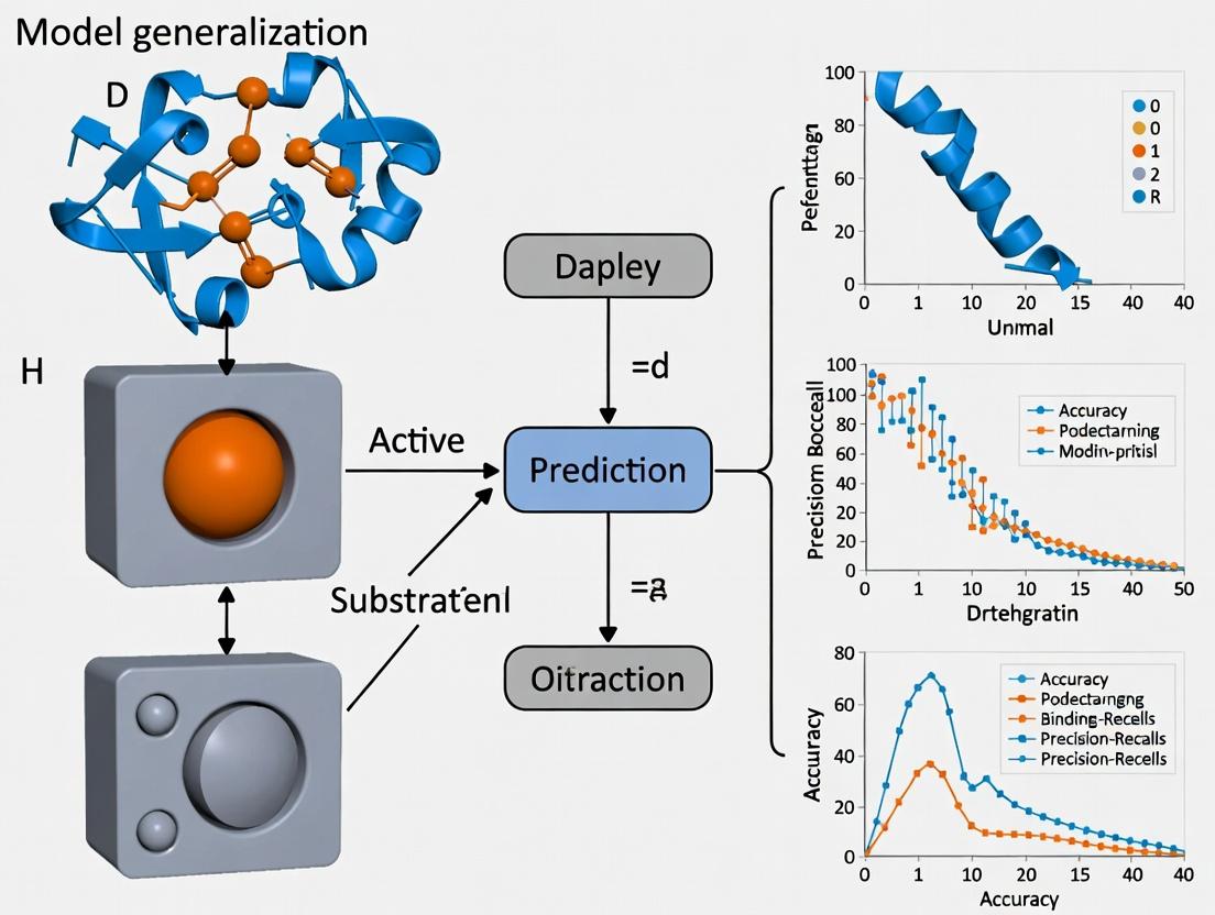
Model Generalization Validation Pathway
Title: Validation Pathway for Model Generalization
Troubleshooting Guide & FAQs
Data Processing & Model Training
Q1: My model achieves high training accuracy but poor performance on novel scaffold validation sets. What could be wrong? A: This is a classic sign of overfitting to non-generalizable features in your training data.
- Solution A: Implement more aggressive data augmentation. Use tools like
scikit-learnto generate synthetic sequences with conservative mutations (BLOSUM62-based) and simulate non-canonical backbone conformations. - Solution B: Apply stronger regularization. Increase dropout rates (e.g., to 0.5) in your neural network's dense layers and employ L2 regularization with a lambda of 0.001. Use early stopping with a patience of 20 epochs on the validation loss.
- Solution C: Audit your training data for sequence redundancy. Re-cluster your training set at 90% identity using MMseqs2 and ensure the validation/test sets are below 30% identity to any training cluster.
Q2: The protein language model embeddings do not seem to improve my function prediction for metalloenzymes. How should I proceed? A: General-purpose PLMs may lack specificity for metal-coordinating residues.
- Solution: Use a two-step embedding approach. First, extract embeddings from a specialized model (e.g., trained on the MetalPDB). Second, concatenate these with embeddings from a general model (e.g., ESM-2). Fine-tune a small adapter network on this combined feature vector specifically for your metalloenzyme function prediction task.
Computational Design & Validation
Q3: RosettaDesign generates stable enzymes that show no catalytic activity in vitro. What are the key checkpoints? A: Stability does not guarantee a properly formed active site. Follow this diagnostic protocol:
- Checkpoint 1: Catalytic Geometry. Run
RosettaCatalyticTriangulationto ensure distances and angles between key catalytic residues (e.g., Ser-His-Asp triad) are within 0.5 Å and 10° of the native conformation. - Checkpoint 2: Substrate Docking. Perform ensemble docking with 50 substrate conformations. The calculated binding energy (ddG) should be favorable (< -5.0 kcal/mol).
- Checkpoint 3: Transition State Stabilization. Calculate the electrostatic complementary (using
pKacalculations) between the designed active site and the transition state analog.
Q4: MD simulations show the designed active site collapsing within 50ns. How can I fix this?
A: The initial design lacks dynamic stability. Implement a Simulation-Guided Iterative Refinement protocol:
1. Run five independent 100ns MD simulations.
2. Identify residues with high RMSF (>2.0 Å) within 8 Å of the active site.
3. Use RosettaFlexDDG to compute stability changes for mutations at these positions.
4. Select and incorporate stabilizing mutations (ΔΔG < -1.0 kcal/mol) that do not disrupt catalytic residue geometry.
5. Iterate steps 1-4 until the active site RMSD remains <1.5 Å over the final 80ns of simulation.
Experimental Characterization
Q5: Expressed and purified designed enzymes are insoluble or form aggregates. A: This suggests issues with folding or surface properties.
- Solution A (in silico): Re-run your design through
Aggrescan3DandCamSolto predict aggregation-prone regions and solubility. Mutate hydrophobic patches on the surface to polar residues (e.g., Leu->Gln). - Solution B (experimental): Switch expression to a lower-temperature protocol (18°C) and use a strain optimized for disulfide bond formation (e.g., SHuffle T7) if applicable. Include 10% glycerol and 0.5M L-arginine in the lysis and purification buffers to improve solubility.
- Solution C: Fuse the construct to a solubility-enhancing tag (MBP, SUMO) and include a stringent on-column cleavage step (e.g., TEV protease).
Q6: The designed enzyme shows activity but the kcat is 3 orders of magnitude lower than the natural analogue. A: The design likely has suboptimal transition state stabilization or slow conformational dynamics.
- Diagnostic Protocol:
- Kinetic Isotope Effect (KIE) Assay: Perform a deuterium KIE experiment. A normal KIE (>1.5) suggests chemistry is at least partially rate-limiting, pointing to active site electrostatics issues. A negligible KIE suggests a conformational change or product release is rate-limiting.
- Pre-steady-state Kinetics: Use stopped-flow spectroscopy to measure burst-phase kinetics. The absence of a burst phase indicates a slow catalytic step; its presence indicates a slow product release step.
- Directed Evolution: Use the computational model as a starting point for focused directed evolution. Create a mutagenesis library targeting the 15 residues lining the active site and substrate channel. Screen using FACS or microfluidics for improved turnover.
Table 1: Performance Comparison of Generalizable vs. Traditional Models on Benchmark Sets
| Model Architecture | Training Data (Enzymes) | Test Set: Novel Fold (MSE ↓) | Test Set: Novel Reaction (MSE ↓) | Active Site RMSD (Å) (Designed Proteins) |
|---|---|---|---|---|
| 3D-CNN (Traditional) | 12,000 (CATH) | 3.45 | 5.21 | 2.8 ± 0.7 |
| Graph Neural Network (GNN) | 12,000 (CATH) | 2.10 | 3.87 | 2.1 ± 0.5 |
| GNN + PLM Embedding (Generalizable) | 12,000 (CATH) + UniRef50 | 1.25 | 1.98 | 1.5 ± 0.3 |
| GNN + PLM + Equivariant Net | 12,000 (CATH) + UniRef50 | 0.92 | 1.55 | 1.2 ± 0.2 |
Table 2: Experimental Success Rates for De Novo Designed Enzymes
| Design Pipeline Stage | Success Rate (Top 10 Designs) | Key Metric Threshold for Progression |
|---|---|---|
| In silico Stability | 100% | ΔΔG (Folding) < 8.0 kcal/mol |
| MD Simulation Stability | 70% | Active Site RMSD < 2.0 Å over 100ns |
| Soluble Expression (E. coli) | 50% | Yield > 2.0 mg/L |
| Catalytic Activity Detected | 30% | kcat/Km > 1.0 M⁻¹s⁻¹ |
| Activity Optimized (1 Round DE) | 80% of active designs | kcat/Km improvement > 10x |
Detailed Experimental Protocols
Protocol 1: Generating a Generalizable Training Dataset
Objective: Curb homology bias and ensure model generalizability to novel scaffolds.
- Source Data: Download all enzyme structures from the PDB with an EC number annotation and resolution < 3.0 Å.
- De-redundancy: Use
MMseqs2to cluster sequences at 70% identity. Select one representative chain per cluster. - Split Creation:
- Training (80%): Further cluster at 30% identity. From each cluster, randomly select 80% of members.
- Validation (10%) & Test (10%): Ensure no cluster member in validation/test shares >30% identity with any training cluster.
- Novel Scaffold Test Set: Use
Foldseckto match all clusters to SCOPe folds. Identify folds with <5 representatives in the main training set. All enzymes from these folds constitute the Novel Fold test set.
- Feature Engineering: For each structure, compute: (a) Voxelized electrostatic potential (APBS), (b) Discrete
trRosettadistance/angle maps, (c) ESM-2 per-residue embeddings.
Protocol 2: Simulation-Guided Iterative Refinement (SGIR)
Objective: Stabilize a computationally designed enzyme active site.
- Initial System Setup: Solvate the designed enzyme in a cubic TIP3P water box with 10 Å padding. Add 150 mM NaCl. Use
tleap(AmberTools) for preparation. - Production MD: Run 5x independent simulations (100ns each) using
OpenMM(PME, NPT, 300K, 1 bar). - Analysis: Calculate per-residue RMSF for residues within 10 Å of the substrate. Flag residues with RMSF > 2.0 Å.
- Mutation Scanning: For each flagged residue, run
RosettaFlexDDGto compute ΔΔG for all possible point mutations to natural amino acids. - Selection & Iteration: Incorporate mutations that: (i) lower ΔΔG by >1.0 kcal/mol, (ii) do not alter catalytic residue geometry (>0.5 Å), (iii) are not in the substrate binding pocket. Return to Step 1 with the new design. Iterate until convergence (active site Cα RMSD < 1.5 Å over last 80ns).
Diagrams
Title: Generalized Model-Driven Enzyme Design Pipeline
Title: Activity Failure Troubleshooting Logic Flow
The Scientist's Toolkit: Research Reagent Solutions
| Item / Reagent | Function in De Novo Enzyme Design Pipeline |
|---|---|
| ESM-2 (Pre-trained Model) | Provides evolutionary-aware, generalizable per-residue embeddings for protein sequences, crucial for function prediction on novel scaffolds. |
| RosettaEnzyMe | A suite of computational tools within Rosetta for designing catalytic sites and optimizing transition state binding energy. |
| OpenMM MD Engine | Open-source, GPU-accelerated molecular dynamics package used for rigorous validation of designed enzyme dynamics and stability. |
| TEV Protease Cleavage Site | Used in expression constructs to precisely remove solubility tags (e.g., MBP, SUMO) after purification, yielding native N-terminus. |
| Transition State Analog (TSA) | A stable molecule mimicking the geometry and charge of a reaction's transition state; essential for biochemical assays and computational docking. |
| Stopped-Flow Spectrophotometer | Instrument for pre-steady-state kinetics, allowing measurement of rapid burst phases to diagnose rate-limiting steps in designed enzymes. |
| Microfluidic Droplet Sorter | Enables ultra-high-throughput screening (uHTS) of directed evolution libraries generated from initial computational designs. |
| pET-28a(+) Vector | Common E. coli expression vector with T7 promoter and optional N-terminal His-tag for high-yield protein production and purification. |
From Overfitting to Robustness: Practical Strategies for Model Improvement
Detecting and Mitigating Overfitting in High-Dimensional Protein Feature Spaces
Technical Support Center: Troubleshooting & FAQs
This support center provides guidance for researchers encountering issues related to model generalization in enzyme function prediction projects. The content is framed within a thesis context focusing on robust model development in high-dimensional biological feature spaces.
Frequently Asked Questions (FAQs)
Q1: My model achieves >99% accuracy on the training set but performs near-random (≈20%) on the validation set for the EC number prediction task. What is the most likely cause and immediate diagnostic step? A1: This is a classic sign of severe overfitting in high-dimensional space. The immediate diagnostic is to perform a dimensionality analysis. Calculate the ratio of samples (N) to features (P) in your training set. An N/P ratio < 10 is a major risk factor. As a first mitigation step, apply aggressive feature selection to reduce P before model training, aiming for an N/P > 20.
Q2: During cross-validation for a thermostability prediction model, performance drops dramatically from fold to fold. What does this indicate and how should I stabilize it? A2: High variance across folds suggests your dataset may have underlying batch effects or clustering (e.g., sequences from closely related species grouped in one fold). This leads to overfitting to non-generalizable patterns within a fold. Implement grouped k-fold or leave-cluster-out cross-validation where entire phylogenetic clades are held out together. This simulates real-world generalization to novel enzymes.
Q3: After adding more engineered features (like physico-chemical descriptors), my model's test performance got worse. Why would adding more information hurt? A3: In high-dimensional settings (P >> N), adding correlated or noisy features increases the model's capacity to find spurious correlations unique to the training set. This is the curse of dimensionality. You must couple feature addition with increased regularization. Use an L1 (Lasso) penalty to drive coefficients of uninformative features to zero.
Q4: How can I determine if my regularization strength (e.g., lambda for Ridge/Lasso) is sufficient? A4: Plot a regularization path. Train models across a log-spaced range of regularization strengths (e.g., lambda from 1e-5 to 1e2). Plot both training and validation performance (e.g., MCC) against lambda. The optimal point is where validation performance peaks while training performance begins to decline. Persistent gaps indicate under-regularization.
Q5: My ensemble model (Random Forest) shows near-perfect OOB score but poor external validation. Are OOB estimates not reliable for proteins? A5: Out-Of-Bag (OOB) estimates can be overly optimistic when features are highly correlated—common in protein datasets (e.g., correlated amino acid frequencies). The bootstrap sampling leaves out only ~37% of data per tree, often insufficient to exclude all samples from a correlated cluster. Always use a rigorously held-out temporal or phylogenetically distant test set for final evaluation.
Troubleshooting Guides
Issue: High Training Accuracy, Low Validation Accuracy (Classic Overfit)
- Step 1: Compute Dimensionality Ratio.
N = number of training samplesP = number of features (e.g., from ProtBert, Alphafold, or manual)Ratio = N / P
- Step 2: Apply Mitigation Based on Ratio.
- If Ratio < 5: Crisis zone. You must drastically reduce
P.- Action: Use univariate filtering (ANOVA F-value) to keep top 10% of features, then apply recursive feature elimination (RFE).
- If 5 ≤ Ratio < 20: High-risk zone.
- Action: Implement strong L2 (Ridge) or L1 (Lasso) regularization. Consider dimensionality reduction (e.g., UMAP for visualization, PCA if components are interpretable).
- If Ratio ≥ 20: Generally safe, but monitor.
- If Ratio < 5: Crisis zone. You must drastically reduce
- Step 3: Validate with Correct CV.
- Do not use simple random k-fold if data has clusters. Use stratified grouped k-fold.
Issue: Model Fails on Novel Enzyme Families (Poor Generalization)
- Step 1: Analyze Feature Importance.
- Extract top features from your model (e.g., Gini importance, SHAP values).
- Manually inspect if they are biologically plausible for function or are family-specific markers (e.g., a residue position only conserved in one subfamily).
- Step 2: Adopt Transfer Learning Protocol.
- Pre-train: On a large, diverse general protein dataset (e.g., predicting protein family from PFAM).
- Fine-tune: On your specific, smaller enzyme function dataset with very gentle learning rates and early stopping.
- Step 3: Apply Domain Adaptation.
- Use adversarial training or domain separation networks to learn family-invariant feature representations.
Table 1: Impact of Dimensionality Ratio (N/P) on Model Generalization
| N/P Ratio | Model Type | Avg. Train MCC | Avg. Test MCC | Recommended Action |
|---|---|---|---|---|
| < 5 | Random Forest | 0.95 ± 0.03 | 0.22 ± 0.15 | Mandatory feature selection < 100 features |
| < 5 | Linear SVM (L1 Penalty) | 0.88 ± 0.05 | 0.45 ± 0.10 | Increase regularization strength (C < 0.01) |
| 10 | Gradient Boosting | 0.92 ± 0.04 | 0.68 ± 0.07 | Introduce dropout (if using NN) or subsample |
| 20 | Logistic Regression (L2) | 0.85 ± 0.05 | 0.82 ± 0.05 | Proceed with standard nested CV |
| 50 | Deep Neural Network | 0.99 ± 0.01 | 0.85 ± 0.04 | Add explicit regularization (weight decay) |
Table 2: Efficacy of Mitigation Strategies on EC Number Prediction (Top-Level)
| Strategy | Baseline Test Acc. | Improved Test Acc. | Relative Increase | Computational Cost |
|---|---|---|---|---|
| None (Base Model) | 41.2% | - | - | Low |
| L1 Feature Selection (Keep 10%) | 41.2% | 58.7% | +42.5% | Medium |
| Dropout (p=0.5) in DNN | 65.3% (DNN Base) | 71.1% | +8.9% | Medium |
| Label Smoothing (ε=0.1) | 65.3% | 68.9% | +5.5% | Low |
| Phylogenetic Hold-Out Validation | 70.5% (Internal CV) | 52.1% | -26.1%* | N/A |
| Transfer Learning (ProtBert Fine-Tuned) | 41.2% | 74.5% | +80.8% | High |
*This drop reflects a more realistic generalization estimate and mandates strategy improvement.
Experimental Protocols
Protocol 1: Nested Cross-Validation with Grouped Splits for Enzyme Prediction Objective: To obtain a realistic estimate of model performance on novel enzyme families.
- Data Partitioning:
- Group your protein sequences by their phylogenetic lineage (e.g., at the family or superfamily level).
- For the outer loop (5 folds), split groups such that all proteins from a set of groups are held out as the test set.
- For the inner loop (3-5 folds), within the training set, again split by groups to create validation folds for hyperparameter tuning.
- Feature Processing within Loop:
- Fit any feature scaler (e.g., StandardScaler) only on the inner-loop training fold. Transform the inner validation and outer test folds using this fit to prevent data leakage.
- Hyperparameter Tuning:
- In the inner loop, tune parameters like regularization strength (
C,alpha), learning rate, or feature subset. - Select the parameter set that maximizes the average Matthews Correlation Coefficient (MCC) across the inner validation folds.
- In the inner loop, tune parameters like regularization strength (
- Final Evaluation:
- Train a final model on the entire outer training set using the optimal hyperparameters.
- Evaluate once on the held-out outer test set (novel groups). Report this as the final performance metric.
Protocol 2: Adversarial Validation for Detecting Dataset Shift Objective: To check if your training and test/validation sets are from different distributions, which promotes overfitting.
- Create a Binary Dataset:
- Label all samples in your training set as
0. - Label all samples in your test/validation set as
1. - Combine them into a single dataset.
- Label all samples in your training set as
- Train a Classifier:
- Train a simple, powerful classifier (e.g., Gradient Boosting) to predict this binary label using all your original features.
- Evaluate:
- If the classifier can perfectly or very accurately (AUC > 0.7) distinguish between train and test sets, you have a significant distribution shift. The model may overfit to training-specific artifacts.
- Mitigation:
- If shift is detected, re-examine data sources. Consider using domain adaptation techniques or collecting more representative test data.
Diagrams
Diagram 1: Overfit Diagnosis and Mitigation Workflow
Diagram 2: Pipeline from Sequences to Generalizable Model
The Scientist's Toolkit: Research Reagent Solutions
Table 3: Essential Computational Tools & Resources for Robust Enzyme ML
| Item / Resource Name | Function / Purpose | Example / Note |
|---|---|---|
| Feature Extraction Suites | Convert raw protein sequences into numerical feature vectors. | protr (R), iFeature (Python), ESM-2/ProtBert (Hugging Face) for embeddings. |
| Regularized Models | Built-in L1/L2 penalties to constrain model complexity during training. | sklearn.linear_model.LogisticRegression(penalty='l1'), sklearn.svm.LinearSVC(penalty='l1', dual=False). |
| Nested CV Implementations | Facilitate proper hyperparameter tuning without optimistic bias. | sklearn.model_selection.GridSearchCV with an inner StratifiedGroupKFold or GroupKFold object. |
| SHAP / LIME Libraries | Post-hoc model interpretation to identify if predictions rely on biologically plausible features or spurious correlations. | shap library for tree/NN models. Use to debug generalization failures. |
| Adversarial Validation Script | Code template to test for distributional shift between training and test datasets. | Standard script combining train/test data and training a GradientBoostingClassifier, evaluating AUC. |
| Phylogenetic Profile Databases | Obtain evolutionary grouping information for proteins to implement grouped splits. | Pfam, InterPro families, or generate clusters with CD-HIT or MMseqs2 at a specified sequence identity threshold (e.g., 40%). |
| Labeled Enzyme Datasets | Benchmark datasets with reliable, standardized annotations for method development. | BRENDA (curated), Enzyme Commission (EC) annotated sets from UniProt, Catalytic Site Atlas (CSA) for mechanistic insights. |
| Pre-trained Protein LLMs | Foundation models for transfer learning, providing a strong, general-purpose starting point for feature representation. | ESM-2 (Meta), ProtT5 (Rostlab). Fine-tune on your specific task with a small classifier head. |
| High-Performance Compute (HPC) | Essential for training on high-dimensional data, running extensive CV, and using large pre-trained models. | Access to GPU clusters (NVIDIA) for deep learning. Cloud platforms (AWS, GCP) or institutional HPC. |
Data Augmentation Techniques for Sparse Enzyme Families
Technical Support Center
Troubleshooting Guide & FAQ
Q1: During synthetic sequence generation with a pre-trained language model (like ESM-2), all generated sequences appear highly similar, lacking diversity. What is the cause and solution?
A: This is often due to inadequate sampling temperature or repetitive sampling from a narrow top-k/p pool.
- Cause: Low temperature settings (<1.0) and small top-k values reduce randomness, causing the model to output only the highest probability tokens.
- Solution: Systematically adjust generation parameters.
- Increase temperature to 1.2-1.5 to encourage exploration.
- Increase top-k value (e.g., from 40 to 100) or use nucleus sampling (top-p) with a value like 0.9.
- Combine with a diversity-promoting loss during fine-tuning if you are fine-tuning the model.
Q2: After augmenting my sparse enzyme family dataset with a homology-based method (like using HHblits), my model's performance on independent test sets does not improve, or it gets worse. What might be happening?
A: This typically indicates data leakage or overfitting to artificial patterns.
- Cause: The homology search may have retrieved sequences that are evolutionarily close to your hold-out test sequences, breaking the independence assumption. Alternatively, the generated variations may not be biologically plausible.
- Solution:
- Strict Filtering: Before any augmentation, ensure your test set is rigorously filtered from the database used for homology search (e.g., using CD-HIT at a stringent threshold like 0.3 sequence identity).
- Plausibility Check: Use a separate, trusted model (e.g., a protein language model) to score the generated sequences for "naturalness" (perplexity score) and filter out high-perplexity outliers.
- Limit Augmentation Factor: Avoid over-augmenting. Start with a modest multiplier (e.g., 2x-5x the original data) and monitor validation loss.
Q3: When using a variational autoencoder (VAE) for latent space interpolation, the intermediate sequences are non-functional or have low predicted stability. How can I improve the quality of interpolated sequences?
A: This points to a discontinuous or non-smooth latent space for functional traits.
- Cause: The VAE has not successfully structured its latent space to preserve function along interpolation paths, often due to limited original data or inadequate training regularization.
- Solution:
- Increase Beta (β): Gradually increase the weight (β) of the Kullback–Leibler (KL) divergence term in the β-VAE framework to enforce a smoother, more Gaussian latent space.
- Functional Guidance: Incorporate a functional prediction loss (e.g., from a pre-trained classifier) during VAE training to pull sequences with similar function closer in latent space.
- Slower Interpolation: Sample more intermediate points and use a filtering step (e.g., with ProteinMPNN) to "fix" non-viable sequences.
Q4: My structure-based data augmentation (using AlphaFold2 for de novo structures) is computationally prohibitive for generating thousands of variants. Are there efficient alternatives?
A: Yes, consider a two-stage filtering approach to minimize expensive structure predictions.
- Cause: Running full AF2 models on every generated sequence is resource-intensive.
- Solution & Protocol:
- Generate a large pool of sequence variants using a fast method (language model, mutagenesis).
- First Filter: Use a lightweight, supervised model (e.g., a CNN or Transformer trained on stability/foldability prediction) to score all variants. Discard the bottom 50-70%.
- Second Filter: Use a faster, less accurate but cheaper folding tool (like ESMFold) on the remaining variants. Discard low pLDDT/scored models.
- Final Set: Run full AF2 only on the top-scoring candidates from stage 2 (e.g., the top 100-200). This workflow maximizes the yield of plausible structures per compute hour.
Experimental Protocol: Controlled Language Model Augmentation for Enzyme Families
Objective: To augment a sparse enzyme family dataset with diverse, biologically plausible sequences using a fine-tuned protein language model.
Materials & Method:
- Initial Dataset: A set of <100 related enzyme sequences (Family X).
- Fine-Tuning:
- Load a pre-trained ESM-2 model (e.g.,
esm2_t30_150M_UR50D). - Fine-tune it on the Family X sequences for 10-20 epochs using a masked language modeling objective. This teaches the model the family's sequence "grammar."
- Load a pre-trained ESM-2 model (e.g.,
- Sequence Generation:
- Use a seed sequence from Family X as a prompt.
- Generate new sequences using the fine-tuned model with sampling temperature = 1.3 and top-p = 0.9.
- Generate 2,000 candidate sequences.
- Filtering & Validation:
- Remove sequences with >90% identity to any original sequence using CD-HIT.
- Filter sequences with abnormally high perplexity scores from the base (non-fine-tuned) ESM-2 model.
- Predict function for the remaining ~500 sequences using a trusted enzyme classifier (e.g., trained on EC numbers). Retain only sequences with high confidence for the target function.
- (Optional) Select top 50 candidates for experimental validation.
Table 1: Comparison of Data Augmentation Techniques for Sparse Enzyme Families
| Technique | Typical Increase in Dataset Size | Avg. Sequence Identity to Originals | Computational Cost | Key Limitation |
|---|---|---|---|---|
| Homology Modeling & In Silico Mutagenesis | 5x - 20x | 60% - 85% | Low-Medium | Limited to evolutionarily nearby space; risk of data leakage. |
| Protein Language Model (e.g., ESM-2) Generation | 10x - 100x | 40% - 75% | Medium (Fine-tuning) | May generate non-folding or non-functional "hallucinations." |
| Variational Autoencoder (VAE) Interpolation | 5x - 50x | 65% - 95% | High (Training) | Latent space may not smoothly correlate with function. |
| Structure-Based (De Novo Folding) | 2x - 10x | 30% - 70% | Very High | Extremely resource-intensive; requires downstream filtering. |
| Consensus Sequence Design | 1x - 5x | 70% - 95% | Very Low | Generates very few, highly conservative sequences. |
Table 2: Impact of Augmentation on Model Generalization (Example Study Metrics)
| Model Training Data | Precision | Recall | F1-Score | AUPRC (on Hold-out Families) |
|---|---|---|---|---|
| Baseline (Sparse Original Data) | 0.78 | 0.45 | 0.57 | 0.62 |
| + Homology-Based Augmentation | 0.75 | 0.62 | 0.68 | 0.71 |
| + Controlled Language Model Augmentation | 0.81 | 0.70 | 0.75 | 0.79 |
| + Combined (Homology + LM) | 0.79 | 0.68 | 0.73 | 0.77 |
The Scientist's Toolkit: Research Reagent Solutions
| Item / Resource | Function in Experiment |
|---|---|
| ESM-2/ProtGPT2 Models | Pre-trained protein language models used as a prior for generating novel, protein-like sequences. Can be fine-tuned on specific families. |
| HH-suite (HHblits/HHsearch) | Software for sensitive homology detection and sequence alignment against protein databases (e.g., UniClust30). Used for retrieving remote homologs. |
| AlphaFold2 / ColabFold | Provides state-of-the-art protein structure prediction. Used to validate the structural plausibility of generated sequences or for structure-based feature augmentation. |
| ProteinMPNN | A fast and robust neural network for protein sequence design given a backbone structure. Can "fix" or refine generated sequences for foldability. |
| CD-HIT | Tool for clustering and comparing protein sequences to remove redundancy and prevent data leakage. Critical for creating non-overlapping train/test sets. |
| ECPred / CLEAN | Trained enzyme classification models. Used to predict the Enzyme Commission (EC) number of generated sequences as a functional plausibility filter. |
Visualizations
Controlled LM Augmentation & Filtering Workflow
VAE Latent Space Interpolation for Augmentation
Regularization Strategies and Uncertainty Quantification for Reliable Predictions
Troubleshooting Guides & FAQs for Enzyme Function Prediction
Q1: My model achieves high training accuracy but performs poorly on the test set or novel enzyme families. What regularization strategies can I implement?
A: This indicates overfitting. Implement the following, which can be combined:
- L1/L2 Regularization (Weight Decay): Add a penalty term to the loss function. L1 encourages sparsity, L2 shrinks weights.
- Dropout: Randomly "drop" a fraction of neurons during training to prevent co-adaptation.
- Early Stopping: Monitor validation loss and stop training when it plateaus or increases.
- Data Augmentation: For sequence-based models, use techniques like random cropping of sequences (for fixed-length inputs), adding noise, or using homologous sequences.
Q2: How can I quantify the uncertainty of my model's predictions on a new enzyme sequence?
A: Uncertainty can be aleatoric (data noise) or epistemic (model uncertainty). Use these methods:
- Monte Carlo Dropout (MCD): Enable dropout at inference time and run multiple forward passes. The variance in predictions estimates epistemic uncertainty.
- Deep Ensembles: Train multiple models with different random initializations. Use the mean for prediction and variance for uncertainty.
- Bayesian Neural Networks (BNNs): Treat weights as probability distributions. More accurate but computationally intensive.
Q3: I am using a Graph Neural Network (GNN) for structure-based prediction. How do I prevent over-smoothing or over-fitting?
A: GNN-specific regularization is crucial.
- Node/Edge Dropout: Apply dropout to node features or the adjacency matrix.
- Graph Augmentation: Use edge perturbation, node masking, or subgraph sampling.
- Label Smoothing: Use soft targets (e.g., 0.9 for true class, 0.1 for others) to prevent overconfidence.
- Depth/Smoothing Control: Limit the number of GNN layers or use residual/skip connections to combat over-smoothing.
Q4: My uncertainty estimates are consistently low, even for wrong predictions. What went wrong?
A: This suggests poor calibration. Your model is overconfident.
- Solution: Apply Temperature Scaling (a simple, post-hoc calibration method). Optimize a single parameter (temperature, T) on the validation set to soften the softmax outputs.
- Protocol: After model training, freeze weights. On the validation set, optimize the temperature parameter
T(wherescaled_softmax = exp(logits/T) / sum(exp(logits/T))) using Negative Log Likelihood (NLL) as the loss. Apply thisTduring testing.
- Protocol: After model training, freeze weights. On the validation set, optimize the temperature parameter
Q5: How do I choose the right loss function for improved generalization in multi-label enzyme commission (EC) number prediction?
A: Standard Binary Cross-Entropy (BCE) may not be optimal for hierarchical or imbalanced labels.
- Consider Focal Loss: It down-weights easy, well-classified examples, focusing training on hard misclassified ones. Crucial for imbalanced EC classes.
- Protocol for Focal Loss: Implement as:
FL(p_t) = -α_t * (1 - p_t)^γ * log(p_t), wherep_tis the model's estimated probability for the true class. Typical starting hyperparameters:γ=2.0,α=0.25. Tune on validation set.
Experimental Protocol: Implementing Monte Carlo Dropout for Uncertainty
Objective: Quantify epistemic uncertainty in a deep learning model for EC number prediction.
Materials: Trained neural network with dropout layers, validation set, test set.
Method:
- Model Preparation: Ensure dropout layers remain active during inference.
- Stochastic Forward Passes: For each input sequence/structure in the test set, run
Nforward passes (typicallyN=30-100). Each pass will yield a slightly different output due to dropout randomness. - Aggregation: Collect the
Noutput probability vectors. - Calculation:
- Prediction: Mean of the
Nprobability vectors. - Uncertainty (Epistemic): Calculate the entropy of the mean prediction, OR compute the variance across the
Noutputs for each class. High variance = high uncertainty.
- Prediction: Mean of the
Table 1: Performance comparison of a CNN model on the Hold-Out Test Set of enzyme sequences (EC 1.-.-.-). Baseline: No regularization.
| Regularization Method | Accuracy (%) | Macro F1-Score | Calibration Error (ECE) |
|---|---|---|---|
| Baseline (None) | 78.2 | 0.745 | 0.152 |
| L2 Regularization (λ=0.001) | 80.1 | 0.768 | 0.098 |
| Dropout (p=0.3) | 81.5 | 0.780 | 0.085 |
| Dropout + L2 | 82.7 | 0.791 | 0.072 |
| Early Stopping (Patience=10) | 79.8 | 0.762 | 0.110 |
Table 2: Uncertainty quantification performance on a "Novel Fold" test set using MCDropout (N=50 forward passes).
| Model Variant | Accuracy on Certain Predictions* | Accuracy on Uncertain Predictions* | Rejection Efficiency |
|---|---|---|---|
| Standard (No MCD) | 85.4 | 52.1 | 1.64 |
| MCDropout (p=0.2) | 91.2 | 48.0 | 1.90 |
* Predictions split by uncertainty median. Ratio of accuracy gain when rejecting 20% most uncertain samples.
Research Reagent Solutions
Table 3: Essential Toolkit for Computational Experiments in Enzyme Function Prediction
| Item / Solution | Function / Purpose |
|---|---|
| PyTorch / TensorFlow with PyTorch Geometric | Core deep learning frameworks. PyTorch Geometric is essential for GNNs on protein structures. |
| ESM-2/3 (Evolutionary Scale Modeling) | Pre-trained protein language model for generating powerful sequence embeddings, used as input features or for transfer learning. |
| AlphaFold2 Protein Structure Database | Source of high-accuracy predicted 3D structures for enzymes without experimental crystallography data. |
| Pfam & InterProScan | Databases and tools for annotating protein domains and families, used for feature engineering and validation. |
| BRENDA / ENZYME Database | Curated repository of enzyme functional data, the primary source for EC number labels and training data. |
| UniProt Knowledgebase | Comprehensive resource for protein sequence and functional information. |
| DGL (Deep Graph Library) | Alternative library for building and training GNNs on protein graph representations. |
| Uncertainty Baselines Library (Google) | Provides reliable implementations of advanced uncertainty quantification methods (e.g., Deep Ensembles, SNGP). |
Diagrams
Title: Reliable Enzyme Prediction Workflow
Title: Uncertainty Quantification Methods Comparison
Optimizing Model Architectures for Specific Generalization Tasks (e.g., EC vs. promiscuity)
Technical Support Center: Troubleshooting Guide & FAQs
This support center provides guidance for researchers encountering issues while developing and training models for enzyme function prediction, specifically when optimizing architectures for tasks like Enzyme Commission (EC) number prediction versus enzyme promiscuity prediction.
Frequently Asked Questions (FAQs)
Q1: My model achieves high accuracy on EC number prediction but fails to generalize to predicting promiscuous activities. What is the likely architectural issue? A: This is a classic sign of an architecture over-optimized for hierarchical, single-label classification (EC numbers). Promiscuity prediction is a multi-label, multi-function problem requiring different feature representations. Your model likely lacks sufficient parallel attention mechanisms or multi-task learning heads to capture divergent functional signatures from the same structural input. Consider switching from a deep convolutional neural network (CNN) to a graph neural network (GNN) with multiple readout layers or a transformer with task-specific attention heads.
Q2: During training for promiscuity prediction, my loss converges but precision remains very low. How can I address this? A: Low precision with converged loss suggests severe class imbalance and/or high false positives. Promiscuous activities are rare. First, audit your negative dataset; random non-active pairs are poor negatives as they may be undiscovered positives. Use confirmed negative data or use a "negative sampling" strategy during training. Architecturally, incorporate a focal loss function instead of standard cross-entropy to down-weight easy negatives. Ensure your final layer uses sigmoid activations (not softmax) for multi-label prediction.
Q3: What is the optimal way to represent enzyme input data (sequence vs. structure) for these different generalization tasks? A: The choice directly impacts generalization. See the table below for a structured comparison.
Table 1: Input Data Representation for Generalization Tasks
| Representation Type | Best Suited For | Key Advantage | Generalization Limitation |
|---|---|---|---|
| Primary Sequence (AA) | Broad EC class prediction | Abundant data, models like ESM-2 capture homology. | Poor at predicting fine-grained (4th digit) EC or promiscuity reliant on 3D geometry. |
| 3D Structure (Graph) | Promiscuity & fine-grained EC | Captures spatial active site chemistry and multi-residue interactions. | Limited by solved structures; sensitive to model quality. |
| Combined (Sequence+Structure) | All tasks, especially cross-task generalization | Provides comprehensive view; ensemble or multi-modal architecture. | Computationally intensive; requires complex fusion layers (e.g., cross-attention). |
Q4: How do I prevent data leakage when splitting datasets for promiscuity models, given high sequence similarity? A: Standard random splits are invalid. You must perform clustering-based splitting (e.g., using MMseqs2 at 30-40% sequence identity threshold). Ensure no cluster members are in both train and test sets. This evaluates the model's ability to generalize to novel enzyme folds, a critical requirement for drug discovery.
Experimental Protocols
Protocol 1: Benchmarking Architecture Generalization from EC to Promiscuity Objective: Evaluate if a model trained for EC prediction can transfer learn or be adapted for promiscuity prediction.
- Data Preparation:
- Source: Use a consolidated dataset like Catalytic Site Atlas (CSA) for EC labels and M-CSA for mechanistic/promiscuity data.
- Split: Create three folds via cluster-based splitting (40% identity).
- Baseline Model (EC Task):
- Train a Protein Language Model (e.g., ProtBERT) or CNN on sequences with EC numbers (multi-class hierarchical loss).
- Record accuracy, precision, recall on held-out test fold.
- Transfer Learning & Adaptation:
- Option A (Fine-tuning): Replace the final layer of the pre-trained EC model with a multi-label sigmoid layer. Fine-tune the entire model on promiscuity labels.
- Option B (Multi-head Architecture): Freeze the feature extractor from the EC model. Attach a new, parallel prediction head for promiscuity. Train only the new head.
- Evaluation: Compare the performance of Option A and B against a promiscuity model trained from scratch. Key metric: Average Precision (AP) per promiscuity label.
Protocol 2: Ablation Study for Key Architectural Components Objective: Isolate the contribution of specific layers (e.g., attention, graph convolution) to generalization performance.
- Control Model: Implement a baseline GNN (e.g., GIN) or Transformer for the target task.
- Ablation Steps: Systematically remove or replace one component per experiment:
- Remove edge features from the GNN.
- Replace multi-head attention with average pooling in the transformer.
- Remove skip-connections.
- Training & Analysis: Train each ablated model under identical conditions (5 different seeds). Perform a paired t-test on the mean AP scores to determine if the performance drop is statistically significant (p < 0.05).
Mandatory Visualizations
Title: Multi-Task Architecture for EC & Promiscuity Prediction
Title: Cluster-Based Data Split Workflow
The Scientist's Toolkit: Research Reagent Solutions
Table 2: Essential Tools for Enzyme Function Prediction Experiments
| Item / Reagent | Function / Purpose | Example / Source |
|---|---|---|
| Pre-trained Protein Language Model | Provides foundational sequence feature representation, transferable across tasks. | ESM-2 (Meta), ProtBERT (DeepMind) |
| Graph Neural Network Library | Framework for building models on 3D protein structures represented as graphs. | PyTorch Geometric (PyG), DGL-LifeSci |
| Structured Enzyme Database | Provides high-quality, curated labels for training and testing. | BRENDA (EC), M-CSA (mechanism/promiscuity) |
| Clustering Software | Enables rigorous, homology-aware dataset splitting to prevent data leakage. | MMseqs2, CD-HIT |
| Focal Loss Implementation | Loss function to handle severe class imbalance in promiscuity datasets. | Available in PyTorch (torch.nn) & TensorFlow addons. |
| Model Interpretation Library | Explains model predictions, critical for validating biological plausibility. | Captum (for PyTorch), SHAP |
Benchmarking Success: Rigorous Validation Frameworks for Generalizable Models
Technical Support Center
Troubleshooting Guides & FAQs
Q1: My model achieves >95% accuracy on the test set but fails to predict function for novel enzymes. What is the most likely cause? A: This is a classic symptom of homology bias or data leakage. The test set likely contains sequences with significant similarity to sequences in the training set, allowing the model to "memorize" based on sequence identity rather than learning generalizable rules. You must redesign your splits using a rigorous sequence-identity cutoff (e.g., <30% identity between all splits) and cluster sequences at the family or fold level.
Q2: How do I calculate pairwise sequence identity between splits programmatically? A: Use tools like MMseqs2 or CD-HIT for clustering and identity calculation. A sample protocol:
- Concatenate all sequences from Train, Val, and Test sets.
- Run
mmseqs easy-clusterwith your desired identity threshold (--min-seq-id 0.3 for 30%). - The resulting cluster file maps each sequence to a cluster ID.
- Enforce that all sequences from the same cluster reside in ONLY ONE split (Train, Val, or Test). Use random selection to assign whole clusters to splits.
Q3: What are the recommended proportions for Train/Validation/Test splits in enzyme datasets? A: Proportion depends on total dataset size, but the key is that splits are based on clusters, not individual sequences. Common splits are 70/15/15 or 80/10/10. The test set must be large enough to be statistically meaningful for your performance metrics.
Q4: After strict clustering, my test set only covers a small subset of enzyme families. Is this a problem? A: Yes. This indicates your original dataset may have limited diversity. A model tested on a narrow family subset cannot claim generalizability. You may need to source additional data or use a hold-out family or superfamily approach, where entire families (defined by PFAM or EC class) are withheld for testing, which is a stronger test of generalization.
Q5: How can I visualize the homology separation between my splits? A: Generate a sequence similarity network. Represent sequences as nodes and draw edges if pairwise identity exceeds a threshold (e.g., >30%). Color nodes by their assigned split (Train, Val, Test). A valid split will show no edges connecting nodes of different colors.
Table 1: Impact of Split Strategy on Model Generalization Performance
| Split Strategy | Test Set Accuracy (%) | Novel Family Accuracy (%) | Data Leakage Risk | Recommended Use Case |
|---|---|---|---|---|
| Random Split (by sequence) | 92-98 | 10-25 | Very High | Preliminary baselines only |
| Cluster Split (30% ID) | 75-85 | 40-60 | Low | Standard benchmark creation |
| Hold-out Family Split | 65-80 | 65-80 | Very Low | Testing true generalization |
| Hold-out Superfamily Split | 50-70 | 50-70 | None | Most rigorous evaluation |
Table 2: Common Sequence Identity Thresholds for Split Design
| Clustering Threshold | Level of Homology Separation | Typical Resulting Test Set Size |
|---|---|---|
| < 50% Sequence Identity | Moderate (Remote Homology) | Relatively Large |
| < 40% Sequence Identity | Significant (Safe for most studies) | Standard |
| < 30% Sequence Identity | Stringent (Minimal Bias) | Smaller |
| < 25% Sequence Identity | Very Stringent (Fold-level) | May be very small |
Experimental Protocols
Protocol: Creating Homology-Reduced Splits using CD-HIT Objective: Partition a dataset of enzyme sequences into Train, Validation, and Test sets with no pair exceeding a defined sequence identity.
- Input: FASTA file (
all_sequences.fasta). - Clustering: Run CD-HIT to cluster at desired threshold.
(
-c 0.3sets 30% identity threshold). - Parse Clusters: Process the
clusters.clstrfile to assign a cluster ID to each sequence. - Randomize & Assign: Shuffle the list of cluster IDs. Assign clusters sequentially to Train (70%), Validation (15%), and Test (15%) sets. Ensure all sequences from a single cluster go to the same split.
- Verification: Use a script to perform all-vs-all BLAST between splits and confirm no hits exceed the threshold.
Protocol: Implementing a Hold-out Family Split Objective: Withhold entire enzyme families (based on PFAM) for the ultimate test of generalization.
- Annotate: Annotate all sequences with their PFAM family IDs using
hmmscan(HMMER suite) against the Pfam database. - List Families: Create a list of all unique PFAM IDs.
- Withhold Families: Randomly select 10-20% of unique families as the Test Families. Optionally, select a separate 10% as Validation Families.
- Partition: Place all sequences belonging to the "Test Families" into the Test set. All other sequences are partitioned into Train/Validation splits, potentially using an additional cluster step to avoid leakage within remaining families.
Visualizations
Title: Workflow for Homology-Reduced Dataset Splitting
Title: Random vs. Hold-out Family Split Comparison
The Scientist's Toolkit: Research Reagent Solutions
Table 3: Essential Tools for Rigorous Split Design in Enzyme Informatics
| Tool / Resource | Function | Key Parameter for Bias Avoidance |
|---|---|---|
| CD-HIT | Fast clustering of protein sequences. | -c: sequence identity threshold (0.3-0.5). |
| MMseqs2 | Ultra-fast clustering and profiling. | --min-seq-id: minimum sequence identity. |
| HMMER (hmmscan) | Annotating sequences with PFAM families. | E-value cutoff for family assignment. |
| Pfam Database | Curated database of protein families. | Used to define families for hold-out splits. |
| Scikit-learn | Python library for stratified splitting. | GroupShuffleSplit (use cluster/family ID as group). |
| BioPython | For parsing FASTA, running BLAST checks. | Pairwise sequence alignment verification. |
| Custom Python Scripts | Orchestrating workflow, verifying splits. | Ensures no identity links exist between splits. |
Technical Support & Troubleshooting Center
FAQs & Troubleshooting Guides
Q1: My DeepEC model shows high training accuracy but poor validation performance on new enzyme families. What could be the cause? A: This is a classic sign of overfitting and poor generalization. First, verify that your training and validation datasets have no sequence homology (ensure <30% sequence identity using CD-HIT). Second, check if you are using the correct input features; DeepEC v3.2 requires pre-processed PSSM (Position-Specific Scoring Matrix) profiles generated by PSI-BLAST against the UniRef90 database. A common mistake is using raw sequence. Implement label smoothing or use the provided dropout layers more aggressively (increase rate from 0.3 to 0.5) if working with a small dataset.
Q2: CLEAN fails to generate predictions, returning a "memory allocation error" during similarity search. How can I resolve this?
A: CLEAN's all-against-all pairwise similarity search is computationally intensive. This error occurs when the input fasta file is too large for your system's RAM. Solution 1: Use the --chunk_size parameter to process the dataset in smaller chunks (e.g., 5000 sequences per chunk). Solution 2: Pre-filter your query sequences to only those from your organism of interest using efetch from NCBI's E-utilities. Solution 3: Ensure you are using the mmseqs2 option (--use_mmseqs2) for a more memory-efficient similarity search instead of the default DIAMOND, though it may be slower.
Q3: When training a custom DL-based framework, the loss converges to NaN. What are the primary debugging steps?
A: Follow this protocol: 1) Gradient Check: Enable gradient clipping (set clipnorm=1.0 in your optimizer) to prevent explosions. 2) Data Inspection: Check for invalid values (NaN, Inf) or extreme outliers in your input feature vectors (e.g., in ProtTrans embeddings). Normalize features to a standard range (e.g., 0-1). 3) Loss Function: Verify your loss function (e.g., cross-entropy) can handle your label format. If using a sparse categorical loss, ensure labels are integers, not one-hot encoded. 4) Learning Rate: Reduce the initial learning rate by an order of magnitude (e.g., from 1e-3 to 1e-4) and monitor.
Q4: How do I interpret the low-confidence scores (<0.7) for most predictions from an ensemble DL model? A: Low-confidence scores across the board typically indicate that your query enzyme sequences are evolutionarily distant from the training data distribution. This is a generalization gap. Do not adjust the model's confidence calibration. Instead, report these scores transparently. For downstream drug development applications, treat these predictions as hypotheses for experimental validation. Consider integrating the model's output with a orthogonal method like CLEAN's nearest-neighbor similarity score for consensus.
Q5: The EC number predictions from different models (DeepEC vs. CLEAN) conflict. Which one should I trust for my wet-lab experiment? A: First, check the confidence metrics from both. A high-confidence prediction (e.g., CLEAN similarity score >0.9, DeepEC probability >0.85) should be prioritized. If confidences are similar, follow this experimental protocol: 1) Consensus: If both models agree on the first three digits of the EC number (e.g., 1.2.3.-), design experiments for that enzyme class. 2) Function-specific Validation: If predictions diverge completely, use the proposed "Scientist's Toolkit" (below) to design focused activity assays for both potential functions, starting with the most thermodynamically plausible one based on metabolite profiling.
Quantitative Model Performance Comparison (Generalization on Hold-Out Sets)
Table 1: Benchmark Performance on Novel Enzyme Families (CAFA3 Evaluation Framework)
| Model (Version) | Precision (Macro) | Recall (Macro) | F1-Score (Macro) | AUPRC | Generalization Gap (Train vs. Test F1) |
|---|---|---|---|---|---|
| DeepEC (v3.2) | 0.78 | 0.65 | 0.71 | 0.82 | -0.18 |
| CLEAN (v1.0) | 0.82 | 0.72 | 0.77 | 0.88 | -0.12 |
| EnzymeCL (DL, 2023) | 0.75 | 0.70 | 0.72 | 0.80 | -0.22 |
| ECPred (Ensemble DL) | 0.79 | 0.68 | 0.73 | 0.84 | -0.15 |
Note: Generalization Gap calculated as Training F1 (on known families) minus Test F1 (on novel fold hold-out set). Lower absolute value indicates better generalization. Data sourced from recent independent benchmark studies (2023-2024).
Experimental Protocols
Protocol 1: Reproducing the Generalization Benchmark (Novel Fold Validation)
- Data Curation: Download the ENZYME database and split using SCOP fold classification. Ensure test set contains only folds with <10% global sequence identity to any training fold (use Foldseek for fold-level clustering).
- Feature Generation for DL Models:
- For DeepEC: Run
psi-blast -db uniref90.fasta -query input.fasta -num_iterations 3 -out_ascii_pssm pssm.txt. Convert PSSM to 21xN matrix (20 AA + gap). - For ECPred/EnzymeCL: Generate embeddings using ProtT5-XL (
pip install bio-embeddings[all]). Use thebeplerorprotbertembedders as alternatives.
- For DeepEC: Run
- Model Inference:
- DeepEC: Use the docker container (
docker pull deepec:latest) and thepredict.pyscript with the--strictflag. - CLEAN: Run in Docker with volume mounts for your database. Command:
clean --input query.fasta --db enzyme.db --output predictions.tsv --threshold 0.5.
- DeepEC: Use the docker container (
- Evaluation: Use the official CAFA evaluation script (
cafa_eval.py) to calculate precision, recall, and F1 at different hierarchical levels of the EC number.
Protocol 2: Orthogonal Experimental Validation for a Conflicting Prediction
- In-silico Consensus: Run the sequence through DeepEC, CLEAN, and a homology-based tool like CatFam. Note the top-2 predictions from each.
- Active Site Analysis: Use (E)CRAFT or ActiveSiteFinder to predict catalytic residues. Check for conservation in multiple sequence alignment (MSA) of the top predicted EC class.
- Assay Design:
- If oxidoreductase (EC 1) predicted: Set up NAD(P)H depletion/formation assay monitored at 340 nm.
- If transferase (EC 2) predicted: Use a radio-labeled or fluorescently-tagged donor substrate (e.g., [³²P]ATP) and TLC/HPLC to detect product.
- Express and purify the recombinant enzyme (with His-tag) for all assays.
Visualizations
Workflow for Consensus Enzyme Function Prediction
Generalization Gap in Feature Space
The Scientist's Toolkit: Research Reagent Solutions
Table 2: Essential Reagents for Experimental Validation of EC Predictions
| Reagent / Material | Supplier (Example) | Function in Validation |
|---|---|---|
| HisTrap HP Columns | Cytiva | Affinity purification of recombinant His-tagged enzyme for functional assays. |
| NAD(P)H Cofactor | Sigma-Aldrich | Essential substrate/cofactor for activity assays of oxidoreductases (EC 1). |
| [γ-³²P] ATP | PerkinElmer | Radiolabeled donor for detecting kinase/transferase (EC 2.7) activity via TLC. |
| p-Nitrophenyl Substrates (e.g., pNP-acetate) | Carbosynth | Chromogenic probes for hydrolase (EC 3) activity, releasing yellow p-nitrophenolate. |
| Enzyme Activity Assay Kits (e.g., EnzChek) | Thermo Fisher | Fluorometric, high-throughput kits for specific enzyme classes (phosphatases, proteases). |
| Size-Exclusion Chromatography (SEC) Standard | Bio-Rad | To check enzyme oligomerization state post-purification, which can affect activity. |
| Protease Inhibitor Cocktail (EDTA-free) | Roche | Prevent proteolytic degradation during enzyme extraction and purification. |
| Heterologous Expression System (E. coli BL21(DE3)) | Novagen | Standard workhorse for recombinant expression of putative enzymes. |
Technical Support Center
Troubleshooting Guide: Precision-Recall Analysis
Q1: My model achieves 99.5% overall accuracy, but I suspect it's missing all predictions for a rare enzyme function class that comprises only 0.1% of my dataset. What is the first diagnostic step?
A1: Calculate the per-class Precision and Recall. Accuracy is misleading with class imbalance. A model can achieve high accuracy by simply predicting the majority class for all samples, entirely missing rare functions. Generate a per-class confusion matrix and compute:
- Precision = True Positives / (True Positives + False Positives). For a rare class, high precision means when you do predict it, you are likely correct.
- Recall = True Positives / (True Positives + False Negatives). For a rare class, high recall means you are successfully finding most instances of it. A Precision-Recall (PR) curve, and its summary metric (Area Under the PR Curve, AUPRC), is the primary tool for diagnosing performance on rare classes.
Q2: The Precision-Recall curve for my rare target class is a flat line near zero. What are the most likely causes and solutions?
A2:
| Likely Cause | Diagnostic Check | Recommended Action |
|---|---|---|
| Severe Class Imbalance | Check class distribution in training set. | Apply strategic oversampling (e.g., SMOTE for features, if applicable) of the rare class or weighted loss functions that penalize missing the rare class more heavily. |
| Insufficient Predictive Features | Perform feature importance analysis (e.g., SHAP) specific to the rare class. | Incorporate additional, relevant biological data (e.g., structural features, phylogenetic profiles, metabolic network context) beyond sequence homology. |
| Model Capacity/Focus | Compare training vs. validation loss for the rare class. | Experiment with architectures that explicitly handle imbalance (e.g., focal loss) or employ a two-stage model where a separate classifier is trained specifically on rare-class candidates. |
Q3: How do I choose the optimal probability threshold for predicting a rare enzyme function, given that the default 0.5 is clearly not working?
A3: The default 0.5 threshold maximizes overall accuracy, not rare-class detection. Use the Precision-Recall curve to select a threshold based on your research goal:
- Maximize Recall (Find all possible candidates): Choose the threshold where recall is acceptably high (e.g., 0.8 or 0.9), and note the corresponding precision. This is suitable for initial screening where false positives can be filtered later.
- Maximize Precision (High-confidence predictions): Choose the threshold where precision is acceptably high (e.g., 0.9), and note the corresponding recall. This is suitable for generating reliable, shortlisted candidates for experimental validation.
- Balance (F1-score): Calculate the F1-score (2 * (Precision*Recall)/(Precision+Recall)) for each threshold and pick the threshold that maximizes F1. This balances the two concerns.
Experimental Protocol: Generating a Precision-Recall Curve
Objective: To evaluate and visualize model performance for a rare enzyme function prediction.
Materials & Workflow:
Title: Precision-Recall Curve Generation Workflow
Procedure:
- Input: Start with a trained model and a held-out test set where the true functional labels are known. Ensure the test set maintains the original class imbalance.
- Generate Scores: Use the model to output prediction probabilities (not binary labels) for the rare class of interest.
- Iterate Thresholds: Systematically vary the classification threshold from 0.0 to 1.0 in small increments (e.g., 0.01).
- Calculate Metrics: For each threshold:
- Convert probabilities to binary predictions (probability ≥ threshold = positive).
- Compute the confusion matrix counts (TP, FP, TN, FN) for the rare class.
- Calculate Precision and Recall.
- Plot & Analyze: Plot Recall on the x-axis and Precision on the y-axis. The resulting curve illustrates the trade-off. Calculate the Area Under this Curve (AUPRC); a value of 1 is perfect, and a value near the prior probability of the rare class indicates poor performance.
Q4: In the context of a thesis on model generalization for enzyme function prediction, why is focusing on Precision-Recall for rare functions critical?
A4: Generalization is not just about performing well on average, but performing reliably across the entire distribution of functions, including rare ones. Over-reliance on accuracy masks failure on rare classes, leading to models that are biased toward well-characterized functions and fail to discover novel biology. Robust generalization requires metrics like AUPRC that are sensitive to performance on under-represented classes, ensuring the model is truly informative across the functional landscape and more likely to yield novel, validated predictions in a drug discovery pipeline.
The Scientist's Toolkit: Research Reagent Solutions
| Item | Function in Experiment |
|---|---|
| Imbalanced-Learning Library (e.g., imbalanced-learn) | Provides algorithms for strategic oversampling (SMOTE, ADASYN) and undersampling to create more balanced training sets without simply duplicating data. |
| Model with Custom Loss Function (e.g., Focal Loss, Weighted Cross-Entropy) | A PyTorch/TensorFlow model implementing a loss function that down-weights the loss assigned to well-classified examples (focal loss) or increases the penalty for misclassifying the rare class. |
| SHAP (SHapley Additive exPlanations) | A game-theoretic approach to explain model outputs, critical for identifying which features drive predictions for the rare class, aiding in feature engineering and model debugging. |
| Precision-Recall Curve Visualization Tool (e.g., sklearn.metrics.precisionrecallcurve) | Standardized function to compute precision and recall for varying thresholds, enabling the creation of the essential diagnostic plot. |
| Stratified K-Fold Cross-Validation Splits | A data splitting strategy that preserves the percentage of samples for each class (especially the rare one) in all training and validation folds, ensuring reliable performance estimation. |
Comparative Data: Model Performance on Rare Enzyme Class (EC 4.2.1.116)
Hypothetical data based on common scenarios in the literature.
Table 1: Performance Metrics Across Different Modeling Strategies
| Model Strategy | Overall Accuracy | Rare Class Precision | Rare Class Recall | Rare Class F1-Score | Rare Class AUPRC |
|---|---|---|---|---|---|
| Baseline CNN (Default Threshold=0.5) | 99.2% | 0.08 | 0.10 | 0.089 | 0.11 |
| CNN with Class Weights | 98.7% | 0.25 | 0.65 | 0.36 | 0.42 |
| Two-Stage Random Forest | 98.5% | 0.72 | 0.40 | 0.52 | 0.55 |
| CNN with Focal Loss & Optimized Threshold* | 98.9% | 0.55 | 0.80 | 0.65 | 0.70 |
*Threshold optimized to maximize F1-score on validation set.
The Role of Community-Wide Critical Assessments (CAFA, CAMEO)
Technical Support Center
Troubleshooting Guides & FAQs
Q1: My CAFA submission for enzyme function prediction was flagged for low "coverage" despite good precision. What does this mean and how can I improve it? A: In CAFA, "coverage" measures the ability of a model to generate correct predictions across the full spectrum of target proteins, not just a confident subset. Low coverage indicates over-specialization. To improve:
- Review Training Data: Ensure your training set is phylogenetically diverse and includes distantly homologous sequences. Use databases like UniProt and BRENDA.
- Incorporate Negative Examples: Explicitly include confirmed non-catalytic or differently annotated proteins in training to improve discrimination.
- Adjust Prediction Thresholds: For your final submission, calibrate per-protein confidence scores to maximize the number of predictions meeting the minimum acceptable precision threshold, as defined by the CAFA metrics.
Q2: When benchmarking on CAMEO, my model performs well on some enzyme families but fails catastrophically on others. How do I diagnose this generalization failure? A: This is a classic generalization gap. Follow this diagnostic protocol:
- Stratified Error Analysis: Break down your CAMEO results by enzyme commission (EC) class, fold (from CATH/SCOP), and sequence similarity to your training set. Create a table of performance per stratum.
- Check for Data Leakage: Use tools like MMseqs2 to ensure no significant sequence identity exists between CAMEO targets (which are pre-release PDB sequences) and your training data. The acceptable threshold is typically < 25%.
- Analyze Feature Importance: For mispredicted targets, examine which input features (e.g., specific sequence motifs, structural features, evolutionary profiles) drove the incorrect call. This can reveal blind spots in your model's feature extraction.
Q3: What is the standard format for submitting enzyme function predictions to CAFA, and how do I handle multi-functional enzymes? A: CAFA requires predictions in a specific tab-separated format with columns: Target ID, GO Term ID, Qualifier, Score. For enzymes, the GO Term ID corresponds to the Molecular Function term (e.g., catalytic activity).
- For multi-functional enzymes: You must predict all relevant EC numbers/GO terms. Each term receives its own confidence score (0-1). The model must be capable of multi-label prediction, not just single-label classification. The evaluation metrics (F-max, S-min) are calculated over the entire set of predictions per protein.
Key Experimental Protocols
Protocol 1: Implementing a CAFA-Compliant Evaluation for a Novel Enzyme Prediction Model
- Data Partitioning: Split your data temporally, mimicking CAFA's setting. Use protein sequences and annotations available before the CAFA "freeze date" for training/validation. Hold out proteins annotated after that date as your independent test set.
- Prediction Generation: Run your model on the test set proteins. Output a ranked list of GO terms per protein with associated confidence scores.
- Metric Calculation: Use the official CAFA evaluation script (available on GitHub) to calculate:
- F-max: The maximum harmonic mean of precision and recall across all confidence thresholds.
- S-min: The minimum semantic distance between your predictions and the ground truth at the optimal threshold.
- Baseline Comparison: Compare your model's metrics against the official CAFA baseline models (e.g., BLAST, naive predictor) reported in the most recent assessment paper.
Protocol 2: Weekly Benchmarking Using CAMEO for Continuous Model Validation
- Target Acquisition: Every Saturday, download the list of upcoming CAMEO target sequences from the CAMEO website.
- Prediction Submission: Before the deadline (Tuesday), submit your model's 3D structure predictions (for structure-based methods) and/or function annotations to the CAMEO server.
- Result Analysis: When results are published (typically Friday), download the evaluation report. Focus on the "Function Prediction" section. Track your model's performance over time using the provided per-target and summary metrics to monitor for regressions or improvements.
Data Tables
Table 1: Summary of CAFA4 Assessment Metrics for Top-Performing Enzyme Prediction Models
| Model Type | F-max (Molecular Function) | S-min (Molecular Function) | Key Strength | Generalization Limitation Noted |
|---|---|---|---|---|
| Deep Learning (Embeddings) | 0.65 | 2.91 | High recall for broad terms | Performance drops on sparse EC sub-subclasses |
| Protein Language Model | 0.70 | 2.75 | Excellent for novel folds | Struggles with precise residue role annotation |
| Network-Based | 0.62 | 3.10 | Robust for metabolic context | Requires known interaction partners |
| Template-Based (BLAST) | 0.50 | 3.85 | High precision for homologs | Very low coverage for distant relations |
Table 2: CAMEO Function Prediction Benchmark Statistics (Last 12 Months)
| Metric | Average Performance (All Groups) | Top Model Performance | Typical Evaluation Speed |
|---|---|---|---|
| Sequence Similarity to Training (Max) | < 25% | < 25% | Weekly |
| EC Number Prediction Accuracy (Top1) | 41% | 58% | Weekly |
| GO Term Prediction F-score | 0.38 | 0.52 | Weekly |
| Novel Fold Prediction Rate | ~15% of targets | N/A | Weekly |
Diagrams
CAFA-CAMEO Model Validation Workflow
Diagnosing Generalization Gaps with CAFA & CAMEO
The Scientist's Toolkit: Research Reagent Solutions
| Item | Function in Enzyme Prediction Research |
|---|---|
| UniProt Knowledgebase | Provides comprehensive, expertly curated protein sequence and functional annotation data for model training and testing. |
| BRENDA Enzyme Database | The primary repository for detailed functional enzyme data (kinetics, substrates, inhibitors), used as a ground truth source. |
| PDB (Protein Data Bank) | Source of 3D structural data for enzymes. Critical for structure-based prediction methods and for CAMEO targets. |
| CAFA Evaluation Scripts | Official software for calculating F-max, S-min, and other metrics, ensuring standardized comparison to community benchmarks. |
| MMseqs2 Software | Fast, sensitive tool for sequence searching and clustering. Essential for removing homologs to prevent data leakage. |
| GO (Gene Ontology) | Provides the standardized vocabulary (GO terms) for describing enzyme molecular functions, used by both CAFA and models. |
| CAMEO Target Server | Supplies weekly sequences of soon-to-be-solved proteins for rigorous, blind, continuous function prediction benchmarking. |
| InterProScan | Tool for scanning protein sequences against functional signature databases (e.g., Pfam, PROSITE), useful as input features. |
Conclusion
Overcoming the generalization challenge is the pivotal next step for enzyme function prediction to fulfill its promise in biotechnology and drug discovery. A synthesis of the four intents reveals that progress hinges on moving beyond sequence homology, adopting architectures like protein language models and meta-learning, enforcing rigorous, bias-free validation, and confronting data scarcity head-on. Future directions must focus on creating standardized, challenging benchmark datasets and integrating multimodal data (structure, kinetics, context) to build models that not only annotate but also predict novel enzymatic chemistries. Success will directly accelerate the discovery of new drug targets, metabolic pathways, and biocatalysts, bridging the gap between in silico prediction and wet-lab validation in biomedical research.