Broadening the Horizon: Strategies to Overcome Limited Substrate Scope in Ene-Reductase Catalyzed Reactions
This article provides a comprehensive analysis of the persistent challenge of limited substrate scope in ene-reductase (ERED)-catalyzed alkene reduction.
Broadening the Horizon: Strategies to Overcome Limited Substrate Scope in Ene-Reductase Catalyzed Reactions
Abstract
This article provides a comprehensive analysis of the persistent challenge of limited substrate scope in ene-reductase (ERED)-catalyzed alkene reduction. Aimed at researchers, scientists, and drug development professionals, we explore the structural and electronic roots of enzyme selectivity before detailing cutting-edge methodologies to expand substrate acceptance. We cover practical troubleshooting for reaction failures and present rigorous validation frameworks for comparing engineered enzymes and non-natural catalytic systems. The content synthesizes current research to offer a strategic roadmap for deploying EREDs in the synthesis of complex, chiral molecules for pharmaceutical and fine chemical applications.
Understanding the Bottleneck: Why Ene-Reductases Exhibit Narrow Substrate Specificity
Technical Support Center: Troubleshooting Limited Substrate Scope in Ene-Reductase (ERED) Experiments
Introduction: This technical support resource is framed within the ongoing research thesis aimed at overcoming the limited substrate scope in ene-reductase (ERED, Old Yellow Enzyme family) catalyzed asymmetric hydrogenation reactions. Understanding the structural basis of substrate recognition—specifically active site architecture and steric constraints—is critical for engineering enzymes with broader utility in pharmaceutical synthesis.
FAQs & Troubleshooting Guides
Q1: My target prochiral alkene shows no conversion with the wild-type ERED. What are the primary structural factors to investigate? A: Low or no conversion typically indicates a substrate recognition failure. Investigate these active site features:
- Steric Bottleneck: Measure the distance between the catalytic tyrosine (Y) and the FMN cofactor. Substrates with bulky substituents near the reacting alkene may not fit.
- Substrate-Binding Loop Conformation: Closed conformations often create a narrow, specific pocket. Check if your substrate clashes with residues in loops 296-310 (in Thermus scotoductus OYE1 numbering).
- Polar Interaction Mismatch: The substrate's electron-withdrawing group (EWG) must form hydrogen bonds with conserved histidine (H) and asparagine (N). Ensure your substrate's EWG geometry allows this.
Q2: How can I quickly assess if steric constraints are the issue for my novel substrate? A: Perform a computational docking and clash analysis. Use the following protocol:
- Obtain a crystal structure (e.g., PDB ID: 1OYB) or a high-quality homology model of your ERED.
- Prepare your substrate molecule (MOL2/SDF format) and the enzyme (add hydrogens, assign charges using a tool like UCSF Chimera or AutoDock Tools).
- Define a docking grid centered on the FMN cofactor in the active site.
- Run rigid or flexible docking (software: AutoDock Vina, Glide).
- Analyze top poses for:
- Distance between the reactive alkene carbon and the FMN N5 atom (<3.5 Å is ideal).
- Van der Waals clashes (overlap >0.4 Å) with side chains, particularly the "gatekeeper" residues like Phe/Tyr above the FMN plane.
Q3: My engineered variant improves activity for one substrate but drastically reduces it for the native one. Why does this happen? A: This is a classic trade-off in altering active site architecture. Mutations that enlarge the pocket to accommodate bulky substrates often weaken essential polar interactions or precise orientation mechanisms for smaller, native substrates. Conduct steady-state kinetics to quantify the changes (see Table 1).
Table 1: Representative Kinetic Parameters for Wild-Type vs. Engineered ERED
| Enzyme Variant | Substrate | kcat (s⁻¹) | KM (mM) | kcat/KM (M⁻¹s⁻¹) | Primary Structural Change |
|---|---|---|---|---|---|
| Wild-Type OYE1 | (S)-Carvone | 12.5 ± 0.8 | 0.18 ± 0.03 | 69,400 | Baseline |
| F296A Mutant | (S)-Carvone | 1.2 ± 0.1 | 0.95 ± 0.12 | 1,260 | Enlarged pocket, loss of π-stacking |
| Wild-Type OYE1 | Bulky Substrate X | <0.01 | N/D | <10 | Steric clash with F296 |
| F296A Mutant | Bulky Substrate X | 5.7 ± 0.4 | 0.32 ± 0.05 | 17,800 | Clash relieved, substrate fits |
Data is illustrative, compiled from recent literature. N/D: Not determinable.
Q4: What is a reliable experimental protocol to probe active site architecture and steric limits? A: Protocol for Substrate Scope Profiling and Steric Mapping. Objective: Systematically evaluate the activity of an ERED against a series of substrates with incremental increases in steric bulk near the reactive alkene. Materials: Purified ERED enzyme, NAD(P)H, substrate library (e.g., cyclohexenone derivatives with varying N-alkyl or N-aryl substituents), anaerobic cuvettes, UV-Vis spectrophotometer. Procedure:
- Prepare 1 mL reaction mixtures in quartz cuvettes: 50 mM potassium phosphate buffer (pH 7.0), 0.1 mM NADPH, 0.1-1.0 mM substrate (from DMSO stock, keep [DMSO] ≤ 2% v/v).
- Pre-incubate the mixture at 30°C. Initiate the reaction by adding enzyme to a final concentration of 100 nM.
- Monitor the decrease in absorbance at 340 nm (NADPH consumption) for 2-5 minutes.
- Calculate initial velocity. Perform assays in triplicate.
- Plot relative activity (%) vs. substrate steric parameter (e.g., Taft's Es constant). A sharp drop indicates a strict steric constraint.
Q5: Are there specific signaling or regulatory pathways I should consider when expressing EREDs in heterologous hosts for engineering studies? A: While EREDs themselves are not typically part of complex signaling pathways, their functional expression is. Key considerations involve cellular stress responses to protein overexpression and cofactor (FMN) availability.
Diagram Title: Cellular Factors Influencing Functional ERED Expression
The Scientist's Toolkit: Key Research Reagent Solutions
Table 2: Essential Materials for ERED Substrate Recognition Studies
| Item | Function & Rationale |
|---|---|
| Wild-Type & Library of ERED Plasmids (e.g., pET-based) | Templates for protein expression and site-saturation mutagenesis targeting the active site. |
| FMN (Flavin Mononucleotide) Cofactor | Essential prosthetic group. Must be supplemented in vitro or ensured via host riboflavin pathway for holoenzyme formation. |
| NAD(P)H Regeneration System (e.g., Glucose-6-Phosphate/G6PDH) | Maintains cofactor supply for continuous kinetic assays or preparative biotransformations. |
| Prochiral Alkene Substrate Library | Includes compounds with varying EWG strength (ketones, aldehydes, nitro), ring sizes, and steric bulk to map enzyme constraints. |
| Chiral GC/HPLC Columns (e.g., Cyclodextrin-based) | Critical for determining enantiomeric excess (ee) of products, quantifying stereoselectivity. |
| Molecular Docking Software Suite (e.g., AutoDock Vina, Schrödinger Suite) | For in silico modeling of substrate binding and identifying steric clash points prior to mutagenesis. |
| Site-Directed Mutagenesis Kit | For creating targeted point mutations to test hypotheses on specific residue roles in steric constraints. |
Experimental Protocol: Determining Enantioselectivity (E-value) Objective: Quantify the stereochemical preference of an ERED for a given substrate. Method: Biased Michaelis-Menten analysis or direct conversion method.
- Perform asymmetric reduction on 10-100 mg scale. Monitor conversion via TLC/GC.
- At partial conversion (20-50%), extract the product and remaining substrate.
- Analyze the enantiomeric excess of the product (eeP) and the remaining substrate (eeS) using chiral chromatography.
- Calculate the E-value using the formula: E = ln[(1 - C)(1 - eeS)] / ln[(1 - C)(1 + eeS)], where C = conversion.
- An E > 20 is considered highly selective for practical synthesis.
Technical Support Center: Troubleshooting Limited Substrate Scope in Ene-Reductase Catalysis
FAQs & Troubleshooting Guides
Q1: My ene-reductase (ER) shows no activity with a new, structurally similar substrate. What are the primary electronic factors to check? A: The absence of activity is frequently due to insufficient electrophilicity of the activated alkene. Confirm the substrate meets the core electronic requirement: the alkene must be conjugated to an electron-withdrawing group (EWG). Check the substrate's calculated LUMO energy. ERs typically require substrates with LUMO energies below -1.5 eV for efficient hydride transfer. If the EWG is too weak (e.g., a single ester), consider substrates with stronger EWGs (see Table 1). Also, verify that the alkene is in the trans configuration where applicable, as cis-alkenes often have poor activity.
Q2: The reaction proceeds but enantioselectivity is poor. How can I improve it? A: Poor enantioselectivity often stems from a mismatch between the substrate's steric profile and the enzyme's active site topology. Troubleshooting steps: 1) Screen homologous ERs from different organism sources (e.g., Yersia vs. Thermus). 2) Employ site-saturation mutagenesis at predicted substrate-binding residues (e.g., positions 244 and 245 in OYE1) to reshape the binding pocket. 3) Experiment with cofactor engineering: switching from NADPH to NADH (or using mimics) can alter the binding geometry and influence stereocontrol. Use the protocol below for cofactor specificity screening.
Q3: The reaction stalls prematurely. Is this a cofactor regeneration issue? A: Likely yes. Native NADPH cofactor is expensive and unstable. Implement a cofactor regeneration system. For lab-scale, the glucose-6-phosphate (G6P)/G6P dehydrogenase system is most common. For preparative scale, consider a phosphite dehydrogenase (PTDH) system, which is more cost-effective. Ensure your reaction buffer contains Mg²⁺ (5 mM), which is essential for dehydrogenase activity. Monitor reaction pH, as hydride transfer can alter local pH and inhibit enzymes.
Q4: I want to test a substrate with an unconventional EWG. What predictive tools can I use? A: Use computational chemistry to predict activity. Calculate the substrate's LUMO energy (Gaussian or ORCA with B3LYP/6-31G*). Substrates with LUMO energies between -1.5 and -4.0 eV are likely candidates. Also, calculate the Michaelis constant (KM) and Maximum velocity (Vmax) from a kinetic assay. Compare these to known good substrates. A high K_M indicates poor binding affinity, suggesting the need for enzyme engineering.
Table 1: Correlation of EWG Strength, Calculated LUMO Energy, and Observed ER Activity
| Substrate Core (C=C-C-X) | EWG (X) | Hammett σp Constant | Calculated LUMO (eV) | Relative Activity (%) | Typical ee (%) |
|---|---|---|---|---|---|
| Maleimide | -N-CO- | 0.36 | -3.82 | 100 (Reference) | >99 |
| Nitroalkene | -NO₂ | 0.78 | -3.15 | 95 | 98 |
| Cyanoacrylate | -CN | 0.66 | -2.89 | 85 | 95 |
| Chalcone (enone) | -CO-Ph | 0.44 | -2.10 | 70 | 88 |
| Cinnamate | -COOCH₃ | 0.45 | -1.95 | 45 | 75 |
| Unsaturated Aldehyde | -CHO | 0.42 | -1.78 | 25 | Variable |
| Unsaturated Acid | -COOH | 0.45 | -1.65 | <5 | N/A |
Table 2: Cofactor Dependence & Regeneration Efficiency in Common Systems
| Regeneration System | Cofactor | Turnover Number (TON) | Required Cofactor (mM) | Additional Enzymes/Cost | Optimal Scale |
|---|---|---|---|---|---|
| G6P/G6PDH | NADPH | 500-1000 | 0.05 | G6PDH / High | Lab (mL) |
| FDH (Formate) | NADH | >10,000 | 0.02 | FDH / Medium | Pilot (L) |
| PTDH (Phosphite) | NADPH | >50,000 | 0.01 | PTDH / Low | Industrial |
| Whole Cell | NAD(P)H | Cell-dependent | Endogenous | None / Very Low | All scales |
Experimental Protocols
Protocol 1: Kinetic Assay for Determining Cofactor Preference (KM, kcat)
- Prepare Assay Mixture: In a quartz cuvette, add 980 µL of 50 mM potassium phosphate buffer (pH 7.0), 10 µL of 10 mM NAD(P)H, and 5 µL of purified ER enzyme (0.1 mg/mL).
- Initiate Reaction: Rapidly add 5 µL of substrate stock solution (in DMSO or EtOH) to yield a final concentration of 0.1-1.0 mM. Mix immediately.
- Monitor Absorbance: Record the decrease in absorbance at 340 nm (ε₃₄₀ = 6.22 mM⁻¹cm⁻¹ for NAD(P)H) for 2 minutes using a spectrophotometer.
- Data Analysis: Plot initial velocity (v₀) against substrate concentration. Fit data to the Michaelis-Menten equation using software (e.g., GraphPad Prism) to extract KM and Vmax. Calculate kcat = Vmax / [E], where [E] is the molar enzyme concentration.
Protocol 2: High-Throughput Screening for ER Variants with Altered Substrate Scope
- Library Preparation: Generate an ER mutant library via error-prone PCR or site-saturation mutagenesis.
- Expression & Lysis: Express variants in 96-deep-well plates. Lyse cells chemically or via freeze-thaw.
- Activity Screening: In a 96-well UV-transparent plate, combine 180 µL of lysate, 10 µL of 10 mM NADPH, and 10 µL of 50 mM novel substrate (in 10% DMSO). Include positive (known substrate) and negative (no enzyme) controls.
- Detection: Immediately monitor A₃₄₀ every 30 seconds for 10 minutes. Calculate initial rates normalized to total protein (Bradford assay). Hits show significant NADPH consumption compared to background.
The Scientist's Toolkit: Key Research Reagent Solutions
| Reagent / Material | Function & Rationale |
|---|---|
| OYE1 (Old Yellow Enzyme 1) | Model ene-reductase from Saccharomyces pastorianus. Benchmark for mechanistic and structural studies. |
| Glucose-6-Phosphate Dehydrogenase (G6PDH) | Most common lab-scale cofactor regeneration enzyme. Oxidizes G6P to regenerate NADPH from NADP⁺. |
| NADPH Tetrasodium Salt | Native hydride donor cofactor. Essential for all in vitro assays. Store aliquots at -80°C in neutral buffer. |
| Cyclopentenone | Standard substrate for activity checks. Strong EWG (enone) ensures high activity across many ERs. |
| D-Glucose-6-Phosphate | Substrate for G6PDH in the cofactor regeneration cycle. Provides driving force for catalytic turnover. |
| Hydrophobic Resin (e.g., XAD-16) | Used in situ to adsorb product and prevent inhibition, especially for substrates with poor aqueous solubility. |
| Site-Directed Mutagenesis Kit | For creating targeted mutations in the ER active site to alter stereoselectivity or accommodate bulky substrates. |
Visualizations
Title: ER Substrate Scope Expansion Workflow
Title: NADPH Cofactor Regeneration Cycle
Technical Support Center
Troubleshooting Guides & FAQs
Q1: Our ene-reductase (ER) screening shows no activity for a novel pharma-relevant nitroalkene. What are the primary limitations? A: The inactivity is likely due to steric hindrance around the reactive alkene and/or electronic effects. ERs from the "Old Yellow Enzyme" family typically favor electron-deficient alkenes (e.g., activated α,β-unsaturated carbonyls). Nitroalkenes, while electron-deficient, can present different steric and electronic profiles. First, verify that your substrate can fit into the canonical binding pocket by comparing its dimensions to known substrates (see Table 1). Second, consider using ERs from alternative clades (e.g., thermophilic or fungal sources) known for broader substrate acceptance.
Q2: How can I predict if a sterically demanding α,β-unsaturated lactam will be reduced by my ER library? A: Direct prediction remains challenging, but a systematic profiling workflow can be implemented. Key parameters to check:
- Steric Map: Calculate the steric footprint (Topological Polar Surface Area, molecular volume) and compare to successful substrates.
- Electronic Profile: Calculate the LUMO energy of the alkene; values more positive than -1.5 eV are often poor.
- Rapid Microscale Screening: Use a high-throughput, NADPH-depletion assay (UV-vis at 340 nm) to test against a diverse, pre-expressed ER panel.
Q3: We observe low enantioselectivity with a β-alkyl substituted cyclic enone. How can this be improved? A: Low enantioselectivity often stems from insufficient steric differentiation in the substrate's prochiral faces within the enzyme's active site. Troubleshooting steps:
- Enzyme Engineering: Target residues in the "specificity loop" (e.g., positions 295-305 in Saccharomyces pastoris OYE1) for saturation mutagenesis.
- Co-solvent Screening: Add 10-20% v/v of a biocompatible co-solvent (e.g., DMSO, tert-butanol) which can subtly alter active site dynamics and improve selectivity.
- Double Bond Isomerization: Ensure your substrate is not isomerizing under reaction conditions (pH 7-8), which can create a mixture.
Q4: What are common functional group incompatibilities in ER biocatalysis? A: While ERs are robust, certain functionalities can cause failure:
- Thiols & Heavy Metals: Can denature the enzyme or poison the flavin cofactor.
- Strong Electrophiles (e.g., acyl chlorides, epoxides): May react nonspecifically with active site residues.
- Aldehydes: Can be competitive inhibitors or undergo side-reactions.
- Conjugated Dienes: May bind non-productively. See Table 2 for a compatibility chart.
Experimental Protocols
Protocol 1: High-Throughput ER Activity Screen (NADPH Depletion Assay) Purpose: To rapidly assess activity of multiple ERs against novel substrates. Method:
- Prepare assay buffer: 50 mM potassium phosphate, pH 7.0, containing 0.1 mg/mL bovine serum albumin.
- In a 96-well UV-transparent plate, add 280 µL of assay buffer.
- Add 10 µL of substrate stock solution in DMSO (final concentration 1-2 mM).
- Add 10 µL of purified ER enzyme (final concentration 0.05-0.1 mg/mL).
- Initiate the reaction by adding 10 µL of NADPH (final concentration 0.2 mM).
- Immediately monitor the decrease in absorbance at 340 nm (ε = 6220 M⁻¹ cm⁻¹) for 1-5 minutes using a plate reader.
- Calculate initial velocity. A negative control without enzyme and a positive control with a known good substrate (e.g., cyclohex-2-enone) must be included.
Protocol 2: Preparative-Scale Bioreduction and Product Isolation Purpose: To perform gram-scale asymmetric reduction and isolate the chiral product. Method:
- Set up a reaction mixture in a stirred bioreactor: 50 mM Tris-HCl buffer (pH 7.5), 10 mM substrate (from a 1 M stock in DMSO or tert-butanol), 5 mM NADP⁺, 20% v/v glucose, and 1 mg/mL purified ER.
- Add a cofactor recycling system: 1 U/mL glucose dehydrogenase (GDH).
- Incubate at 30°C with moderate agitation. Monitor reaction completion by TLC or HPLC.
- Upon completion, extract the reaction mixture 3x with ethyl acetate.
- Dry the combined organic layers over anhydrous MgSO₄, filter, and concentrate in vacuo.
- Purify the crude product by flash chromatography. Determine enantiomeric excess by chiral HPLC or GC.
Data Presentation
Table 1: Historical Substrate Scope Metrics for Wild-Type OYE1
| Substrate Class | Example | Conversion (%)* | ee (%)* | Typical Limitation |
|---|---|---|---|---|
| Cyclic Enones | Cyclohex-2-enone | >99 | >99 (R) | Benchmark substrate |
| Nitroalkenes | (E)-1-Nitroprop-1-ene | 95 | 98 (R) | Sensitive to decomposition |
| Maleimides | N-Ethylmaleimide | >99 | N/A | Excellent activity |
| α,β-Unsaturated Acids | Cinnamic Acid | <5 | N/A | Low electron deficiency |
| β,β-Disubstituted Alkenes | (E)-3-Methylpent-2-en-4-one | 15 | 30 (R) | Severe steric hindrance |
*Representative data from standard assays (pH 7.0, 25°C).
Table 2: Functional Group Compatibility Guide
| Functional Group | Tolerance in ER Rxn | Common Issue | Mitigation Strategy |
|---|---|---|---|
| Ketone | High | May compete for binding | Use neutral pH |
| Ester | High | None | - |
| Aldehyde | Low | Enzyme inhibition | Use < 1 mM or mask as acetal |
| Nitrile | Moderate | Can be poorly reduced | Screen thermophilic ERs |
| Halide (Cl, Br) | Moderate | Potential for SN2 | Keep pH < 8.0 |
| Thioether | Moderate | Potential oxidation | Use anaerobic conditions |
| Free Alcohol | High | None | - |
| Free Amine | Low | Alters local pH, non-productive binding | Protect as ammonium salt |
Visualizations
Title: Troubleshooting Workflow for Substrate Scope Limitations
Title: Key Interactions in Ene-Reductase Catalysis
The Scientist's Toolkit: Research Reagent Solutions
| Item | Function & Rationale |
|---|---|
| OYE1 (S. pastoris) | Benchmark ene-reductase; essential positive control for activity assays. |
| Glucose Dehydrogenase (GDH) | For efficient, in situ NADPH cofactor recycling in preparative reactions. |
| NADPH Tetra-Lithium Salt | The essential redox cofactor for all ERs. Use fresh, high-purity stocks. |
| Cyclohex-2-enone | Standard reference substrate for validating enzyme activity and stereoselectivity. |
| DMSO, tert-Butanol | Biocompatible co-solvents for dissolving hydrophobic pharma-scaffolds. |
| Chiral HPLC Column (e.g., Chiralpak IA/IB) | Critical for determining enantiomeric excess of reduced products. |
| Glucose | Inexpensive driving force for GDH-based cofactor recycling systems. |
| Potassium Phosphate Buffer (pH 7.0) | Standard assay buffer; maintains optimal pH for most ERs. |
Technical Support Center
Troubleshooting Guides & FAQs
Q1: My ene-reductase (ERED) shows no activity with a non-native, sterically hindered alkene substrate. What could be the cause and how can I address it? A: This is a classic kinetic barrier due to poor substrate fit in the active site. The primary cause is steric clash between the substrate and the enzyme's binding pocket, preventing proper orientation for hydride transfer from the flavin cofactor.
- Troubleshooting Steps:
- Run a computational docking simulation (if possible) to visualize the clash.
- Switch to a "bulkier-tolerating" ERED variant. Consider homologs like Thermus scotoductus SA-01 ERED (TsER) or engineered variants of Old Yellow Enzyme 1 (OYE1) with expanded active sites (e.g., Trp116 mutations).
- Modify reaction conditions: Increase enzyme loading (e.g., from 1 mg/mL to 5 mg/mL), extend reaction time (24-72 hours), and consider adding cosolvents (e.g., 10-20% DMSO) to improve substrate solubility.
- Employ a substrate walking strategy. Test the enzyme on a structurally similar but less hindered alkene first to establish baseline activity.
Q2: I get low conversion (<20%) for an electron-deficient alkene that is not an "ideal" OYE substrate (e.g., β,β-disubstituted α,β-unsaturated ester). Is this a thermodynamic issue? A: Likely yes. While the reaction is irreversible overall, the initial binding equilibrium and the electronic stabilization of the substrate-enzyme complex can be unfavorable. Non-ideal substrates may have higher dissociation constants (Kd).
- Troubleshooting Steps:
- Verify the redox potential. Ensure your nicotinamide cofactor regeneration system (e.g., glucose/glucose dehydrogenase, GDH) is functioning. Check NADPH concentration spectrophotometrically (A340).
- Adjust the thermodynamic driving force. Increase the NADPH/NADP⁺ ratio by increasing the concentration of the sacrificial substrate (e.g., glucose) in the regeneration system.
- Lower the reaction temperature. Shifting from 30°C to 4°C can sometimes favor binding for entropically challenged substrates, though reaction rate will slow.
- Consider photobiocatalysis. Using an engineered photoredox partner (e.g., a light-activated NADPH regeneration system) can provide a greater thermodynamic push.
Q3: My reaction with a non-native alkene yields a mixture of stereoisomers, while the enzyme is known to be stereoselective with native substrates. Why? A: This indicates a loss of enantio- or diastereocontrol due to poor substrate positioning. The kinetic barrier to proper binding allows the substrate to adopt multiple, non-productive orientations in the active site.
- Troubleshooting Steps:
- Confirm the stereochemistry of the product(s) via chiral HPLC or GC.
- Tighten binding constraints. Try mutagenesis to introduce targeted residues that can form weaker, guiding interactions (e.g., a serine to a phenylalanine for π-π stacking) with the non-native substrate.
- Use enzyme immobilization. Immobilizing the ERED on a support can sometimes restrict conformational dynamics, leading to improved stereoselectivity with challenging substrates.
- Screen ERED homologs. Different natural homologs may have intrinsically better pre-organized active sites for your specific substrate class.
Experimental Protocol: Directed Evolution to Overcome Kinetic Barriers for a Bulky Substrate
This protocol outlines a high-throughput screening method to evolve an ERED for activity on a sterically hindered alkene.
Objective: Evolve OYE1 from Saccharomyces pastorianus for activity on (E)-1-(4-(trifluoromethyl)phenyl)-2-(4-methoxyphenyl)prop-1-en-1-ol.
Materials:
- Plasmid: pET-28a(+) containing oye1 gene.
- Host: E. coli BL21(DE3) competent cells.
- Substrate: Target non-native alkene (100 mM stock in DMSO).
- Screening Reagent: 50 mM Potassium Phosphate Buffer, pH 7.0, containing 200 µM NADP⁺, 100 mM glucose, and 5 U/mL GDH.
- Method: Whole-cell absorbance-based screening for NADPH depletion (A340).
Procedure:
- Library Creation: Perform error-prone PCR (epPCR) on the oye1 gene using a mutagenesis kit (e.g., Genemorph II). Use conditions to achieve 1-3 amino acid substitutions per gene.
- Transformation & Expression: Transform the epPCR product into E. coli BL21(DE3). Plate on LB-kanamycin agar. Pick individual colonies into 96-deep-well plates containing 1 mL TB-Kan media per well. Grow at 37°C, 220 rpm to OD600 ~0.6, induce with 0.1 mM IPTG, and express at 25°C for 18 hours.
- Cell Harvest: Centrifuge plates at 3000 x g for 15 min. Discard supernatant and resuspend cell pellets in 200 µL of Screening Reagent per well.
- Primary Screening: To each well, add 2 µL of the 100 mM substrate stock (final conc. ~1 mM). Immediately start kinetic measurement of A340 in a plate reader at 30°C for 60 minutes.
- Hit Selection: Identify clones where the rate of A340 decrease (indicative of NADPH consumption) is >3 standard deviations above the average of the negative control (cells with empty vector).
- Validation & Sequencing: Re-test hits in small-scale (5 mL) liquid cultures. Purify the expressed variant enzyme and measure kinetic parameters (kcat, KM) compared to wild-type. Sequence hit genes to identify beneficial mutations.
Table 1: Kinetic Parameters of Wild-Type vs. Engineered OYE1 on Challenging Substrates
| Enzyme Variant | Substrate (Non-Native Alkene) | kcat (s⁻¹) | KM (mM) | kcat/KM (M⁻¹s⁻¹) | Conversion (%)* | Reference (Year) |
|---|---|---|---|---|---|---|
| OYE1 (WT) | (E)-1,3-Diphenylprop-2-en-1-one | 0.5 ± 0.1 | 0.8 ± 0.2 | 625 | 12 | Bench Data |
| OYE1 W116I | (E)-1,3-Diphenylprop-2-en-1-one | 1.8 ± 0.3 | 0.5 ± 0.1 | 3600 | 95 | Toogood et al. (2022) |
| OYE1 (WT) | (R)-Carvone | 2.1 ± 0.2 | 0.3 ± 0.05 | 7000 | >99 | Bench Data |
| OYE1 W116F | (R)-Carvone | 0.05 ± 0.01 | 2.5 ± 0.5 | 20 | 5 | Toogood et al. (2022) |
| TsER (WT) | (E)-Methyl 2-methyl-3-phenylacrylate | 0.07 ± 0.01 | 0.05 ± 0.01 | 1400 | 45 | Walton et al. (2023) |
| OYE1 (WT) | (E)-Methyl 2-methyl-3-phenylacrylate | No Activity | N/A | N/A | <1 | Walton et al. (2023) |
*Conversion after 24h under standard conditions (1 mM substrate, 1 mg/mL enzyme, NADPH regeneration, 30°C).
Table 2: Thermodynamic and Physical Properties of Problematic Alkene Classes
| Alkene Class | Example | Predicted ΔGbind (kcal/mol)* | LogP | Common Issue |
|---|---|---|---|---|
| β,β-Disubstituted | (E)-2-Methyl-1-nitropent-1-ene | +2.1 | 2.5 | Severe steric clash in active site |
| Tetrasubstituted | (E)-2,3-Dimethylbut-2-enedinitrile | +5.3 | 0.8 | Extreme steric & electronic barrier |
| Macrocyclic | (E)-Cyclododec-2-en-1-one | -1.8 | 4.1 | Conformational rigidity prevents binding |
| Styryl | (E)-1,2-Diphenylethene | +0.7 | 4.5 | Lack of polarization; poor binding affinity |
*Estimated by docking to OYE1 (WT) active site; positive values indicate unfavorable binding.
Research Reagent Solutions
Table 3: Essential Toolkit for Overcoming ERED Substrate Scope Barriers
| Reagent/Material | Function & Rationale |
|---|---|
| OYE Homolog Kit (e.g., OYE1-3, YqjM, PETNR, TsER) | Provides a panel of enzymes with different innate active site volumes and electronic preferences for initial substrate scoping. |
| Site-Saturation Mutagenesis Kit (e.g., NNK codon library for residues W116, I244, Y375 in OYE1) | Enables targeted engineering of the active site "ceiling" and "floor" to accommodate bulky substituents. |
| Glucose Dehydrogenase (GDH) from Bacillus subtilis | Robust, inexpensive NADPH regeneration system crucial for maintaining thermodynamic drive in high-throughput screens and preparative reactions. |
| Deuterated NADPH (NADPD) | Diagnostic tool to probe mechanism and identify if hydride transfer is rate-limiting for a new substrate via kinetic isotope effect (KIE) studies. |
| Chiral Stationary Phase GC/HPLC Columns (e.g., Cyclosil-B, Chiralcel OD-H) | Essential for accurate determination of enantiomeric excess (ee) and diastereomeric ratio (dr) when stereoselectivity breaks down with non-native substrates. |
| Cosolvent Panel (DMSO, MeCN, iPrOH, MTBE) | Used to solubilize hydrophobic non-native alkenes in aqueous buffer; can subtly modulate enzyme activity and selectivity. |
| Flavin Analogues (e.g., 8-Cl-FMN, 5-Deaza-FMN) | Replacing native FMN cofactor can alter the redox potential and electronic landscape of the active site, potentially activating unreactive alkenes. |
Visualizations
Diagram Title: Troubleshooting Workflow for ERED Substrate Scope Failure
Diagram Title: Kinetic Barrier in Non-Native Alkene Reduction
Diagram Title: Directed Evolution Cycle to Bypass Barriers
Engineering Solutions: Directed Evolution, Rational Design, and System Tuning to Expand Scope
Technical Support Center
Troubleshooting Guides & FAQs
Q1: Our HPLC/GC screening shows minimal conversion for all variants in the library. What are the primary causes? A1: This is often due to loss of enzyme cofactor (NAD(P)H) or incorrect assay conditions. Verify the following:
- Cofactor Regeneration: Ensure your coupled regeneration system (e.g., glucose/glucose dehydrogenase, formate/formate dehydrogenase) is active. Check the pH and temperature stability of the regeneration enzyme.
- Anaerobic Conditions: EREDs are often oxygen-sensitive. Confirm your assay is properly degassed and maintained under an inert atmosphere (N₂ or Ar) during setup and incubation.
- Substrate Solubility: The substrate may be precipitating. Increase co-solvent concentration (e.g., DMSO, iso-propanol) stepwise, but do not exceed 5% (v/v) to avoid enzyme denaturation.
- Cell Lysate Issues: If using lysates, confirm lysis efficiency and that the enzyme is expressed in soluble form. Re-run SDS-PAGE.
Q2: We observe high background (non-enzymatic) reduction in our UV-Vis kinetic screen. How can we mitigate this? A2: High background is common with highly reactive substrates (e.g., α,β-unsaturated aldehydes).
- Lower Assay Temperature: Reduce from 30°C to 25°C or 20°C.
- Adjust pH: Slightly shift pH away from the optimum of the non-enzymatic reaction (test +/- 0.5 pH units).
- Immediate Measurement: Initiate the reaction in the plate reader and begin kinetic reads within 10-30 seconds.
- Include Control: Always run a "no-enzyme" control for every substrate to quantify and subtract background.
Q3: Our high-throughput sequencing data reveals a loss of library diversity early in the campaign. What went wrong? A3: This indicates a selection or screening bottleneck.
- Screening Stringency: Your initial screening pressure (e.g., substrate concentration, reaction time) may have been too high, eliminating all but a few functional clones. Use a more permissive first round.
- Expression Bias: The expression host may selectively propagate certain sequences. Use a low-copy number plasmid and ensure robust, uniform cell growth before induction.
- PCR Bottleneck: Review your gene assembly or library amplification protocol. Use high-fidelity polymerase and minimize amplification cycles.
Q4: During whole-cell biocatalysis, we see excellent conversion for model substrates but failure with new, bulky substrates. What strategies can we try? A4: This points to substrate access or cellular efflux issues.
- Permeabilization: Add low concentrations of detergents (e.g., 0.1% CTAB) or solvents (e.g., 2% DMSO) to the reaction buffer.
- Membrane Transport: Co-express putative transporter proteins or switch to cell-free lysates to bypass transport limitations.
- Substrate Pre-treatment: For hydrophobic substrates, pre-incubate with cyclodextrins or liposomes to improve aqueous dispersion.
Experimental Protocols
Protocol 1: High-Throughput UV-Vis Kinetic Screen for ERED Activity Principle: Monitors NAD(P)H consumption at 340 nm (ε = 6220 M⁻¹cm⁻¹) in a 96- or 384-well plate format. Method:
- In each well, add 175 µL of assay buffer (50 mM potassium phosphate, pH 7.0).
- Add 10 µL of cell lysate or purified enzyme variant.
- Add 5 µL of substrate stock (in DMSO, final concentration 1-5 mM).
- Initiate reaction by adding 10 µL of NAD(P)H (final concentration 200 µM).
- Immediately place plate in a pre-warmed (30°C) plate reader.
- Shake for 5 seconds and measure absorbance at 340 nm every 15 seconds for 5 minutes.
- Calculate initial velocity from the linear decrease in absorbance. Include controls with no enzyme and no substrate.
Protocol 2: Solid-Phase Colorimetric Prescreen for ERED Expression & Folding Principle: Uses NBT/BCIP to detect soluble, folded enzymes with an N-terminal alkaline phosphatase fusion on colony lifts. Method:
- Plate transformed library on LB-agar with appropriate antibiotic.
- Grow colonies at 30°C for 24-36 hours.
- Carefully place a nitrocellulose membrane on the plate for 1 minute to lift colonies.
- Remove membrane and lyse cells by placing it (colony-side up) in a closed container with a few mL of chloroform vapor for 15 minutes.
- Develop the membrane by submerging in developing solution (100 mM Tris-HCl pH 9.5, 100 mM NaCl, 5 mM MgCl₂, 330 µg/mL NBT, 165 µg/mL BCIP).
- Incubate in the dark at room temperature until purple precipitate forms (10-60 minutes).
- Identify colonies expressing soluble fusion protein (dark purple) and pick the corresponding colonies from the master plate for further screening.
Data Presentation
Table 1: Performance Summary of Evolved ERED Mutants Against Challenging Substrate Classes
| Mutant ID (Parent) | Substrate Class (Example) | kcat (s⁻¹) | KM (mM) | kcat/KM (M⁻¹s⁻¹) | Reference WT Ratio (kcat/KM) |
|---|---|---|---|---|---|
| ERED-AA1 (OYE1) | β,β-Disubstituted Nitroalkene (2-Methyl-1-nitropropene) | 0.85 ± 0.04 | 0.21 ± 0.03 | 4.05 × 10³ | 210x |
| ERED-BB7 (NER) | Macrocyclic Enone (Testosterone) | 1.42 ± 0.11 | 0.08 ± 0.01 | 1.78 × 10⁴ | >1000x* |
| ERED-CC3 (XenA) | α,β-Unsaturated Acid (Cinnamic Acid) | 0.12 ± 0.01 | 5.50 ± 0.80 | 2.18 × 10¹ | 15x |
| ERED-DD5 (YqjM) | Bulky trans-Stilbene Derivative | 0.05 ± 0.005 | 0.05 ± 0.008 | 1.00 × 10³ | Detected |
No activity detected for WT NER. *Activity below quantifiable limits for WT YqjM.
Visualization
Title: Directed Evolution and Screening Workflow for EREDs
Title: ERED Catalytic Reduction Mechanism
The Scientist's Toolkit
Research Reagent Solutions for ERED Directed Evolution
| Item | Function/Benefit |
|---|---|
| Glucose Dehydrogenase (GDH) | Robust NADPH regeneration system. Uses inexpensive glucose, driving reactions to completion. |
| NADPH Tetrasodium Salt | Preferred cofactor for most EREDs. Higher stability in solution compared to NADH for screening. |
| Cyclodextrins (HP-β-CD) | Enhances solubility of hydrophobic substrates in aqueous screening buffers, preventing precipitation. |
| Nitroblue Tetrazolium (NBT) / BCIP | Colorimetric reagents for colony-based prescreens of soluble protein expression (alkaline phosphatase fusions). |
| Cetyltrimethylammonium Bromide (CTAB) | Mild detergent for whole-cell permeabilization, allowing bulky substrate entry during biocatalysis. |
| Chiral GC Column (e.g., Cyclosil-B) | Essential for validating enantioselectivity of evolved mutants towards prochiral substrates. |
| Liquid Handling Robot | Enables reproducible setup of 100s of micro-scale reactions for kinetic screening of mutant libraries. |
| Site-Saturation Mutagenesis Kits | For systematic exploration of active site residues identified from sequencing hits (e.g., NNK codon libraries). |
Technical Support Center
Troubleshooting Guides & FAQs
Q1: During FRESCO virtual screening for ene-reductase variants, I encounter the error "Ligand parameterization failed for non-standard substrate." What are the steps to resolve this?
A: This is common when screening non-natural alkene substrates. Follow this protocol:
- Pre-process the ligand: Use the
antechamberandparmchk2modules from AMBERTools to generate GAFF2 parameters and frcmod files for your non-standard substrate. - Create a library file: In your FRESCO setup directory, create a
substrate.libfile. The format should be: - Modify the FRESCO configuration: In your
fresco.infile, point to the library: - Re-run the docking phase before initiating the full screening.
Q2: My Rosetta enzyme design simulation fails with a "packing failure" error. How can I improve computational efficiency and success rate?
A: Packing failures often arise from clashes in the designed active site. Use this optimized protocol:
- Pre-relax the structure: Run a fast relaxation before the design run.
- Adjust design parameters: In your RosettaScripts XML, tighten the packing tolerance and limit design space.
- Use a more conservative score function like
ref2015_cartfor the final design step to ensure stereochemical合理性.
Q3: In MD simulations of enzyme-substrate complexes, the substrate dissociates from the binding pocket within the first 10 ns. How can I enhance binding pose stability?
A: This indicates inadequate equilibration or weak initial docking pose. Implement this enhanced equilibration and restraint protocol:
- Apply positional restraints: Use a stepwise reduction of restraints on protein backbone and substrate heavy atoms during equilibration.
- Perform Hamiltonian Replica Exchange MD (HREMD): If resources allow, set up 8-16 replicas with scaling factors (λ) between 0.05 and 1.0 for the non-bonded interactions of the substrate. This enhances sampling of bound states.
- Analyze binding quantitatively: Post-simulation, calculate the Ligand Root Mean Square Deviation (RMSD) relative to the crystallographic or docked pose and the protein-ligand interaction energy over time to diagnose instability.
Table 1: Comparison of Computational Tools for Ene-Reductase Engineering
| Tool (Version) | Primary Use Case | Typical Runtime (CPU hrs) | Success Metric (Typical Range) | Key Limitation for Non-Standard Substrates |
|---|---|---|---|---|
| FRESCO (2.9) | Virtual screening of mutant libraries | 24-72 per 1000 variants | Enrichment Factor (EF₁%): 5-25 | Requires pre-parameterized ligand force fields |
| Rosetta (2024.08) | De novo active site design | 48-120 per design | ddG (ΔΔG) of binding: -2.5 to -8.0 kcal/mol | Packing failures with bulky non-natural substrates |
| GROMACS (2024.2) | MD Simulations & Binding Affinity | 96-240 per 1µs simulation | RMSD of bound substrate: 0.5-2.0 Å | High computational cost for large mutant screens |
Table 2: Troubleshooting MD Simulation Instability: Impact of Protocol Modifications
| Protocol Modification | Avg. Substrate RMSD in Pocket (Å) at 100 ns | % Simulation Time Substrate Remains Bound | Estimated Computational Cost Increase |
|---|---|---|---|
| Standard Minimization & NVT/NPT | 4.5 ± 2.1 | 35% | Baseline |
| + Stepwise Restraint Equilibration | 1.8 ± 0.6 | 78% | +15% |
| + HREMD (12 replicas) | 1.5 ± 0.4 | >95% | +1100% |
| + Targeted Substrate Restraints (50 kJ/mol/nm²) | 1.2 ± 0.3 | 100% | +5% |
Experimental Protocols
Protocol 1: Integrated Workflow for Evaluating Ene-Reductase Mutants with a Novel Substrate
Objective: To computationally assess the activity and stability of ene-reductase variants against a non-natural alkene substrate.
Methodology:
- Initial Pose Generation:
- Use FRESCO's
docking_setupmodule with the parameterized non-standard substrate (see FAQ 1). - Run
fresco.dockingto generate 50 candidate binding poses. Cluster poses by RMSD and select the top 3 for further analysis.
- Use FRESCO's
Rosetta-Based Active Site Refinement:
- For each selected pose, run Rosetta Enzyme Design using a fixed-backbone protocol focused on residues within 8Å of the substrate.
- Apply a
Resfileto allow repacking and design only of catalytic and lining residues (e.g., Tyr, His, Asn, Ser). - Score designs using the
ref2015_cartscore function and filter for total score and substrate shape complementarity (sc > 0.65).
MD Simulation for Validation:
- Solvate the top-scoring design in a TIP3P water box with 150 mM NaCl.
- Follow the enhanced equilibration protocol from FAQ 3, using soft positional restraints on the substrate.
- Run a production simulation of 100-200 ns in triplicate.
- Quantitative Analysis: Calculate:
- Substrate binding pose RMSD.
- Distance between substrate reactive carbon and FMN N5 atom.
- Hydrogen bond occupancy between substrate and key active site residues.
Protocol 2: Generating a Focused Mutant Library for Experimental Testing
Objective: To move from in silico designs to a manageable, experimentally testable mutant library.
Methodology:
- Consensus Analysis: From MD trajectories of 5-10 top designs, identify residues with >80% conservation of a specific sidechain conformation or interaction.
- FRESCO Mutant Scan: Use FRESCO's
scan_directoriesfunction to perform a focused single-point mutant scan (Ala, Val, Leu, Ile, Phe, Tyr) on the 6-8 most flexible lining residues identified in Step 1. - Ranking & Library Design:
- Rank all single mutants by predicted binding energy (ΔGbind).
- Use a combination rule: Select all single mutants with ΔGbind < -5.0 kcal/mol. Combine mutations that are >12Å apart in the structure and have additive predicted effects.
- Final Library: Output a list of 20-30 single, double, and triple mutants for site-saturation mutagenesis or ordered synthesis.
Visualizations
Title: Computational Enzyme Design Workflow for Expanded Substrate Scope
Title: Troubleshooting MD Simulation Instability Decision Tree
The Scientist's Toolkit: Research Reagent Solutions
Table 3: Essential Computational Reagents for Ene-Reductase Design
| Item / Software | Function & Relevance | Typical Source / Version |
|---|---|---|
| AMBERTools / antechamber | Generates force field parameters for non-standard substrates, essential for accurate MD and docking. | UC San Diego, ambertools.org |
| PyMOL or ChimeraX | Visualization of docking poses, analysis of active site geometry, and preparation of publication figures. | Schrödinger / UCSF |
| PyRosetta | Python interface to Rosetta, allowing for scripting of custom design protocols and high-throughput analysis. | Rosetta Commons, version 2024.08 |
| GROMACS | High-performance MD engine for validating designs and calculating binding free energies (MM/PBSA). | gromacs.org, version 2024.2 |
| PLIP (Protein-Ligand Interaction Profiler) | Automated detection and analysis of non-covalent interactions from MD trajectories or static structures. | Universität Hamburg |
| Jupyter Notebook / Python (BioPython, MDAnalysis) | Custom analysis scripts for integrating data from FRESCO, Rosetta, and MD simulations into unified metrics. | Project Jupyter |
Troubleshooting Guides & FAQs
Q1: My ene-reductase (ER) shows no activity with a new, bulky substrate. Should I focus on modifying the substrate or the enzyme first? A: This is a classic scope limitation. A complementary approach is recommended. First, perform substrate engineering by synthesizing analogues with subtly modified steric or electronic profiles (e.g., adding/removing protecting groups, halogen atoms). Test these. In parallel, initiate enzyme engineering via site-saturation mutagenesis (SSM) at predicted active-site residues (e.g., Tyr, Trp, Asn) known to gate substrate access. The table below summarizes recent data on success rates:
Table 1: Success Rates for Addressing Inactive Bulky Substrates
| Approach | Typical Timeframe (Weeks) | Approximate Success Rate* | Primary Outcome |
|---|---|---|---|
| Substrate Engineering (Analogues) | 2-4 | ~40% | Identifies tolerable substituents; yields immediate, albeit sometimes inferior, products. |
| Enzyme Engineering (SSM Loop Residues) | 6-10 | ~25% | Generates a variant with activity on the original target substrate. |
| Combined Approach | 8-12 | ~70% | Optimal variant identified for engineered substrate, often with improved kinetics. |
*Success rate defined as achieving >10% conversion under standard screening conditions. Data compiled from recent literature (2022-2024).
Experimental Protocol: Rapid Substrate Analogue Screening
- Design: Synthesize or procure 5-10 substrate analogues with variations in R-group size (methyl to isopropyl) and polarity (H, F, OMe).
- Assay Setup: In a 96-well plate, add 195 µL of 100 mM potassium phosphate buffer (pH 7.0).
- Reaction Mix: Add 2 µL of 10 mM substrate (or analogue) in DMSO (final 0.1 mM), 1 µL of 10 mM NADP⁺ (final 0.05 mM), and 2 µL of purified ER (final 0.1 mg/mL).
- Initiation: Start reaction by adding 10 µL of 100 mM glucose-6-phosphate (final 5 mM) and 1 µL of 10 U/mL glucose-6-phosphate dehydrogenase (for cofactor recycling).
- Analysis: Monitor absorbance decrease at 340 nm (NADPH consumption) for 5 minutes. Calculate initial velocity.
Q2: I have an enzyme variant with improved activity, but enantioselectivity (ee) dropped. How can I recover it? A: This common trade-off indicates your engineering altered the substrate's binding pose. Use substrate engineering to "match" the new active site. Introduce a small, strategic steric bump (e.g., methyl group) on the pro-chiral face of the substrate. This bump can synergistically clash with a residue in the mutated enzyme, forcing the productive orientation and restoring high ee.
Protocol: Investigating Enantioselectivity Drop
- Kinetic Analysis: Perform Michaelis-Menten kinetics for both wild-type and variant with the original substrate. Calculate kₐₜₜ and Kₘ.
- Chiral Analysis: Scale up reactions for both enzymes to 1 mL. Extract product, and analyze by chiral GC or HPLC to determine ee.
- Substrate Remodeling: Based on the enzyme's mutated residues, model or dock substrate analogues with added methyl groups at the α or β position. Synthesize the top 2-3 predicted candidates.
- Test: Repeat kinetic and chiral analysis with the new substrate analogues. The goal is to find a pair where the variant's kₐₜₜ/Kₘ and ee are both high.
Q3: My directed evolution library yielded no improved hits. What's a complementary substrate-focused strategy? A: Your substrate may be inherently incompatible. Employ substrate engineering to create a "smart probe" substrate containing a chemical handle (e.g., alkyne, azide) at a non-reacting position. Use this to perform Focused Mechanism-Based Enzyme Engineering:
Experimental Protocol: Creating a "Smart Probe" Substrate
- Synthesis: Synthesize your target substrate core with an alkynyl group attached via a short linker (e.g., -CH₂C≡CH) at a position distant from the alkene being reduced.
- Screening: Use this probe to screen your mutant library under standard conditions.
- Click Chemistry Tagging: Post-reaction, treat wells with an azide-biotin reagent via copper-catalyzed azide-alkyne cycloaddition (CuAAC).
- Detection: Use a streptavidin-HRP conjugate and colorimetric assay to identify clones that bound and processed the probe substrate. These hits are primed for activity on the core scaffold.
The Scientist's Toolkit: Key Research Reagent Solutions
Table 2: Essential Materials for Complementary Engineering
| Reagent/Material | Function in Research |
|---|---|
| Site-Saturation Mutagenesis Kit (e.g., NNK codon) | Enables systematic replacement of a single amino acid with all 20 possibilities for enzyme engineering. |
| Glucose-6-Dehydrogenase (G6DH) & NADP⁺ | Essential components for efficient, continuous NADPH recycling in high-throughput activity screens. |
| Chiral Stationary Phase GC/HPLC Columns | Critical for accurate determination of enantiomeric excess (ee) of reduced products. |
| Alkyne/Azide-containing Building Blocks | Used in substrate engineering to synthesize "clickable" probe substrates for mechanistic and screening studies. |
| Computational Docking Software (e.g., AutoDock Vina) | To model substrate-enzyme interactions, guiding both rational substrate design and enzyme mutation planning. |
| Phosphate Buffer (pH 7.0) w/ 10% Glycerol | Standard assay and storage buffer for maintaining ER stability and activity. |
Visualization: Complementary Workflow
Title: Complementary Substrate & Enzyme Engineering Cycle
Title: Two Paths to Overcome Steric Hindrance
Technical Support Center: Troubleshooting Ene-Reductase Catalyzed Reactions
Frequently Asked Questions (FAQs)
Q1: My ene-reductase (ERED) reaction shows poor conversion even with an optimized cofactor recycling system. What could be the primary issue? A: Poor conversion often stems from limited substrate solubility or enzyme inhibition by the organic cosolvent. First, measure the logP (partition coefficient) of your substrate. If logP > 4, solubility in aqueous buffer is likely limiting. Implement solvent engineering by screening a panel of water-miscible organic cosolvents (e.g., DMSO, tert-butanol, acetone) at incremental concentrations (5-15% v/v). Use the following protocol.
Q2: How do I diagnose if cofactor (NAD(P)H) depletion is the bottleneck in my reaction? A: Monitor the reaction spectrophotometrically at 340 nm (absorbance of NAD(P)H) over time. A rapid drop and plateau of absorbance early in the reaction, coupled with stalled product formation, indicates inefficient recycling. Compare initial rates with varying concentrations of the recycling substrate (e.g., glucose, isopropanol). A linear increase in initial rate with more recycling substrate confirms a cofactor recycling limitation.
Q3: My chosen cosolvent improves substrate solubility but completely inactivates the enzyme. What are my alternatives? A: Consider switch to a "water-in-organic" biphasic system or use deep eutectic solvents (DES). For biphasic systems, use a hydrophobic ionic liquid (e.g., [Bmim][Tf2N]) or n-octane as the bulk phase. The aqueous phase (<10% v/v) contains the enzyme and cofactor. This dramatically increases solubility of hydrophobic substrates while protecting the enzyme. A recommended protocol is provided below.
Q4: I am observing unwanted side-products (e.g., alcohols from ketones) in my ERED reaction. How can I improve selectivity? A: This indicates promiscuous activity from alcohol dehydrogenases (ADHs) present in crude enzyme preparations or from the ERED itself under non-optimal conditions. To mitigate:
- Use purified ERED instead of cell lysate.
- Optimize the cosolvent: tert-butanol often suppresses ADH activity better than DMSO.
- Fine-tune the cofactor recycling: A glucose/glucose dehydrogenase (GDH) system maintains a lower, more controlled NADPH level than a whole-cell isopropanol system, potentially reducing side-reactions.
Q5: How can I predict which cosolvent will work best for my novel substrate? A: While empirical screening is best, use the logP-based guideline:
- Substrate logP < 2: Use pure aqueous buffer or ≤5% DMSO.
- Substrate logP 2-4: Screen 10-20% v/v of tert-butanol, acetone, or DMSO.
- Substrate logP > 4: Use biphasic systems (water/n-octane) or DES (e.g., Choline Chloride:Glycerol).
Troubleshooting Guides & Experimental Protocols
Protocol 1: High-Throughput Cosolvent & Cofactor Recycling Screen Objective: Identify optimal reaction conditions for a new hydrophobic substrate.
- Prepare Stock Solutions: Substrate (100 mM in neat cosolvent), ERED (5 mg/mL in phosphate buffer 50 mM, pH 7.0), NADP+ (10 mM in buffer), Glucose (1 M in buffer), GDH (250 U/mL in buffer).
- Set Up Plate: In a 96-well deep well plate, mix (per well): 100 µL buffer, 50 µL cosolvent (varying type and volume for % v/v), 10 µL substrate stock, 10 µL NADP+ stock, 20 µL glucose stock.
- Initiate Reaction: Add 10 µL ERED and 10 µL GDH. Seal and incubate at 30°C with shaking.
- Analyze: At t=0, 1, 2, 4, 24h, extract 50 µL for HPLC/GC analysis. Calculate conversion and initial rate.
Protocol 2: Establishing a Biphasic Reaction System
- Prepare Phases: Organic phase: Dissolve substrate in n-octane (10-50 mM). Aqueous phase (5% of total volume): Contains ERED, NADP+, GDH, and glucose in 50 mM phosphate buffer pH 7.0.
- Reaction Assembly: In a sealed vial, add 950 µL organic phase and 50 µL aqueous phase. The aqueous phase forms discrete droplets.
- Reaction Execution: Incubate at 30°C with vigorous agitation (1000 rpm). This creates a large interface area.
- Sampling & Analysis: Periodically, remove a sample of the organic phase directly for analysis. The aqueous phase can be re-used for further batches.
Data Presentation
Table 1: Performance of Common Cosolvents in ERED Reactions
| Cosolvent (20% v/v) | Log P of Solvent | Relative Activity (%)* | Substrate Solubility Increase (Fold) | Common Issues |
|---|---|---|---|---|
| None (Aqueous Buffer) | - | 100 (Baseline) | 1.0 | Low solubility of hydrophobic substrates. |
| Dimethyl Sulfoxide (DMSO) | -1.3 | 45 - 85 | 25 - 100 | Can denature some enzymes; promotes ADH side-reactions. |
| tert-Butanol | 0.35 | 70 - 95 | 10 - 50 | Good balance; often suppresses ADH activity. |
| Acetone | -0.24 | 10 - 60 | 15 - 30 | Highly denaturing at higher concentrations. |
| Ethylene Glycol | -1.4 | 30 - 70 | 5 - 20 | Viscous, can reduce mass transfer. |
*Enzyme-dependent, range from literature surveys. *Highly substrate-dependent.
Table 2: Comparison of Cofactor Recycling Systems
| Recycling System | Components | Max Turnover Number (TON)* | Required Cofactor | Best Used With |
|---|---|---|---|---|
| Glucose/GDH | Glucose, GDH, NAD(P)+ | >100,000 | NADP+ preferred | Aqueous & cosolvent systems; high stability. |
| Isopropanol/ADH | Isopropanol, ADH, NAD(P)+ | ~50,000 | NAD+ or NADP+ | Low-water systems; can cause side-reduction. |
| Formate/FDH | Sodium Formate, FDH, NAD+ | >50,000 | NAD+ only | Aqueous systems; cost-effective, volatile by-product. |
| Whole-Cell (Engineered) | Glucose, Metabolic Pathways | >10,000 | NADPH | In vivo biotransformations; complex optimization. |
*Theoretical maximum moles product per mole cofactor.
The Scientist's Toolkit: Research Reagent Solutions
| Item | Function in ERED Reactions |
|---|---|
| Old Yellow Enzyme (OYE) Homologues (e.g., YqjM, NemA) | Model ene-reductases with well-characterized structures and broad substrate ranges for method development. |
| Glucose Dehydrogenase (GDH, Bacillus subtilis) | Robust, NAD(P)+-regenerating enzyme; inexpensive substrate (glucose), produces inert gluconolactone. |
| Choline Chloride:Urea or Choline Chloride:Glycerol DES | Non-volatile, tunable solvent medium that can drastically enhance substrate solubility while maintaining enzyme stability. |
| NADP⁺ Regeneration Beads (Immobilized GDH) | Solid-phase recycling system simplifies downstream purification and allows for continuous flow applications. |
| Hydrophobic Ionic Liquids (e.g., [Bmim][Tf₂N]) | As a bulk phase in biphasic systems, offers ultra-high solubility for aromatics and excellent enzyme stability. |
| Substrate LogP Calculator (e.g., ChemAxon, Molinspiration) | Software/online tool to predict substrate hydrophobicity and guide initial solvent choice. |
Visualizations
Title: Troubleshooting Workflow for ERED Reaction Environment
Title: Cofactor Recycling Core Mechanism
Title: Logical Framework of the Research Thesis
Technical Support Center: Troubleshooting Ene-Reductase Catalyzed Reactions
Welcome to the technical support center. This resource is framed within ongoing research to address the limited substrate scope of ene-reductases (EReds), or Old Yellow Enzymes (OYEs), in asymmetric synthesis. Below are common experimental issues, their solutions, and key protocols.
Frequently Asked Questions (FAQs)
Q1: My ene-reductase shows no activity with a new, bulky substrate. What are my primary options? A: This is a core substrate scope limitation. Your options are:
- Enzyme Engineering: Use directed evolution or rational design to mutate the active site (e.g., residues like Trp66, His167, Asn181 in Thermus scotoductus OYE) to accommodate bulkier groups.
- Screening: Test homologous EReds from different organisms (plant, bacterial, fungal) as they have naturally varying binding pockets.
- Reaction Engineering: Increase reaction temperature to improve substrate flexibility, or use cosolvents (e.g., DMSO, tert-butanol) up to 20% v/v to enhance substrate solubility.
Q2: I am getting poor enantioselectivity (e.e.) in the hydrogenation of a prochiral α,β-unsaturated lactone. What could be the cause? A: Poor e.e. often indicates incorrect substrate binding orientation.
- Check Substrate Stereoelectronics: Ensure your substrate's "large" and "small" substituents relative to the olefin are well-defined. EReds bind via a "slide-and-flip" mechanism; bulkier groups should orient away from the catalytic residues.
- Adjust Cofactor Regeneration: An inconsistent NAD(P)H supply can lead to non-enzymatic background reduction. Optimize your regeneration system (e.g., glucose/glucose dehydrogenase ratio).
- Mutate the "Switch Residue": In some OYEs, a residue like Phe296 can act as a stereochemical switch. Saturation mutagenesis at this position can invert or improve e.e.
Q3: My biotransformation yield plateaus at ~50%. How can I drive the reaction to completion? A: This suggests an equilibrium or enzyme inhibition issue.
- Cofactor Driving Force: Ensure a vast excess (typically 10-100:1) of the sacrificial donor (e.g., glucose) for NADPH regeneration to push equilibrium.
- Product Inhibition: Implement an in situ product removal (ISPR) strategy. For volatile products like chiral terpenoids, consider mild vacuum distillation. For acids/lactones, use a biphasic system with resin adsorption.
- Substrate/Product Toxicity: Monitor enzyme stability. Add an inert polymer (e.g., PEG) or use immobilized enzymes to enhance stability against inhibitory compounds.
Q4: What is the best method to scale up an ERed reaction from mg to gram scale? A: Focus on cofactor efficiency and enzyme robustness.
- Switch to Lyophilized Whole Cells: Using recombinant whole cells (e.g., E. coli overexpressing the ERed and GDH) is cost-effective and simplifies catalyst separation.
- Immobilize the Enzyme: Immobilize the ERed on a carrier (e.g., EziG beads, chitosan). This improves recyclability (often 5-10 cycles) and stability.
- Fed-Batch Substrate Addition: Add the substrate gradually to minimize inhibition and toxicity effects.
Troubleshooting Guide: Common Problems & Solutions
| Problem Symptom | Potential Cause | Diagnostic Test | Recommended Solution |
|---|---|---|---|
| No Conversion | Inactive enzyme, no cofactor, incorrect pH | Run assay with known good substrate (e.g., cyclohexenone). Check NADPH generation spectrophotometrically at 340 nm. | Prepare fresh enzyme/cofactor solutions. Adjust pH to optimal range (typically 6.5-8.0). |
| Low Yield | Poor substrate solubility, product inhibition, enzyme denaturation | Check for precipitate. Measure enzyme activity over time. | Introduce cosolvent (DMSO, <20%). Use ISPR. Switch to whole-cell biocatalyst. |
| Low e.e. | Incorrect substrate binding, competing non-enzymatic reduction | Run reaction with heat-denatured enzyme as control. Test homologous enzymes. | Redesign substrate blocking groups. Perform directed evolution on active site. |
| Reaction Stalls | Cofactor depletion, oxygen inhibition (for some OYEs) | Measure NAD(P)H concentration. Run under anaerobic conditions. | Increase regenerating enzyme/substrate concentration. Sparge reaction with N₂. |
Detailed Experimental Protocols
Protocol 1: Directed Evolution to Expand Substrate Scope Objective: Evolve TsOYE for accepting β,β-disubstituted nitroalkenes.
- Library Creation: Perform error-prone PCR on the TsOYE gene. Target residues Trp66 and Tyr78.
- High-Throughput Screening: Clone library into E. coli. Express in 96-well plates. Assay with target nitroalkene. Quench with HCl and add FeCl₃ solution; active clones yield a red complex (λ_max ~540 nm).
- Characterization: Purify positive hits. Determine kinetic parameters (kcat, KM) and enantioselectivity (e.e.) via chiral HPLC.
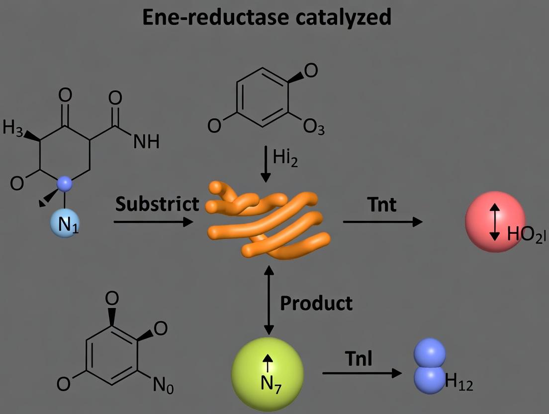
Protocol 2: Gram-Scale Synthesis of (R)-Dihydrocarvone Objective: Asymmetric reduction of (R)-(-)-Carvone to (R)-Dihydrocarvone, a valuable terpenoid precursor.
- Reaction Setup: In a 250 mL bioreactor, combine 1 g (6.6 mmol) of (R)-(-)-Carvone, 100 mg of purified OYE1 from Saccharomyces pastorianus, 0.1 mM NADP⁺, 10 mM glucose, and 5 U glucose dehydrogenase in 100 mL 50 mM phosphate buffer (pH 7.0).
- Process: Stir at 30°C and 200 rpm for 8 hours. Monitor by TLC or GC.
- Extraction & Analysis: Extract with ethyl acetate (3 x 50 mL). Dry over Na₂SO₄, concentrate in vacuo. Determine yield by NMR and e.e. by chiral GC (e.g., Cyclosil-B column).
The Scientist's Toolkit: Key Research Reagent Solutions
| Item | Function in ERed Research |
|---|---|
| Glucose Dehydrogenase (GDH) | Robust, irreversible NADPH regeneration; drives reaction to completion. |
| NADP⁺ Sodium Salt | Cofactor precursor. More stable than NADPH. Used with a regeneration system. |
| EziG Carrier (Epoxy) | Robust immobilization support for enzymes via covalent binding, enabling reuse. |
| Chiral GC/HPLC Columns | Essential for determining enantiomeric excess (e.g., Cyclosil-B, Chiralcel OD-H). |
| Error-Prone PCR Kit | Creates random mutagenesis libraries for directed evolution campaigns. |
| Phosphate Buffer (pH 7.0) | Standard reaction medium for most OYEs, maintaining enzyme stability. |
Visualization: Experimental Workflows
Diagram 1: Substrate Scope Expansion Workflow
Diagram 2: ERed Biocatalytic Reaction Mechanism
Diagnosing and Solving Reaction Failures: A Practical Guide for Chemists
FAQs & Troubleshooting Guide
Q1: My ene-reductase (ERED) reaction shows no conversion. What should I check first? A1: First, verify the activity of your enzyme preparation. Use a known, high-activity substrate like 2-cyclohexen-1-one (control substrate) under standard assay conditions (see Protocol 1). If conversion is high (>95%), the enzyme is active, and the problem likely lies with your target substrate. If conversion is low, the issue is with the enzyme or system.
Q2: The enzyme is active with control substrates, but not my target alkene. What does this indicate? A2: This is the core challenge of limited substrate scope. It suggests either:
- Substrate Inaccessibility: The substrate cannot bind to the active site due to steric or electronic mismatches.
- Insufficient Activation: The alkene may not be sufficiently electrophilic for the mechanism. Check the substrate's reduction potential (Eº'). EREDs typically reduce alkenes with Eº' between -0.5 and -1.5 V vs. SHE.
- Unproductive Binding: The substrate binds in a non-productive orientation.
Q3: I see partial conversion but low enantioselectivity. Is this an enzyme or substrate issue? A3: This is typically an enzyme-substrate mismatch. The enzyme's active site is not optimally chiral for that specific substrate scaffold. Protein engineering (saturation mutagenesis) or screening homologous EREDs is the recommended path.
Q4: My reaction stalls. Could the cofactor recycling system be failing? A4: Yes. Monitor the NADPH absorbance at 340 nm over time (see Protocol 2). A steady decrease indicates successful recycling. If it plateaus, the issue could be:
- Cofactor degradation (ensure fresh stocks, avoid light).
- Inefficient recycling enzyme (e.g., glucose dehydrogenase, GDH) activity.
- Insufficient concentration of the recycling substrate (e.g., glucose).
Experimental Protocols
Protocol 1: Standard ERED Activity Assay
- Prepare 1 mL assay mixture in a UV-transparent cuvette: 50 mM potassium phosphate buffer (pH 7.0), 0.2 mM NADPH, 100 µM substrate (e.g., 2-cyclohexen-1-one from a 100 mM stock in DMSO).
- Pre-incubate the mixture at 30°C for 2 minutes.
- Initiate the reaction by adding purified ERED to a final concentration of 1 µM.
- Immediately monitor the decrease in absorbance at 340 nm (ε = 6.22 mM⁻¹cm⁻¹ for NADPH) for 2 minutes.
- Calculate activity: ΔA340/min ÷ 6.22 = mM NADPH consumed/min. Specific activity = (mM/min) / [enzyme] in mg/mL.
Protocol 2: Cofactor Recycling System Efficiency Test
- Set up a 1 mL reaction with your full system: Buffer, target substrate, ERED, NADP⁺ (0.1 mM), GDH (2 U), and glucose (10 mM).
- Monitor A340 continuously for 1 hour.
- A sharp initial drop (ERED using NADPH) followed by a steady, slow decline indicates successful recycling. A quick drop to a stable plateau indicates recycling failure.
Data Presentation
Table 1: Diagnostic Scenarios and Recommended Actions
| Observation (Control vs. Target Substrate) | Likely Problem | Diagnostic Test | Potential Solution |
|---|---|---|---|
| High conv. (Control), No conv. (Target) | Substrate Scope / Activation | Measure substrate Eº' or perform docking. | Use more activated alkene (e.g., cyano, nitro); try ERED from different organism. |
| High conv. (Control), Low conv. (Target) | Substrate Binding / Sterics | Kinetic analysis (Km, kcat). | Enzyme engineering (broadening active site); use solubilizing additives (e.g., 5% DMSO). |
| Low/No conv. (Both) | Enzyme or Cofactor System | Protocol 1 & 2; SDS-PAGE. | Use fresh enzyme/cofactor; check expression/purification; optimize recycling system. |
| Good conv., Low ee (Target) | Enantioselectivity Mismatch | Chiral HPLC/Gas Chromatography of product. | Directed evolution for enantioselectivity; screen homologous EREDs. |
Table 2: Key Reduction Potentials for Diagnostic Comparison
| Alkene Substrate | Typical Eº' (V vs. SHE) | Expected ERED Activity? |
|---|---|---|
| (E)-2-Methyl-1-nitroprop-1-ene | ~ -0.5 | High (Favored) |
| 2-Cyclohexen-1-one | ~ -0.8 | High |
| (R)-Carvone | ~ -1.2 | Moderate to High |
| α-Methylstyrene | ~ -1.8 | Low (Unfavored) |
Visualizations
Title: ERED Problem Diagnosis Flowchart
Title: ERED Catalytic & Recycling Cycle
The Scientist's Toolkit: Research Reagent Solutions
| Reagent / Material | Function in ERED Experiments |
|---|---|
| Old Yellow Enzyme (OYE) homologs (e.g., PETNR, YqjM, OYE1-3) | The core biocatalysts. Different homologs have varying substrate scopes and stereoselectivities. |
| NADPH Tetrasodium Salt | Essential hydride donor cofactor. Light and temperature-sensitive; prepare fresh solutions. |
| Glucose Dehydrogenase (GDH) & D-Glucose | Common enzymatic cofactor recycling system. Regenerates NADPH from NADP⁺, allowing catalytic cofactor use. |
| 2-Cyclohexen-1-one | Standard, high-activity control substrate for diagnosing basic enzyme activity. |
| Chiral GC/HPLC Columns (e.g., cyclodextrin-based) | Essential for determining enantiomeric excess (ee) of reduced products. |
| Enzyme Expression System (E. coli BL21(DE3), pET vector) | Standard platform for recombinant expression of cloned ERED genes. |
| Nickel-NTA Agarose | For affinity purification of His-tagged EREDs, ensuring pure and consistent enzyme preparations. |
| DMSO (anhydrous) | Common, biocompatible solvent for dissolving hydrophobic alkene substrates in aqueous reaction buffers. |
Technical Support Center
Welcome to the technical support center for optimizing ene-reductase (ERED) catalyzed reactions. This resource, framed within our research thesis on addressing the limited substrate scope of EREDs, provides targeted troubleshooting guides and FAQs to help you overcome poor conversion rates.
Troubleshooting Guide & FAQs
Q1: My reaction yield has dropped significantly with a new, bulky substrate. I suspect enzyme inhibition or poor binding. What initial steps should I take? A: Begin by analyzing the reaction microenvironment. Suboptimal pH can alter the ionization states of critical residues in the active site, while incorrect temperature can affect enzyme flexibility and stability. Follow Protocol A to systematically screen pH and temperature.
Q2: I've optimized pH and temperature, but conversion for my hydrophobic substrate remains below 20%. What is the next strategic step? A: Poor aqueous solubility of hydrophobic substrates severely limits access to the active site. This is a common bottleneck in expanding substrate scope. The introduction of cosolvents is the standard approach. Implement Protocol B for a structured cosolvent screen, prioritizing biocompatible options like 2-methyl-2-butanol (2M2B).
Q3: I added 15% (v/v) DMSO as a cosolvent and now see no activity. What went wrong? A: Many cosolvents, including DMSO, methanol, and acetone at high concentrations (>10-20%), can denature the enzyme by disrupting its essential water layer. Refer to Table 1 for cosolvent tolerance thresholds. Switch to a more biocompatible cosolvent like 2M2B or glycerol, and always add the cosolvent to the buffer before introducing the enzyme to avoid localized denaturation.
Q4: How do I balance cosolvent concentration between substrate solubility and enzyme activity? A: This requires an empirical optimization. Perform a cosolvent concentration gradient (e.g., 5%, 10%, 15%, 20% v/v) while keeping other parameters constant. Plot conversion vs. cosolvent concentration; the optimum is typically at the "knee" of the curve before activity plummets. See Diagram 1 for the decision workflow.
Q5: My optimized reaction uses 25% cosolvent and works well in lab-scale. How do I ensure scalability for preparative synthesis? A: Focus on process robustness. Confirm enzyme stability over the full reaction time (e.g., sample aliquots and measure activity). Ensure efficient NADPH cofactor regeneration. For scale-up, consider immobilized EREDs for reusability and continuous processing.
Experimental Protocols
Protocol A: Systematic pH and Temperature Screening for ERED Reactions.
- Prepare a 100 mM buffer series: Citrate-phosphate (pH 5.0-6.5), Potassium phosphate (pH 6.5-8.0), Glycine-NaOH (pH 8.5-9.5).
- In a 96-well plate, add 180 µL of buffer from each pH point.
- Add 10 µL of substrate stock solution (in a minimal, compatible solvent like 2M2B) and 10 µL of ERED enzyme preparation.
- Initiate the reaction by adding 10 µL of NADPH cofactor solution (or your regeneration system).
- Incubate the plate in parallel at multiple temperatures (e.g., 25°C, 30°C, 37°C) with shaking.
- Monitor conversion over time via UV-Vis (decrease in A340 for NADPH) or GC/HPLC analysis.
- Plot conversion vs. pH for each temperature to identify the optimal combination.
Protocol B: Structured Cosolvent Screen for Hydrophobic Substrates.
- Select a panel of cosolvents: e.g., 2-methyl-2-butanol (2M2B), dimethyl sulfoxide (DMSO), tert-butanol, glycerol, propylene glycol.
- Prepare your optimal buffer (from Protocol A) and add the cosolvent to achieve 10% (v/v) final concentration. Mix thoroughly.
- Dissolve your hydrophobic substrate directly in this buffer-cosolvent mixture to the desired concentration (e.g., 10 mM). Vortex/sonicate if needed.
- In a 1 mL reaction, combine 880 µL of the substrate mixture, 100 µL of enzyme, and 20 µL of NADPH/regeneration system starter.
- Incubate at your optimal temperature with agitation. Sample at t=0, 30min, 1h, 2h, 4h.
- Extract samples with an organic solvent (e.g., ethyl acetate) and analyze by GC/HPLC to determine initial reaction rates and final conversion.
- For the top 1-2 cosolvents, repeat at different concentrations (5%, 15%, 20%) to find the activity-solubility optimum.
Data Presentation
Table 1: Cosolvent Tolerance and Performance in ERED Reactions
| Cosolvent | Typical Max Tolerable Concentration (v/v) for ERED Activity | Primary Function | Impact on Hydrophobic Substrate Solubility |
|---|---|---|---|
| 2-Methyl-2-butanol (2M2B) | 20-30% | Biocompatible, log P ~1.3 | High - Excellent for aromatics, alkenes |
| tert-Butanol | 15-25% | Biocompatible, less denaturing | Moderate to High |
| Dimethyl Sulfoxide (DMSO) | 10-15% | Powerful solubilizer | Very High - but often denaturing |
| Glycerol | 20-40% | Protein stabilizer, viscous | Low - primarily used for stabilization |
| Acetone | <10% | Organic solvent | High - but strongly denaturing above 10% |
| Propylene Glycol | 15-25% | Mild solubilizer and stabilizer | Moderate |
Table 2: Example Optimization Results for a Model Bulky Substrate (Chalcone)
| Condition (Buffer, 30°C) | Cosolvent (15% v/v) | Initial Substrate Solubility | Conversion at 4h | Observed Initial Rate (µmol/min/mg) |
|---|---|---|---|---|
| Potassium Phosphate, pH 7.0 | None | < 1 mM | 5% | 0.1 |
| Potassium Phosphate, pH 7.0 | DMSO | > 20 mM | 15% | 0.8 |
| Potassium Phosphate, pH 7.0 | tert-Butanol | > 15 mM | 45% | 2.5 |
| Potassium Phosphate, pH 7.0 | 2M2B | > 25 mM | 92% | 5.2 |
| Glycine-NaOH, pH 9.0 | 2M2B | > 25 mM | 88% | 4.9 |
Mandatory Visualization
Diagram 1: Workflow for Troubleshooting Poor Conversion in ERED Reactions
The Scientist's Toolkit: Research Reagent Solutions
| Item | Function in ERED Optimization |
|---|---|
| Ene-Reductase (ERED) Kit | Contains purified wild-type or mutant EREDs (e.g., OYE1, YqjM) for initial activity screens. |
| NADPH Cofactor Regeneration System | Typically glucose-6-phosphate (G6P) and G6P dehydrogenase; maintains cofactor levels cost-effectively. |
| Biocompatible Cosolvents (2M2B, t-BuOH) | Increases solubility of hydrophobic substrates while preserving enzyme activity. |
| Broad-Range Buffer Salts | For precise pH screening (citrate, phosphate, Tris, glycine). |
| Immobilized ERED Enzyme | For reusability and stability in high cosolvent or preparative-scale reactions. |
| Analytical Internal Standards | For accurate GC-FID or HPLC-UV quantification of substrate and product (e.g., alkyl benzenes). |
| 96-Well Deep Well Plates | For high-throughput screening of pH, temperature, and cosolvent variables. |
Addressing Low Enantioselectivity with Non-Cognate Substrates
Troubleshooting Guides & FAQs
Q1: We are screening an ene-reductase (ER) from a model organism against a new prochiral alkene substrate. The reaction proceeds but yields a racemic product (≈50% ee). What are the primary strategies to improve enantioselectivity?
A1: Low enantioselectivity with non-cognate substrates is a common challenge. The primary troubleshooting strategies are:
- Enzyme Engineering: Perform directed evolution or semi-rational design targeting residues in the substrate-binding pocket. Saturation mutagenesis at positions lining the pocket often improves substrate fit and stereocontrol.
- Enzyme Screening: Test homologs from different organisms or metagenomic libraries. ERs from atypical sources may have inherent complementary substrate scopes.
- Reaction Engineering:
- Solvent Engineering: Switch from aqueous buffer to a water-miscible organic co-solvent (e.g., 20-30% v/v tert-butanol, DMSO) to modulate substrate accessibility and enzyme flexibility.
- pH Optimization: Adjust pH (typically 6.0-8.0 for ERs) to alter protonation states of key residues, potentially influencing substrate orientation.
- Additives: Use crowding agents (e.g., PEG) or low concentrations of chaotropes/ kosmotropes to subtly alter enzyme dynamics.
Q2: During directed evolution for improved ee, we see a trade-off where improved enantioselectivity comes with a drastic drop in conversion. How can we address this?
A2: This indicates your selectivity mutations may be compromising catalytic efficiency. Refine your screening protocol:
- Implement a Primary Screen for Activity: Before high-throughput ee analysis, use a rapid colorimetric assay (e.g., monitoring NADPH depletion at 340 nm) to filter out variants with catastrophically low activity.
- Use Dual-Selection Pressure: In your screening plates, assay for both conversion (via a coupled enzyme assay) and enantioselectivity (via chiral GC/HPLC of extracted samples). Only advance variants passing a minimum conversion threshold (e.g., >30%).
- Consider Backcrossing: Recombine beneficial selectivity mutations with the wild-type background to eliminate potential neutral or negative synergistic mutations affecting activity.
Q3: We aim to perform computational docking of a non-cognate substrate into an ER's active site to guide mutagenesis. What are critical parameters to set for reliable predictions of binding pose?
A3: Accurate docking is crucial for generating rational hypotheses. Use this protocol:
Protocol: Computational Docking Setup for Ene-Reductases
- Protein Preparation: Obtain your ER structure (e.g., PDB ID: 4BXX). Remove water molecules and the co-crystallized ligand. Add hydrogen atoms, and assign protonation states to key residues (e.g., the catalytic tyrosine, histidine, and asparagine). Ensure the flavin mononucleotide (FMN) cofactor is correctly parameterized and present.
- Define the Active Site: The docking search space should encompass the entire substrate-binding tunnel, typically defined by residues within 8-10 Å of the N5 atom of the FMN isoalloxazine ring and the catalytic tyrosine.
- Ligand Preparation: Draw your substrate, minimize its geometry using MMFF94 or similar, and generate probable tautomers/protonation states at the reaction pH.
- Docking Software & Parameters: Use flexible docking software (e.g., AutoDock Vina, GOLD).
- Set exhaustiveness/search parameter to a high value (e.g., 50 for Vina).
- Use the empirical scoring function but prioritize poses where: (a) the reactive alkene is parallel to the FMN isoalloxazine ring (for hydride transfer), and (b) the prochiral face to be reduced is positioned away from the bulky "blocking" residue (often a phenylalanine or leucine).
Diagram: Workflow for Improving ER Enantioselectivity
(Title: ER Enantioselectivity Improvement Workflow)
Diagram: Key Interactions in ER Active Site for Stereocontrol
(Title: Key Active Site Interactions Determining ER Selectivity)
The Scientist's Toolkit: Research Reagent Solutions
| Reagent / Material | Function in Experiment |
|---|---|
| Old Yellow Enzyme (OYE) Homologs (e.g., OYE1-3, YqjM, NerA, XenA) | Benchmark enzymes with well-characterized structures and substrate profiles for initial screening and comparison. |
| Glucose-6-Phosphate (G6P) & Glucose-6-Phosphate Dehydrogenase (G6PDH) | Components of a common NADPH regeneration system, crucial for maintaining cofactor levels during preparative or kinetic assays. |
| NADPH (Tetrasodium Salt) | The essential hydride-donating cofactor for ERs. High-purity stocks are necessary for accurate kinetic measurements. |
| Tert-Butanol (anhydrous) | A preferred water-miscible organic co-solvent for ER reactions; minimizes enzyme denaturation while improving solubility of hydrophobic substrates. |
| Chiral Stationary Phase HPLC Columns (e.g., Chiralpak IA, IB, IC; Chiralcel OD-H) | Essential for analytical-scale separation and accurate quantification of enantiomers to determine enantiomeric excess (ee). |
| Site-Directed Mutagenesis Kit (e.g., Q5 from NEB) | For rapid construction of single-point mutations identified through computational design or evolution campaigns. |
| E. coli BL21(DE3) Competent Cells | Standard heterologous expression host for production of recombinant ER variants. |
Table 1: Impact of Common Solvent Systems on ER Activity & Selectivity Data representative of trends observed across multiple OYE family studies.
| Solvent System (% v/v) | Relative Activity (%)* | Typical Δee Impact | Notes |
|---|---|---|---|
| Potassium Phosphate Buffer (pH 7.0) | 100 (Reference) | 0 | Aqueous baseline. Poor solubility for lipophilic substrates. |
| 20% tert-Butanol | 70 - 95 | +5 to +15 ee | Often the optimal compromise; maintains stability, boosts substrate solubility. |
| 20% Dimethyl Sulfoxide (DMSO) | 40 - 80 | Variable (+/- 10 ee) | Can significantly alter enzyme selectivity; useful for probing. |
| 20% Acetonitrile | 10 - 30 | Often Negative | Generally denaturing; not recommended for ERs. |
| 30% Glycerol* | 50 - 70 | +2 to +8 ee | A crowding agent; can stabilize protein but increase viscosity. |
Relative to aqueous buffer control for a given enzyme-substrate pair. *Direction and magnitude are highly substrate-dependent. *Not a co-solvent but a common additive.
Table 2: Benchmarking ERs Against Challenging Substrate Classes
| Substrate Class (Example) | Wild-Type OYE1 (ee%) | Engineered Variant (ee%) | Key Mutations(s) | Reference Context |
|---|---|---|---|---|
| β,β-Disubstituted Nitroalkene | <10 (S) | >99 (R) | W116I, F296N | Reversed & enhanced selectivity via pocket enlargement. |
| α,β-Unsaturated Lactone | 60 (R) | 98 (R) | F296Y, H164N | Improved π-stacking and H-bonding. |
| Cyclic Enone (bulky) | 20 (S) | 95 (S) | M193N, Y375V | Removed steric clash, improved substrate alignment. |
| Unsaturated Carboxylic Acid | Racemic | 85 (S) | L370A, Y375G | Opened a specific sub-pocket for acid chain. |
Managing Substrate/Product Inhibition and Enzyme Inactivation
Troubleshooting Guide & FAQs
Q1: During screening, my ene-reductase (ER) shows no activity with new, non-native substrates. Is this solely a substrate scope issue, or could inhibition be a factor? A: While limited substrate scope is a primary challenge, rapid inhibition upon initial binding can mask activity. Before concluding the substrate is not accepted, perform a rapid dilution assay. Pre-incubate the enzyme with a low concentration of the new substrate (e.g., 0.1 mM) for 5 minutes, then dilute the mixture 100-fold into a standard assay mixture containing a known good substrate (e.g., 1 mM citral). If activity on the good substrate is significantly lower than a control without pre-incubation, it suggests the new substrate is a reversible inhibitor, blocking the active site.
Q2: My ER reaction progress plateaus at low conversion, despite substrate remaining. How can I diagnose if this is due to substrate inhibition or product inhibition? A: Perform two parallel diagnostic experiments and monitor progress over time.
- Substrate Inhibition Test: Run reactions at varying initial substrate concentrations (e.g., 1, 5, 10, 25 mM). A decrease in initial reaction rate at higher concentrations indicates substrate inhibition.
- Product Inhibition Test: Run standard reactions spiked with varying concentrations of the expected product (0, 2, 5, 10 mM). A reduction in the initial rate with added product confirms product inhibition.
Table 1: Diagnostic Results for Reaction Plateau at 30% Conversion (Hypothetical Data)
| Diagnostic Test | Condition | Observed Initial Rate (µmol/min/mg) | Inference |
|---|---|---|---|
| Substrate Inhibition | [S] = 2 mM | 0.85 | Classic Michaelis-Menten kinetics. |
| [S] = 25 mM | 0.22 | Strong substrate inhibition observed. | |
| Product Inhibition | [P] added = 0 mM | 0.85 | Baseline rate. |
| [P] added = 5 mM | 0.31 | Strong competitive product inhibition. |
Q3: I observe a steady decline in reaction rate over time, not just a plateau. Is this enzyme inactivation, and how can I address it? A: A continuous decline suggests irreversible enzyme inactivation, often from cofactor dissociation, oxidative damage, or thermal instability. To diagnose:
- Cofactor Stability: Supplement the reaction with additional NAD(P)H (e.g., a second pulse at 50% conversion). If the rate is restored, cofactor degradation/dissociation is implicated.
- Oxidative Damage: Run reactions under an inert atmosphere (N₂/Ar) vs. air. Include sacrificial reductants like dithiothreitol (DTT, 1 mM) in the buffer.
- Thermal Inactivation: Perform an activity "half-life" (t₁/₂) assay. Incubate the enzyme at reaction temperature, sampling at intervals and assaying residual activity under standard conditions.
Table 2: Enzyme Inactivation Half-Life (t₁/₂) Under Different Conditions
| Stabilization Strategy | Incubation Condition | Calculated t₁/₂ (min) | Relative Improvement |
|---|---|---|---|
| None (Control) | 30°C, in air | 45 | 1.0x |
| + 1 mM DTT | 30°C, in air | 68 | 1.5x |
| Inert Atmosphere | 30°C, under N₂ | 120 | 2.7x |
| Immobilized Enzyme | 30°C, in air | >300 | >6.7x |
Q4: What are practical experimental strategies to overcome these limitations in preparative-scale biotransformations? A: Implement engineering and process design solutions.
- For Inhibition: Use continuous feeding of substrate to maintain its concentration below inhibitory levels. Alternatively, employ in-situ product removal (ISPR), such as coupling the reaction to resin-based adsorption or extraction, to keep product concentration low.
- For Inactivation: Use enzyme immobilization on solid supports to enhance stability. Employ fed-batch cofactor regeneration using a robust system like glucose dehydrogenase (GDH)/glucose to maintain reducing power.
Detailed Experimental Protocols
Protocol 1: Rapid Dilution Assay for Inhibitor Screening
- Prepare an inhibitor solution (new substrate) at 10x the desired final concentration (e.g., 1 mM).
- Prepare the ER enzyme in assay buffer at 10x its standard assay concentration.
- Mix 10 µL of enzyme with 10 µL of inhibitor solution. Incubate for 5 minutes at room temperature.
- Dilute 2 µL of the enzyme-inhibitor mix into 198 µL of a standard activity assay mixture containing a reporting substrate (e.g., 1 mM citral) and NADPH.
- Immediately monitor the decrease in A₃₄₀ (NADPH consumption) for 60 seconds. Compare the initial rate to a control where buffer replaces the inhibitor solution.
Protocol 2: In-Situ Product Removal (ISPR) Using Resin Adsorption
- Set up a standard ER reaction (e.g., 10 mL scale) in a stirred vessel.
- Add 10% (w/v) of a hydrophobic adsorption resin (e.g., XAD-16N) to the reaction mixture at time zero.
- Initiate the reaction by adding the enzyme.
- Monitor substrate and product concentrations in the aqueous phase by periodic HPLC sampling.
- Upon completion, separate the resin by filtration. Elute the adsorbed product from the resin with an organic solvent (e.g., ethanol or acetone).
Visualizations
Diagnosing Inhibition vs. Inactivation
Strategies to Overcome Inhibition & Inactivation
The Scientist's Toolkit: Research Reagent Solutions
Table 3: Essential Reagents for Managing Inhibition & Inactivation
| Reagent/Material | Function & Application |
|---|---|
| Hydrophobic Resin (XAD-16N) | For in-situ product removal (ISPR); adsorbs hydrophobic products to shift equilibrium. |
| Glucose Dehydrogenase (GDH) | Robust cofactor regeneration enzyme; used with glucose to maintain NADPH supply. |
| Dithiothreitol (DTT) | Reducing agent; protects cysteine residues from oxidation, stabilizing enzyme activity. |
| NADPH (tetrasodium salt) | Essential cofactor; use high-purity, fresh aliquots to prevent spurious inactivation results. |
| Enzyme Immobilization Support (e.g., EziG) | Controlled porosity glass or polymer for covalent enzyme attachment, enhancing stability and reusability. |
| Oxygen-Scavenging System (Glucose Oxidase/Catalase) | Maintains low-O₂ environment in solution to reduce oxidative inactivation. |
When to Screen a New Enzyme Library vs. Re-engineering the Reaction Conditions
Technical Support Center: Troubleshooting Limited Substrate Scope in Ene-Reductase Catalysis
Troubleshooting Guides
Guide 1: Diagnosing Poor Conversion with a New Substrate
- Step 1: Perform a negative control (no enzyme) to confirm the reaction is enzyme-dependent.
- Step 2: Run a positive control (a known good substrate) with the same batch of enzyme and cofactor (NAD(P)H) to verify enzyme activity.
- Step 3: Check cofactor recycling system efficiency (e.g., measure glucose dehydrogenase activity if using GDH/glucose).
- Step 4: Analyze reaction mix by HPLC/GC at T=0 to check for non-enzymatic substrate degradation or binding.
- Step 5: If steps 1-4 are normal, the issue is likely enzyme-substrate incompatibility. Proceed to FAQs.
Guide 2: Systematic Reaction Condition Re-engineering Workflow
- Vary Solvent System: Start with phosphate buffer (pH 7), then test co-solvents (e.g., 10-30% DMSO, MeCN, i-PrOH).
- Adjust pH: Screen pH 5.5 to 9.0 in 0.5 unit increments.
- Modify Temperature: Test from 20°C to 40°C.
- Optimize Cofactor Supply: Increase recycling enzyme concentration or switch system (e.g., from GDH/glucose to phosphite dehydrogenase/phosphite).
- Alter Additives: Introduce low concentrations of surfactants (e.g., Triton X-100) or salts (e.g., KCl).
- Evaluate: If conversion remains <20%, consider library screening.
Frequently Asked Questions (FAQs)
Q1: My target α,β-unsaturated compound shows <10% conversion. Should I screen a new enzyme library immediately? A: Not necessarily. First, re-engineer basic conditions: Ensure your substrate is soluble (try ≤20% DMSO as co-solvent). Test pH 6.0 and 8.5, as ene-reductase activity profiles vary. If conversion does not improve beyond 20% after these steps, a library screen is advisable.
Q2: What quantitative benchmarks indicate that condition optimization is sufficient? A: Refer to the table below. If you meet Tier 2 metrics, condition optimization is likely sufficient. Tier 1 metrics warrant a new library screen.
Table: Decision Matrix for Screening vs. Re-engineering
| Metric | Tier 1 (Screen Library) | Tier 2 (Optimize Conditions) | Measurement Method |
|---|---|---|---|
| Conversion (%) | < 20 | 20 - 80 | HPLC/GC Analysis |
| Apparent ( K_m ) (mM) | > 10 | 1 - 10 | Michaelis-Menten Kinetics |
| Specific Activity (U/mg) | < 0.05 | 0.05 - 0.5 | Initial Rate Assay |
| Enantiomeric Excess (ee%) | < 70% (if chiral) | > 70% | Chiral HPLC |
| Cofactor Recycling Rate (min⁻¹) | < 100 | > 100 | NAD(P)H depletion assay |
Q3: How do I practically test if the enzyme's active site is simply too small for my bulky substrate? A: Perform a homology model docking experiment.
- Obtain the protein sequence of your ene-reductase (e.g., OPR1, YqjM, GRE2).
- Use Swiss-Model (swissmodel.expasy.org) to generate a 3D model.
- Dock your substrate into the flavin-binding active site using AutoDock Vina or a similar tool.
- Analyze the binding pose. If the substrate cannot orient within 4Å of the N5 atom of the flavin cofactor, or shows severe steric clashes, active site engineering or a new library screen is required.
Q4: What is a detailed protocol for a high-throughput ene-reductase library screen? A: Protocol for 96-Well Plate Library Screen
- Materials: Library of ene-reductase lysates (e.g., in E. coli crude lysate), substrate (10 mM in DMSO), NADP⁺ (0.1 mM), glucose (10 mM), GDH (5 U/mL), phosphate buffer (50 mM, pH 7.0).
- Method:
- Dispense 180 µL of buffer + cofactor mix (NADP⁺, glucose, GDH) to each well.
- Add 10 µL of substrate stock.
- Initiate reaction by adding 10 µL of enzyme lysate. Seal and shake at 30°C.
- After 4-16 hours, quench with 100 µL of ethyl acetate and vortex.
- Analyze organic layer by GC-FID or HPLC-UV to determine conversion.
Q5: Are there key reaction additives that can dramatically alter substrate scope without changing the enzyme? A: Yes. The following additives can be crucial:
- Cosolvents: Up to 30% (v/v) DMSO, tert-butanol, or PEG-400 to solubilize hydrophobic substrates.
- Surfactants: 0.1% (w/v) Triton X-100 or CTAB can aid substrate delivery.
- Chaotropic Salts: 100-200 mM KCl can sometimes stabilize protein or improve binding.
- Alternative Electron Donors: Switch from NADPH to cheaper NADH using a stronger recycling system (e.g., 0.5 M i-PrOH), which can alter kinetics.
The Scientist's Toolkit: Key Research Reagent Solutions
| Item | Function in Ene-Reductase Research |
|---|---|
| Glucose Dehydrogenase (GDH) | Robust NAD(P)H recycling enzyme. Coupled with glucose, it drives reactions to completion. |
| NADP⁺ / NAD⁺ | Essential oxidized cofactor. Must be replenished for continuous turnover. |
| Flavin Mononucleotide (FMN) | Prosthetic group for many ene-reductases (e.g., OYEs). May be required for reconstituting apo-enzymes. |
| Phosphate Buffer (pH 7.0) | Standard reaction buffer. Maintains pH and ionic strength. |
| DMSO (Anhydrous) | Common co-solvent to dissolve hydrophobic, poorly water-soluble substrates. |
| Ethyl Acetate (HPLC Grade) | Standard solvent for quenching biocatalytic reactions and extracting products for analysis. |
| Chiral HPLC Column (e.g., Chiralcel OD-H) | Essential for determining enantiomeric excess (ee%) of reduced products. |
| E. coli BL21(DE3) Cells | Standard expression host for recombinant ene-reductase production. |
Visualizations
Decision Pathway for Enzyme Screening vs. Condition Re-engineering
High-Throughput Library Screen Workflow
Benchmarking Success: Validating New ERED Variants and Comparing to Alternative Technologies
Technical Support Center
Troubleshooting Guides & FAQs
Q1: Why is my reaction conversion low despite using a reported ene-reductase? A: Low conversion is often linked to non-optimal reaction conditions for your specific substrate. Key factors to troubleshoot:
- Cofactor Regeneration: Ensure your cofactor regeneration system (e.g., glucose/glucose dehydrogenase) is active. Check for NADPH depletion by HPLC or spectrophotometry.
- Enzyme-Substrate Mismatch: Many ene-reductases have narrow substrate scopes. Perform a homology model of your enzyme's active site to check for steric clashes with your target alkene.
- Oxygen Sensitivity: Some ene-reductases and flavin cofactors are oxygen-sensitive. Degas buffers and run reactions under an inert atmosphere (N₂ or Ar).
Q2: How can I improve the enantiomeric excess (ee) of my product? A: Poor ee indicates insufficient stereocontrol.
- Enzyme Selection: Screen homologous enzymes from different organisms (e.g., OYE1, OYE2, OYE3, YqjM, NemA). Small active site variations drastically impact stereoselectivity.
- Double-Bond Geometry: Confirm the geometry (E/Z) of your substrate. Ene-reductases often show distinct stereopreference based on this.
- Biocatalyst Engineering: If a wild-type enzyme gives moderate ee, consider site-saturation mutagenesis at residues near the binding pocket (e.g., Tyr, Trp, His) to fine-tune steric interactions.
Q3: My Total Turnover Number (TTN) is unexpectedly low. What's the primary cause? A: Low TTN means the enzyme deactivates rapidly. Common culprits:
- Substrate/Product Inhibition: High concentrations can inactivate the enzyme. Titrate substrate concentration (typically 5-20 mM is optimal) and consider in-situ product removal (ISPR).
- Solvent Incompatibility: Even with solvent-tolerant enzymes, >10% (v/v) organic cosolvent can reduce stability. Use biocompatible solvents like MTBE, cyclopentyl methyl ether (CPME), or ionic liquids.
- Shear Stress: If using free enzyme with stirring, consider immobilization on a solid support (e.g., EziG beads) to protect from mechanical degradation.
Q4: My substrate is insoluble in aqueous buffer. How do I balance solvent tolerance with high conversion? A: This is a key challenge in expanding substrate scope.
- Two-Phase Systems: Implement a water-organic solvent biphasic system. Use a logP > 4 solvent (e.g., octane, dodecane) to minimize enzyme phase toxicity. Optimize the phase ratio.
- Cosolvent Grading: Systematically test cosolvents (DMSO, MeOH, iPrOH, acetone, acetonitrile) at increasing concentrations (2%, 5%, 10% v/v) to find the maximum tolerated concentration before activity drops >50%.
- Directed Evolution: Use iterative rounds of error-prone PCR or saturation mutagenesis, selecting mutants for activity in the presence of your target cosolvent.
Data Presentation
Table 1: Benchmarking KPIs for Common Ene-Reductases with Model Substrate (Cyclohex-2-enone)
| Enzyme (Source) | Conversion (%)* | ee (%)* | TTN (NADPH)* | Tolerated Solvent (% v/v)* |
|---|---|---|---|---|
| OYE1 (S. pastorianus) | >99 | >99 (R) | 5.2 x 10⁴ | iPrOH (20%), DMSO (15%) |
| OYE2 (S. pastorianus) | 95 | 95 (R) | 4.1 x 10⁴ | iPrOH (15%), Acetone (10%) |
| YqjM (B. subtilis) | 98 | 99 (S) | 6.0 x 10⁴ | MeOH (30%), DMSO (20%) |
| NCR (Z. mays) | 85 | 88 (R) | 2.8 x 10⁴ | iPrOH (25%) |
| Typical values under standard conditions (5 mM substrate, 0.1-1 mol% enzyme, pH 7.0, 30°C, glucose/GDH regeneration). |
Table 2: Impact of Cosolvent on KPI Degradation
| Cosolvent (15% v/v) | Relative Activity (%) | TTN Retention (%) | Recommended Max % |
|---|---|---|---|
| DMSO | 85 | 80 | 20 |
| 2-Propanol | 90 | 85 | 25 |
| Acetonitrile | 40 | 30 | 5 |
| Methanol | 75 | 70 | 30 |
| Ethyl Acetate | 10 (Biphasic) | 95 (Biphasic) | Biphasic preferred |
Experimental Protocols
Protocol 1: High-Throughput Screening for Solvent Tolerance
- Prepare Stock Solutions: Substrate (200 mM in target solvent), NADP⁺ (10 mM), glucose (1 M), purified ene-reductase (1 mg/mL).
- Set Up Plate: In a 96-well UV plate, mix 85 µL phosphate buffer (100 mM, pH 7.0), 5 µL substrate stock, 5 µL cosolvent (to achieve 2-20% v/v final), 2 µL NADP⁺, 2 µL glucose, and 1 µL GDH (5 U/mL).
- Initiate Reaction: Add 10 µL of enzyme stock. Immediately monitor absorbance at 340 nm (for NADPH consumption) or at the λmax of the alkene substrate for 5 minutes.
- Calculate: Initial velocity (V₀) is proportional to ΔA/min. Normalize activity relative to a no-cosolvent control.
Protocol 2: Determining Total Turnover Number (TTN)
- Reaction Setup: In a sealed vial under N₂, combine substrate (0.1 mmol), NADP⁺ (0.001 mmol), glucose (1 mmol), GDH (10 U), and ene-reductase (0.1 µmol) in 10 mL total volume of optimal buffer.
- Incubate: Stir at 30°C for 24 hours or until conversion plateaus (monitor by TLC/GC).
- Quantify: Measure final product concentration via calibrated HPLC/GC.
- Calculate: TTN = (moles of product formed) / (moles of enzyme used). Use enzyme concentration determined by Bradford assay or active site titration.
Diagrams
Diagram 1: Troubleshooting Low Conversion & ee
Diagram 2: Solvent Tolerance Optimization Workflow
The Scientist's Toolkit: Research Reagent Solutions
| Item | Function & Rationale |
|---|---|
| Glucose Dehydrogenase (GDH) | NADPH regeneration driver. Thermally stable, inexpensive, and prevents cofactor limitation. |
| EziG Immobilization Beads | Controlled porosity glass beads with affinity tags for one-step enzyme immobilization, enhancing solvent stability and reusability. |
| Chiral GC Column (e.g., CyclodexB) | Essential for rapid, accurate determination of enantiomeric excess (ee) of volatile products. |
| NADP⁺/NADPH Cofactor | Redox cofactor for ene-reductases. Use the oxidized form (NADP⁺) with a regeneration system to reduce cost. |
| Deuterated Solvents (e.g., D₂O, d⁸-Toluene) | For NMR reaction monitoring to track conversion and stereochemistry in situ. |
| Oxygen-Scavenging System (Glucose Oxidase/Catalase) | Protects oxygen-sensitive flavin-dependent ene-reductases during long reactions. |
Technical Support Center
Troubleshooting Guide: Engineered Ene-Reductases (EREDs)
Q1: My engineered ERED shows poor activity or no conversion with a new substrate. What are the primary factors to check? A: First, verify the reaction environment.
- pH & Buffer: EREDs are sensitive to pH. Ensure your buffer (e.g., phosphate, Tris) is at the optimal pH (typically 6.0-8.0). Use a pH meter, not paper.
- Cofactor Regeneration: The ERED (Old Yellow Enzyme family) requires reduced nicotinamide cofactor (NADH or NADPH). Check your regeneration system (e.g., glucose/glucose dehydrogenase, isopropanol/ADH). Confirm cofactor concentration spectrophotometrically (A340).
- Solvent Tolerance: If using co-solvents (e.g., DMSO, MeOH) to dissolve hydrophobic substrates, keep concentrations low (<10% v/v). Higher levels denature the enzyme.
- Enzyme Integrity: Run an SDS-PAGE gel to confirm protein purity and lack of degradation. Assay with a known reference substrate (e.g., cyclohexenone) to confirm baseline activity.
Q2: I observe significant background (non-enzymatic) reduction or side-product formation in my Noyori-type ruthenium catalyst reaction. How can I suppress this? A: This often relates to catalyst activation or stability.
- Pre-activation: Ensure your Noyori catalyst (e.g., [RuCl2((R)- or (S)-BINAP)((R,R)- or (S,S)-DPEN)]) is fully activated. The standard protocol involves treatment with base (e.g., KOH or t-BuOK) in the alcohol solvent (e.g., isopropanol) under inert atmosphere (N2/Ar) for 30-60 minutes before substrate addition.
- Inert Atmosphere: Metal catalysts are oxygen-sensitive. Rigorously degas all solvents and perform reactions under an inert atmosphere using Schlenk line or glovebox techniques.
- Substrate Purity: Purify your substrate to remove trace acids or other impurities that can poison the metal center. Pass through a basic alumina plug if necessary.
Q3: My ERED-catalyzed reaction stalls at ~50% conversion. What could cause this? A: This is typical of enzyme deactivation or cofactor depletion.
- Thermal Deactivation: Measure temperature in the reaction vessel. Even at 30°C, prolonged incubations can cause deactivation. Take small aliquots over time; if conversion plateaus, consider adding a second aliquot of fresh enzyme.
- Cofactor Depletion: Check the regeneration system. For isopropanol/ADH systems, ensure a large excess of isopropanol. For GDH/glucose, ensure glucose is not limiting.
- Product/Substrate Inhibition: Some products or high substrate concentrations inhibit EREDs. Dilute the reaction 2-fold and monitor if conversion resumes. Consider fed-batch addition of substrate.
Q4: How do I quickly assess enantioselectivity (ee) for both systems? A:
- For EREDs: Direct analysis via chiral GC or HPLC is standard. For preliminary screening, a chiral shift reagent (e.g., Eu(hfc)3) for ¹H NMR can give an estimate.
- For Noyori Catalysts: Chiral HPLC/GC is definitive. For ketone reductions, the enantiomeric excess of the alcohol product is directly correlated. Monitor the ee over time, as it may erode if the reaction is left after full conversion due to background racemization.
Frequently Asked Questions (FAQs)
Q: What are the key advantages of switching from a traditional Noyori catalyst to an engineered ERED for asymmetric hydrogenation? A: The primary advantages are substrate generality and operational simplicity. Engineered EREDs, via directed evolution, can be tailored to accept structurally diverse substrates beyond the typical activated alkenes (e.g., β,β-disubstituted nitroalkenes, α,β-unsaturated acids). EREDs operate in aqueous buffers at ambient temperature and pressure, eliminating the need for dry solvents, inert atmosphere, and high-pressure H2 gas required for metal catalysts.
Q: For a novel, sterically hindered substrate, which system should I try first? A: Start with a panel of engineered EREDs. Modern libraries (e.g., from ERED mutagenesis studies) contain variants with expanded active sites. Use a high-throughput screening protocol (see below) with NADPH regeneration. Metal catalysts like Noyori's are highly effective but for specific substrate classes (e.g., simple ketones, β-ketoesters); steric hindrance often drastically reduces their performance.
Q: What are the typical Turnover Numbers (TON) and Turnover Frequencies (TOF) for these systems? A:
| Catalyst System | Typical TON Range | Typical TOF Range (h⁻¹) | Key Limiting Factor |
|---|---|---|---|
| Noyori-type Ru Catalyst | 1,000 - 10,000 | 100 - 1,000 | Catalyst deactivation (O2, impurities), substrate scope. |
| Engineered ERED | 5,000 - 50,000+ | 500 - 5,000 | Enzyme stability, product inhibition, cofactor cost. |
Q: How do I handle the cofactor cost issue for large-scale ERED reactions? A: Use an efficient in-situ cofactor regeneration system. A glucose/glucose dehydrogenase (GDH) system is most common. The GDH and glucose are both inexpensive, and the system drives NADP⁺ reduction effectively. For process scale, enzyme immobilization and membrane reactors can be used to recycle the enzyme and cofactor.
Experimental Protocols
Protocol 1: High-Throughput Screening of Engineered ERED Variants Objective: To identify ERED variants with activity on a novel, challenging substrate. Materials: Plate reader (capable of A340), 96-well UV-transparent plates, substrate stock in DMSO. Procedure:
- In each well, add 180 µL of assay buffer (50 mM potassium phosphate, pH 7.0).
- Add 10 µL of purified ERED variant lysate.
- Add 5 µL of substrate stock (final concentration 1-2 mM, DMSO ≤ 2.5%).
- Initiate the reaction by adding 5 µL of cofactor mix (NADPH, final 0.1-0.2 mM).
- Immediately monitor the decrease in absorbance at 340 nm (NADPH consumption) for 5-10 minutes.
- Calculate initial velocity from the linear slope. Include controls: no enzyme, no substrate, wild-type ERED.
Protocol 2: Standard Asymmetric Reduction using a Noyori-type Ru Catalyst Objective: To reduce a prochiral ketone (e.g., acetophenone) to (S)- or (R)-1-phenylethanol. Materials: [RuCl2(p-cymene)]2, (S)-BINAP, (S,S)-DPEN, degassed isopropanol, KOH, Schlenk line. Procedure:
- In a flame-dried Schlenk flask under Ar, combine [RuCl2(p-cymene)]2 (0.005 mmol), (S)-BINAP (0.011 mmol), and (S,S)-DPEN (0.011 mmol).
- Add degassed i-PrOH (5 mL). The solution will turn orange/red.
- Add KOH (1.0 mmol) and stir at 25°C for 30 min to generate the active catalyst.
- Add the ketone substrate (1.0 mmol) via syringe.
- Stir at 25°C under Ar, monitoring by TLC/GC.
- Upon completion, filter through a short silica plug, eluting with EtOAc. Concentrate and purify by flash chromatography.
Visualizations
Title: ERED Variant Screening Workflow
Title: Core Reaction Pathways Compared
The Scientist's Toolkit: Research Reagent Solutions
| Reagent / Material | Function in Context | Key Consideration |
|---|---|---|
| Old Yellow Enzyme (OYE) Library | Panel of starting points for engineering substrate scope. | Choose a library with structural diversity (e.g., OYE1, OYE2, OYE3, PETNR). |
| Glucose Dehydrogenase (GDH) | For NADPH regeneration; oxidizes glucose to gluconolactone. | Thermostable GDH variants allow higher temperature reactions. |
| NADPH (Tetrasodium Salt) | Essential hydride donor cofactor for EREDs. | More stable but costlier than NADH. Use in catalytic amounts with regeneration. |
| Ru Precursors (e.g., [RuCl2(p-cymene)]2) | Source of ruthenium for in-situ Noyori catalyst formation. | Must be handled under inert atmosphere; quality significantly impacts performance. |
| Chiral Ligands (BINAP, DPEN) | Imparts enantioselectivity to the Ru catalyst. | Ensure enantiomeric purity. Store in desiccator, protected from light. |
| Degassed Solvents (i-PrOH, THF) | Aprotic, anhydrous solvents for metal-catalyzed reactions. | Use solvent purification system or purchase anhydrous, degas via freeze-pump-thaw. |
| Phosphate Buffer (pH 7.0) | Standard aqueous milieu for ERED activity and stability. | Prepare fresh; filter sterilize to prevent microbial growth during long incubations. |
| Chiral HPLC Columns (e.g., OD-H, AD-H) | For definitive enantiomeric excess (ee) analysis of products. | Match column chemistry to product polarity; always run racemic standard for calibration. |
Technical Support Center
Troubleshooting Guides & FAQs
FAQ 1: My cascade reaction yields are consistently low after the second enzymatic step. What could be the issue?
- Answer: Low yields in later stages of a multi-step cascade are frequently due to inhibition or instability. Common causes and solutions include:
- Product Inhibition: The product of the first reaction may inhibit the second enzyme. Solution: Introduce a continuous flow setup or use in situ product removal (ISPR) techniques like selective adsorption or extraction.
- Cofactor Regeneration Failure: Insufficient NAD(P)H regeneration will halt ene-reductase (ERED) activity. Solution: Optimize your cofactor regeneration system (e.g., glucose/glucose dehydrogenase, phosphite/phosphate dehydrogenase) concentrations. Ensure the pH and temperature are compatible with both the ERED and the regeneration enzyme.
- Incompatible Reaction Conditions: The optimal pH, temperature, or solvent tolerance for the first and second enzymes may differ. Solution: Run a compatibility matrix. Consider using immobilization to stabilize enzymes or compartmentalization strategies.
FAQ 2: I observe significant side-product formation when scaling up my ERED cascade. How can I improve selectivity?
- Answer: Side products often arise from non-enzymatic background reactions or enzyme promiscuity under stress.
- Action 1: Conduct a control experiment without the ERED under identical conditions to quantify non-enzymatic background.
- Action 2: If the ERED itself is promiscuous, screen homologous EREDs from different organisms; even single mutations can drastically alter selectivity.
- Action 3: Ensure efficient mixing and avoid local pH/temperature gradients during scale-up. Consider fed-batch addition of substrates to maintain low, selective concentrations.
FAQ 3: The stability of my multi-enzyme system drops rapidly after a few hours. How can I extend operational lifetime?
- Answer: Enzyme deactivation is a key bottleneck.
- Strategy A: Immobilization. Co-immobilize enzymes on a shared carrier (e.g., chitosan beads, epoxy-activated resin) to enhance stability and enable reuse. This can also create favorable microenvironments.
- Strategy B: Add Stabilizers. Include compatible additives like glycerol (5-10% v/v), bovine serum albumin (BSA, 0.1 mg/mL), or ionic liquids to protect enzyme structure.
- Strategy C: Engineering. Use whole-cell biocatalysts where the cell membrane provides protection, or employ protein engineering to introduce stabilizing mutations.
FAQ 4: How do I validate that each step in my cascade is proceeding as planned with high conversion?
- Answer: Implement analytical quenching at defined time points.
- Protocol: Withdraw aliquots from the reaction mixture at t=0, 30min, 1h, 2h, 4h, etc.
- Immediately quench the aliquot (e.g., with organic solvent, pH shift, or heat denaturation).
- Analyze via HPLC or GC with authentic standards for the substrate, proposed intermediate, and final product.
- Key Metric: Calculate the stepwise conversion efficiency for each stage. This data is critical for identifying the rate-limiting step.
Validating Stepwise Conversion in a 3-Step Cascade
The table below summarizes ideal conversion data for a successful validated cascade.
Table: Stepwise Conversion Metrics in a Model 3-Step Cascade
| Reaction Step | Enzyme Class | Target Substrate | Target Product | Ideal Conversion at 2h (%) | Key Analytical Method |
|---|---|---|---|---|---|
| Step 1 | Ene-Reductase (ERED) | α,β-Unsaturated ketone (1) | Saturated ketone (2) | >99% | Chiral HPLC |
| Step 2 | Ketoreductase (KRED) | Ketone (2) | Chiral alcohol (3) | >95% | Chiral GC |
| Step 3 | Acyltransferase (AT) | Alcohol (3) | Chiral ester (4) | >90% | LC-MS |
Experimental Protocol: Validating an ERED-KRED Tandem Cascade
Objective: To synthesize a chiral lactol from an α,β-unsaturated lactone and validate each step's conversion.
Materials:
- Substrate: 2 mM α,β-unsaturated lactone in buffer.
- Enzymes: Purified ERED (e.g., from Old Yellow Enzyme 1) and KRED (e.g., from Lactobacillus brevis).
- Cofactors: 0.2 mM NADP+, 20 mM Glucose.
- Regeneration System: 5 U/mL Glucose Dehydrogenase (GDH).
- Buffer: 100 mM Potassium Phosphate, pH 7.0.
- Analytical: HPLC with UV/Vis detector, chiral column.
Procedure:
- Reaction Setup: In a 2 mL vial, combine 975 µL of buffer, 10 µL of 200 mM substrate stock (final 2 mM), 5 µL of 40 mM NADP+ stock (final 0.2 mM), 20 µL of 1M Glucose stock (final 20 mM).
- Initiation: Add 5 µL of GDH solution and 5 µL of ERED solution. Start timer. Incubate at 30°C with shaking at 400 rpm.
- Time-Point Quenching: At t=30, 60, 120 min, withdraw 200 µL aliquot and mix with 400 µL of cold acetonitrile to denature and precipitate proteins. Centrifuge at 14,000 rpm for 5 min. Analyze supernatant by HPLC to monitor depletion of unsaturated lactone and formation of saturated lactone (Step 1 product).
- Second Step Initiation: At t=120 min, add 10 µL of KRED solution directly to the main reaction vial.
- Continued Monitoring: Repeat quenching and analysis at t=150, 180, 240 min to monitor the conversion of the saturated lactone to the final chiral lactol.
- Data Analysis: Calculate conversions using standard curves. Plot concentration vs. time for all species to visualize the cascade progression.
The Scientist's Toolkit: Research Reagent Solutions
Table: Essential Materials for Expanding ERED Substrate Scope
| Reagent/Material | Function in Research | Example Product/Catalog |
|---|---|---|
| ERED Enzyme Kit | Screening multiple ERED homologues to find the most active/selective for a non-standard substrate. | BioCatalytics Ene-Reductase Screening Kit (discontinued, seek from Codexis or Prozomix). |
| NADPH Regeneration System | Sustainable, in situ regeneration of the essential ene-reductase cofactor NADPH. | Glucose Dehydrogenase (GDH) from Bacillus megaterium with D-Glucose. |
| Chiral Analytical Columns | Critical for separating and quantifying enantiomers to determine enantiomeric excess (ee) of products. | Daicel CHIRALPAK IA, IC, or IF-3 columns. |
| Ionic Liquids (e.g., [BMIM][PF6]) | Co-solvents to enhance solubility of hydrophobic substrates, potentially increasing reaction rate and scope. | 1-Butyl-3-methylimidazolium hexafluorophosphate. |
| Epoxy-Activated Immobilization Resin | For enzyme immobilization to improve stability, recyclability, and tolerance to harsh conditions. | ReliZyme HFA403/M (Purolite). |
Diagrams
Diagram Title: ERED Cascade Validation Workflow
Diagram Title: Multi-Step Enzymatic Cascade with Cofactor Recycling
Troubleshooting Guide & FAQs
Q1: During scale-up from 200 µL in a microtiter plate to 100 mL in a bioreactor, my ene-reductase reaction yield drops from >95% to ~60%. What are the most likely causes? A: This is a common scaling challenge. Primary culprits are: 1) Oxygen Mass Transfer Limitation: Ene-reductases (EReds) often require NAD(P)H recycling, typically via an enzymatic cofactor regeneration system (e.g., glucose/glucose dehydrogenase). At larger scales, insufficient oxygen exclusion can lead to oxidase side-reactions or enzyme inactivation. 2) Inhomogeneous Mixing: Poor substrate dispersion at preparative scale leads to localized high concentrations that can inhibit the enzyme. 3) Shear Stress: Magnetic stirring or impellers in larger vessels can denature the enzyme if the shear force is too high. 4) Heat Generation: The exothermic nature of the reaction is dissipated quickly in a microtiter plate but can cause local heating in a stirred tank, deactivating the enzyme.
Protocol for Diagnosis: Conduct a controlled scale-up experiment. Run parallel 10 mL reactions in baffled shake flasks with varying agitation speeds (100, 150, 200 rpm) and monitor yield and enzyme stability over time. Compare to a 1 mL reaction under standard conditions. Use an oxygen sensor to monitor dissolved O₂ in the large-scale vessel; maintain below 10% air saturation by sparging with nitrogen if necessary.
Q2: My NADPH cofactor recycling efficiency decreases drastically upon scale-up, making the process cost-prohibitive. How can I optimize this? A: Cofactor cost is a major scalability barrier. At microtiter scale, cofactor concentrations are often non-limiting. At preparative scale, consider:
- Switch to a Whole-Cell Biocatalyst: Use recombinant microbes expressing both the ERed and a robust internal cofactor recycling system (e.g., yeast with endogenous glucose metabolism). This eliminates exogenous cofactor addition.
- Engineer a Cofactor-Self-Sufficient Enzyme Fusion: Create a fusion protein between your ERed and a phosphite dehydrogenase (PTDH) for NADPH recycling with cheap phosphite as the sacrificial substrate.
- Optimize Cofactor Concentration: Perform a cost-yield analysis. A detailed yield versus [NADPH] study is required.
Protocol for Whole-Cell Biocatalyst Scale-up:
- Transform E. coli BL21(DE3) with plasmid(s) expressing the ERed and GDH.
- Inoculate 5 mL LB with antibiotic, grow overnight (37°C, 220 rpm).
- Transfer to 100 mL TB medium in a 500 mL baffled flask. Grow to OD₆₀₀ ~0.6-0.8.
- Induce with 0.1 mM IPTG, reduce temperature to 25°C, incubate for 16-20h.
- Harvest cells by centrifugation (4000 x g, 10 min). Resuspend in reaction buffer (pH 7.0).
- Use cell suspension directly as biocatalyst in the scaled reaction with glucose (20 mM) for internal cofactor recycling.
Q3: When moving to a stirred-tank reactor, my substrate (an α,β-unsaturated carbonyl) shows inconsistent conversion, with pockets of unreacted material. How do I ensure homogeneous reaction conditions? A: This points to mixing inefficiency. Substrates for EReds are often hydrophobic, leading to poor aqueous solubility and formation of micelles or droplets.
Solution & Protocol: Implement a design of experiments (DoE) approach to optimize stirring parameters and potential use of cosolvents.
- Parameter Ranges: Agitation speed (50-300 rpm), cosolvent (DMSO, i-PrOH) percentage (2-10% v/v), substrate feed rate (if fed-batch).
- Use Computational Fluid Dynamics (CFD) modeling if available to predict dead zones.
- Empirical Test: Perform a reaction with a tracer dye (e.g., methylene blue) in the aqueous phase and visually/spectrophotometrically assess mixing time to homogeneity.
Key Quantitative Data for Scale-up Planning
Table 1: Critical Parameters for ERed Reaction Scale-up
| Parameter | Microtiter Plate (200 µL) | Preparative Scale (100 mL - 1 L) | Optimization Goal for Scale-up |
|---|---|---|---|
| Volumetric Mass Transfer Coefficient (kLa) for O₂) | Not typically controlled; high surface-to-volume ratio. | Must be measured/controlled. Target: <5 h⁻¹ to minimize oxidase activity. | Minimize O₂ ingress via N₂ sparging, sealed headspace. |
| Power Input per Volume (P/V) | Very low (orbital shaking). | 0.1 - 1 kW/m³ for stirred tanks. Critical for mixing & shear. | Find minimum P/V for homogeneous mixing without enzyme shear denaturation. |
| Cofactor (NADPH) Concentration | 0.1 - 0.5 mM often used. | Target <0.05 mM via efficient recycling. | Implement whole-cell or fused enzyme systems to eliminate exogenous cofactor. |
| Space-Time Yield (g·L⁻¹·d⁻¹) | Can be misleadingly high. | The key metric for process viability. | Maximize via high biocatalyst loading, substrate feeding, optimized pH/temp. |
| Enzyme Loading (g enzyme / g substrate) | Often high in screening. | Must be minimized for cost. | Use enzyme immobilization for reuse (e.g., on EziG carriers). |
Table 2: Troubleshooting Checklist for Common Scale-up Issues
| Symptom | Possible Cause | Diagnostic Experiment | Corrective Action |
|---|---|---|---|
| Decreased Yield | Oxygen inhibition, poor mixing, substrate inhibition. | Run under N₂ atmosphere; sample from different vessel locations. | Sparge with N₂; increase agitation; use fed-batch substrate addition. |
| Increased By-product | Protease degradation, pH shifts, side reactions from trace metals. | Run SDS-PAGE of supernatant; monitor pH continuously; run with EDTA. | Add protease inhibitors; use robust buffer (e.g., phosphate); use ultrapure reagents. |
| Loss of Enzyme Activity | Shear force, interfacial denaturation, heating. | Compare activity pre/post pumping through impeller; monitor temperature. | Use low-shear impeller (e.g., anchor); avoid gas-liquid interfaces; implement cooling jacket. |
| Foaming | Released host cell proteins, vigorous agitation. | Observe in sight glass. | Reduce agitation; add anti-foam agents (e.g., polypropylene glycol P-2000). |
The Scientist's Toolkit: Key Research Reagent Solutions
Table 3: Essential Materials for Ene-Reductase Scale-up Experiments
| Item | Function & Relevance to Scalability |
|---|---|
| Baffled Shake Flasks | Increases oxygen transfer and mixing during initial biocatalyst growth and small-scale reaction optimization, mimicking larger-scale turbulence. |
| Oxygen-Sensitive Optodes / Probes | Critical for monitoring dissolved O₂ levels in real-time during preparative reactions to diagnose and prevent oxidase side-reactions. |
| Immobilization Carrier (e.g., EziG, Sepabeads) | Allows enzyme recovery and reuse across multiple batches, dramatically improving process economics at scale. Also stabilizes enzyme against shear and interfaces. |
| Controlled Bioreactor (Stirred-Tank or Bubble Column) | Enables precise control of the most critical scale-up parameters: temperature, pH, agitation, gas sparging (N₂/air), and feeding. |
| Hydrophobic Resin (e.g., XAD-4) | Added in situ to capture hydrophobic products and prevent substrate/product inhibition, which becomes more pronounced at higher substrate concentrations used in scale-up. |
| Cofactor Recycling Enzymes (e.g., Glucose Dehydrogenase, GDH) | Essential for economical large-scale reactions. Fused enzyme systems or immobilized co-immobilized systems are preferred for stability. |
Experimental Workflow & Pathway Diagrams
Title: Workflow for Scaling Ene-Reductase Reactions
Title: Ene-Reductase Catalytic & Cofactor Cycle
Title: Diagnostic Tree for Scale-up Yield Drop
Cost-Benefit Analysis for Industrial Adoption in Pharmaceutical Manufacturing
Troubleshooting & FAQ: Ene-Reductase (ERED) Substrate Scope Expansion
This technical support center is designed within the context of ongoing research to address the limited substrate scope in ene-reductase catalyzed reactions, a key hurdle for industrial biocatalytic adoption.
FAQ 1: My ERED shows no activity with a novel bulky substrate. What could be the primary cause and how can I troubleshoot this? Answer: This is a common issue related to substrate binding. Follow this diagnostic protocol:
- Check Structural Compatibility: Perform in silico docking of your substrate into the active site of a known ERED crystal structure (e.g., Old Yellow Enzyme 1 from Saccharomyces pastorianus, PDB: 1OYA). Look for steric clashes with phenylalanine or tyrosine residues lining the binding pocket.
- Perform a Cofactor Recycling Test: Confirm the enzyme is functional by running a control reaction with a known good substrate (e.g., 2-cyclohexen-1-one). Use a standard protocol:
- Protocol: In 1 mL phosphate buffer (50 mM, pH 7.0), combine 10 mM substrate, 0.1 mg/mL ERED, 100 µM NADP+, and 10 mM glucose with 0.1 mg/mL glucose dehydrogenase (GDH) for cofactor recycling. Monitor at 340 nm (NADPH consumption) or by GC/HPLC for product formation.
- If the control works, the issue is substrate-specific. Proceed with protein engineering (site-saturation mutagenesis) of predicted steric gatekeeper residues or switch to a structurally different ERED from your screening library.
FAQ 2: I observe poor enantioselectivity (e.e.) with an α,β-unsaturated lactone substrate. How can I improve this? Answer: Poor e.e. often stems from inadequate control over the substrate's binding orientation.
- Troubleshooting Steps:
- Analyze Reaction By-Products: Use chiral HPLC to determine if the product is racemic or if there is a minor enantiomer. This informs the mutational strategy.
- Introduce Bulky Residues: Focus mutations near the prochiral carbon of the substrate. Replacing small residues (Ala, Ser) with bulky ones (Phe, Trp) in the "second coordination sphere" can restrict binding modes.
- Consider Double Mutant Variants: Combinatorial mutations (e.g., L296F/Y375W in OYE1) often have synergistic effects on enantioselectivity. Use a site-directed mutagenesis kit (see Toolkit) to create variant libraries.
FAQ 3: The scaled-up biotransformation (1 L) has significantly lower yield than the 1 mL screening reaction. What scale-up factors should I investigate? Answer: This highlights a critical cost-benefit parameter: volumetric productivity. Key factors to check:
| Factor | Investigation Method | Potential Solution |
|---|---|---|
| Oxygen Inhibition | Measure dissolved O₂. EREDs are often inhibited by O₂. | Sparge reaction with N₂/Ar pre- and during reaction. Use sealed vessels. |
| Substrate/Product Inhibition | Run assays with increasing [Substrate]. | Fed-batch addition of substrate. In situ product removal (e.g., resin adsorption). |
| Cofactor Stability | Monitor NADPH fluorescence decay over time. | Optimize GDH concentration or switch to a more stable phosphite dehydrogenase (PTDH) recycling system. |
| Enzyme Stability | Measure activity half-life at process temperature. | Immobilize enzyme on a solid support (e.g., EziG beads) or use a whole-cell catalyst. |
FAQ 4: How do I perform a quick cost analysis to justify further engineering of an ERED for a specific substrate? Answer: Use a simplified model comparing the biocatalytic route to the incumbent chemical synthesis. Key data to gather:
| Cost Component | Biocatalytic Route (Projected) | Chemical Route (Incumbent) | Notes |
|---|---|---|---|
| Catalyst Cost ($/kg product) | [Enzyme production + Cofactor] | [Metal catalyst, e.g., Pd] | Include recycling cycles for both. |
| Efficiency Metric | Space-Time Yield (g/L/h): [Calculated Value] | Turnover Number: [Known Value] | Directly impacts reactor size/capex. |
| Environmental Factor (E-factor) | kg waste/kg product: [Calculated] | kg waste/kg product: [Known] | Includes solvents, purifications. |
| Step Count | [Number of steps] | [Number of steps] | Fewer steps = higher overall yield. |
- Protocol for Estimating Enzyme Cost: If you produce enzyme in-house in E. coli, calculate: (Fermentation cost per liter / enzyme yield per liter) / total enzyme cycles per mg in your process. This "cost per cycle" must be <5% of the product's value to be competitive.
The Scientist's Toolkit: Key Research Reagent Solutions
| Item | Function in ERED Research |
|---|---|
| pET-based OYE Expression Plasmid | Standard vector for high-yield expression of His-tagged ene-reductases in E. coli BL21(DE3). |
| Glucose Dehydrogenase (GDH, Bacillus subtilis) | Robust, inexpensive enzyme for NADPH recycling. Couples glucose oxidation to cofactor reduction. |
| EziG Carrier Beads (e.g., OPAL) | Controlled porosity glass beads with immobilized metal affinity for one-step enzyme immobilization, enhancing stability for scale-up. |
| Site-Directed Mutagenesis Kit (e.g., NEB Q5) | For creating precise point mutations in the ERED gene to alter substrate binding pocket. |
| Chiral HPLC Column (e.g., Chiralpak IA-3) | Essential for separating and quantifying enantiomers of reduced products to determine enantiomeric excess (e.e.). |
| NADP⁺/NADPH Quantification Kit (Fluorometric) | For accurate, sensitive measurement of cofactor concentration and turnover during reaction optimization. |
Experimental Workflows & Pathway Diagrams
Diagram 1: ERED Substrate Scope Troubleshooting Workflow
Conclusion
Overcoming the limited substrate scope of ene-reductases is no longer an insurmountable challenge but a structured engineering problem. By first understanding the foundational constraints (Intent 1), researchers can apply a toolkit of directed evolution, computational design, and system optimization (Intent 2) to develop robust biocatalysts. Practical troubleshooting (Intent 3) ensures reliable performance, while rigorous validation (Intent 4) positions these enzymes as competitive, sustainable alternatives to transition-metal catalysis. The future lies in integrating broad-spectrum EREDs into longer synthetic cascades for the efficient, stereoselective construction of complex active pharmaceutical ingredients (APIs) and natural product analogs, ultimately accelerating green and precise drug development pipelines.