CRISPRi for Dynamic Metabolic Pathway Control: A Guide for Researchers in Biotechnology and Therapeutics
This article provides a comprehensive guide for researchers, scientists, and drug development professionals on implementing CRISPR interference (CRISPRi) for the dynamic, tunable, and reversible regulation of metabolic pathways.
CRISPRi for Dynamic Metabolic Pathway Control: A Guide for Researchers in Biotechnology and Therapeutics
Abstract
This article provides a comprehensive guide for researchers, scientists, and drug development professionals on implementing CRISPR interference (CRISPRi) for the dynamic, tunable, and reversible regulation of metabolic pathways. We begin by exploring the foundational principles of CRISPRi, highlighting its advantages over traditional genetic knockouts and RNAi for metabolic engineering. We then detail methodological steps for designing and applying CRISPRi systems in various organisms, covering vector design, sgRNA targeting strategies, and induction methods. The guide addresses common troubleshooting challenges and optimization techniques to ensure high-efficiency repression with minimal off-target effects. Finally, we discuss validation strategies and compare CRISPRi to alternative metabolic control tools, assessing its efficacy, tunability, and scalability. This resource synthesizes current best practices and emerging trends, empowering researchers to harness CRISPRi for advanced metabolic engineering and therapeutic production.
CRISPRi 101: Understanding the Core Principles for Dynamic Metabolic Control
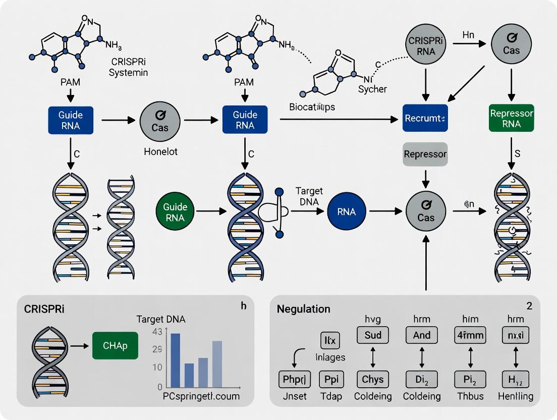
Introduction Within the broader research thesis on CRISPR interference (CRISPRi) for the dynamic regulation of metabolic pathways, the foundational technology is a repurposed CRISPR-Cas9 system. CRISPRi utilizes a catalytically dead Cas9 (dCas9) protein, which lacks endonuclease activity but retains its ability to bind DNA in a guide RNA-programmed manner. When dCas9 is targeted to a genomic locus, it creates a steric block that impedes the progression of RNA polymerase, leading to precise and reversible gene repression without altering the DNA sequence. This application note details the core principles, quantitative performance, and experimental protocols for implementing CRISPRi in metabolic engineering contexts.
Mechanism and Quantitative Performance The efficacy of CRISPRi is determined by the target location within the promoter or coding sequence. Repression is typically measured as a fold-change in mRNA levels or fluorescence for reporter genes. Key performance metrics from recent studies are summarized below:
Table 1: Quantitative Performance of CRISPRi Repression
| Target Gene | Organism | dCas9 Variant | Target Site (Relative to TSS) | Repression Efficiency (% Reduction) | Reference |
|---|---|---|---|---|---|
| yfgA | E. coli | dCas9 | -35 to +1 | 95-99% | Qi et al., 2013 |
| GFP Reporter | HEK293T | dCas9-KRAB | Within -50 to +300 | 85-95% | Gilbert et al., 2013 |
| ldhA (Metabolic) | E. coli | dCas9 | +50 to +150 | 70-85% | 2023, Metab. Eng. |
| titer pathway gene | S. cerevisiae | dCas9-Mxi1 | Promoter (-100 to -1) | 60-80% | 2024, Nat. Comm. |
Detailed Protocol: CRISPRi Knockdown for Metabolic Flux Analysis in E. coli
Objective: To repress a target gene in a central metabolic pathway (e.g., ldhA) and measure the resulting changes in metabolite flux.
Part 1: Vector Construction and Transformation
- Design sgRNAs: Design a 20-nt guide sequence targeting the non-template strand of the gene's promoter or early coding region (e.g., +50 to +150 downstream of the TSS). Use CRISPR design tools (e.g., Benchling) to minimize off-target effects.
- Clone sgRNA: Anneal oligonucleotides encoding the guide sequence and clone them into a CRISPRi expression plasmid (e.g., pCRISPRi) containing a dCas9 gene under an inducible promoter (e.g., pTet) and the sgRNA scaffold.
- Transform: Transform the constructed plasmid into your production E. coli strain via electroporation. Select colonies on appropriate antibiotic plates.
Part 2: Cultivation and Induction
- Inoculation: Pick a single colony and inoculate 5 mL of LB medium with antibiotic. Grow overnight at 37°C, 250 rpm.
- Dilution and Induction: Dilute the overnight culture 1:100 into fresh, pre-warmed minimal medium in a baffled flask. Grow to an OD600 of ~0.3-0.5.
- Induce dCas9 Expression: Add anhydrotetracycline (aTc) to a final concentration of 100 ng/mL to induce dCas9 expression. An uninduced (-aTc) culture serves as the control.
Part 3: Validation and Analysis
- Sampling for qPCR: At 2 and 4 hours post-induction, harvest 1 mL of culture. Isolate total RNA using a commercial kit, followed by DNase I treatment. Synthesize cDNA.
- qPCR: Perform quantitative PCR with primers for the target gene (ldhA) and a reference housekeeping gene (e.g., rpoD). Calculate fold repression using the 2^(-ΔΔCt) method.
- Metabolite Analysis: At mid-exponential phase, centrifuge culture samples. Analyze the supernatant via HPLC or GC-MS to quantify metabolite concentrations (e.g., lactate, acetate, target product). Compare flux distributions between induced and control cultures.
Title: CRISPRi Experimental Workflow for Metabolic Engineering
Title: Mechanism of dCas9-Mediated Transcriptional Interference
The Scientist's Toolkit: Essential Reagents for CRISPRi Experiments
Table 2: Key Research Reagent Solutions
| Reagent/Material | Function/Description | Example/Vendor |
|---|---|---|
| dCas9 Expression Plasmid | Vector encoding catalytically dead Cas9 (D10A, H840A mutations) under an inducible promoter. | Addgene #44249 (pInducer-dCas9) |
| sgRNA Cloning Backbone | Plasmid containing the sgRNA scaffold for inserting target-specific 20nt guides. | Addgene #44251 (pCRISPRi) |
| Inducer Molecule | Small molecule to precisely control dCas9 expression (e.g., aTc, IPTG). | Anhydrotetracycline (aTc) |
| High-Fidelity Polymerase | For accurate amplification of DNA fragments during vector construction. | Q5 High-Fidelity DNA Polymerase |
| RNA Isolation Kit | For pure, DNase-treated total RNA extraction for downstream qPCR validation. | RNeasy Mini Kit |
| Reverse Transcriptase | Enzyme to synthesize cDNA from isolated RNA templates. | SuperScript IV |
| SYBR Green qPCR Master Mix | For quantitative real-time PCR to measure target gene mRNA levels. | PowerUp SYBR Green Master Mix |
| Metabolite Standards | Pure chemical standards for calibrating HPLC/GC-MS analysis of extracellular metabolites. | Sigma-Aldrich Certified Reference Materials |
Within the context of CRISPR interference (CRISPRi) for dynamic regulation of metabolic pathways, the choice between gene knockdown and knockout is pivotal. Permanent knockouts (via CRISPR-Cas9 nuclease) can reveal gene essentiality but lack the nuance to study essential genes or dynamic system responses. Reversible and tunable CRISPRi knockdowns, using a deactivated Cas9 (dCas9) fused to transcriptional repressors, enable precise, dose-dependent gene silencing. This application note details protocols and advantages for employing CRISPRi to reversibly regulate metabolic fluxes, facilitating the study of bottleneck identification, toxicity mitigation, and optimal yield determination in pathway engineering.
Comparative Analysis: Knockdowns vs. Knockouts
Table 1: Core Characteristics and Applications
| Feature | CRISPRi Knockdown (dCas9-Srepressor) | CRISPR Knockout (Cas9 Nuclease) |
|---|---|---|
| Genetic Alteration | Epigenetic/Transcriptional repression | DNA double-strand break, indel mutations |
| Reversibility | Fully reversible upon repressor removal/induction | Typically permanent |
| Tunability | Graded repression via guide RNA design & expression level | Binary (functional vs. non-functional allele) |
| Target Range | Any transcriptional unit (including essential genes) | Non-essential genes only (essential=lethal) |
| Primary Application in Metabolism | Dynamic flux tuning, identifying optimal expression levels, bypassing toxicity | Validating essentiality, removing competing pathways |
| Common Repressors | KRAB, Mxi1, SID4x | N/A |
| Key Readouts | mRNA levels (qRT-PCR), protein levels (Western), metabolite titers (HPLC/MS) | DNA sequencing, phenotypic survival assays |
Table 2: Quantitative Performance Metrics
| Parameter | Typical CRISPRi Efficiency | Typical CRISPR-KO Efficiency | Measurement Method |
|---|---|---|---|
| Max. Transcript Reduction | 80-99% (varies by target) | 100% (functional null) | RNA-seq, qRT-PCR |
| Titration Range | 5-95% of baseline expression | Not applicable | Flow cytometry (reporter) |
| Reversal Kinetics (to 50% original expression) | 24-72 hours (depends on repressor degradation) | N/A | Time-course qRT-PCR |
| Multiplexing Capacity | High (>5 genes simultaneously) | Moderate (limited by repair efficiency) | NGS of target sites |
| Off-target Transcriptional Effects | Low (specified by guide RNA) | Higher (due to off-target DNA cleavage) | RNA-seq, ChIP-seq |
Detailed Protocols
Protocol 1: CRISPRi System Setup for Tunable Knockdown inE. coliorS. cerevisiae
Objective: Construct a CRISPRi platform for tunable, reversible repression of a metabolic pathway gene.
Materials: See "The Scientist's Toolkit" below.
Procedure:
- Design sgRNAs: For your target gene, design 2-3 sgRNAs targeting the non-template strand near the transcriptional start site (TSS, -50 to +300 bp). Use validated algorithms (e.g., CHOPCHOP). Include a negative control sgRNA with no genomic match.
- Clone sgRNAs: Clone annealed oligonucleotides encoding the sgRNA spacer into your CRISPRi expression vector (e.g., pCRISPRi) at the appropriate restriction site (e.g., BsaI).
- Transform: Transform the constructed plasmid into your microbial strain harboring a genomically integrated or plasmid-borne dCas9-repressor (e.g., dCas9-Sso7d for prokaryotes, dCas9-Mxi1 for yeast).
- Induction & Titration:
- For tunable knockdown, induce the dCas9-repressor and/or sgRNA expression with a titratable inducer (e.g., varying concentrations of anhydrotetracycline, aTc).
- For reversibility testing, induce repression for 24-48 hours, then wash cells and plate on media without inducer to restore expression.
- Validation:
- Harvest samples at 0, 6, 12, 24, 48 hours post-induction.
- Quantify mRNA knockdown via qRT-PCR (see Protocol 2).
- Correlate with metabolite production using HPLC-MS (see Protocol 3).
Protocol 2: qRT-PCR for Quantifying Transcriptional Knockdown
Objective: Precisely measure changes in target gene mRNA levels following CRISPRi induction.
Procedure:
- RNA Extraction: Extract total RNA from 1-5 x 10^8 cells using a column-based kit with on-column DNase I treatment.
- cDNA Synthesis: Using 500 ng total RNA, perform reverse transcription with random hexamers and a reverse transcriptase.
- qPCR Setup: Prepare reactions in triplicate with SYBR Green master mix, cDNA template (1:10 dilution), and gene-specific primers. Include a housekeeping gene (e.g., rpoB for bacteria, ACT1 for yeast).
- Run & Analyze: Run on a real-time PCR system. Calculate relative expression (ΔΔCt method) comparing induced samples to uninduced controls and the negative control sgRNA.
Protocol 3: Metabolite Titer Analysis via HPLC-MS
Objective: Link transcriptional knockdown to changes in metabolic pathway output.
Procedure:
- Sample Preparation: After CRISPRi induction, centrifuge culture broth. Filter supernatant (0.22 μm). For intracellular metabolites, perform a rapid methanol/quenching extraction.
- HPLC Conditions: Use a reverse-phase C18 column. Mobile phase A: 0.1% Formic acid in H2O; B: 0.1% Formic acid in acetonitrile. Gradient: 5% B to 95% B over 15 min.
- MS Detection: Use electrospray ionization (ESI) in positive or negative mode. Perform selected ion monitoring (SIM) for your target metabolite(s).
- Quantification: Generate a standard curve with pure metabolite. Integrate peak areas and calculate concentrations from the curve.
Pathway and Workflow Visualizations
Title: CRISPRi Mediated Transcriptional Repression Impacts Metabolic Flux
Title: Experimental Workflow for Dynamic Metabolic Pathway Tuning with CRISPRi
The Scientist's Toolkit
| Research Reagent / Material | Function & Application |
|---|---|
| dCas9-Repressor Plasmid | Constitutively or inducibly expresses the dCas9 protein fused to a transcriptional repression domain (e.g., KRAB). Backbone of the CRISPRi system. |
| sgRNA Expression Vector | Plasmid containing the sgRNA scaffold; allows for cloning of target-specific 20nt spacer sequences. Often includes selectable marker. |
| Titratable Inducer (e.g., aTc, IPTG) | Small molecule used to precisely control the timing and level of dCas9 and/or sgRNA expression, enabling tunable knockdown. |
| RNA Extraction Kit with DNase | For high-quality, genomic DNA-free total RNA isolation, essential for accurate downstream transcriptional analysis (qRT-PCR, RNA-seq). |
| SYBR Green qRT-PCR Master Mix | All-in-one mix for quantitative reverse transcription PCR, enabling sensitive and accurate quantification of target mRNA levels. |
| HPLC-MS System | Analytical platform for separating, identifying, and quantifying target metabolites in complex culture supernatants or cell extracts. |
| Metabolite Standards | Pure chemical standards for target pathway metabolites; required for generating calibration curves for absolute quantification via HPLC-MS. |
| Next-Gen Sequencing Kit | For deep sequencing of sgRNA libraries or whole transcriptomes (RNA-seq) to assess multiplexing efficiency and global off-target effects. |
Within the broader thesis on employing CRISPR interference (CRISPRi) for the dynamic and tunable regulation of metabolic pathways in microbial and mammalian systems, the precise selection and configuration of core components are paramount. This application note details the essential triad—catalytically dead Cas9 (dCas9), single guide RNA (sgRNA) architecture, and repressor domains—that underpin effective transcriptional repression for metabolic engineering and drug target validation.
Key Components: Function and Quantitative Comparison
Catalytically Dead Cas9 (dCas9)
dCas9 is a mutated form of Streptococcus pyogenes Cas9 (commonly D10A and H840A mutations) that retains its ability to bind DNA based on sgRNA complementarity but lacks endonuclease activity. It serves as a programmable DNA-binding scaffold for effector domains.
Table 1: Common dCas9 Orthologs and Properties
| dCas9 Variant | PAM Sequence | Size (aa) | Binding Efficiency (Relative to Wild-Type) | Common Host Systems |
|---|---|---|---|---|
| S. pyogenes (SpdCas9) | 5'-NGG-3' | 1368 | ~100% (Binding) | E. coli, Yeast, Mammalian |
| S. aureus (SadCas9) | 5'-NNGRRT-3' | 1053 | ~95% | Mammalian (AAV delivery) |
| C. jejuni (CjdCas9) | 5'-NNNNRYAC-3' | 984 | ~90% | Mammalian |
sgRNA Architecture
The sgRNA is a chimeric RNA that combines the CRISPR RNA (crRNA) for target recognition and the trans-activating crRNA (tracrRNA) for dCas9 binding. Its architecture, especially the length of the spacer sequence and scaffold stability, dictates targeting specificity and repression efficiency.
Table 2: Impact of sgRNA Spacer Length on Repression Efficiency in E. coli
| Spacer Length (nt) | Target Promoter | Repression Efficiency (%) | Off-Target Score (Predicted) |
|---|---|---|---|
| 20 | Plac | 98.2 ± 1.1 | 75 |
| 18 | Plac | 95.7 ± 2.3 | 85 |
| 17 | Plac | 87.4 ± 3.5 | 90 |
| 20 | Ptet | 99.0 ± 0.5 | 70 |
Repressor Domains
Effector domains fused to dCas9 mediate transcriptional repression by recruiting chromatin-modifying complexes or blocking RNA polymerase. The Krüppel-associated box (KRAB) domain from human KOX1 and the Mxi1 domain are widely used.
Table 3: Comparison of Common Repressor Domains Fused to dCas9
| Repressor Domain | Origin | Size (aa) | Mechanism | Repression Fold-Change (Mammalian Cells)* | Notes |
|---|---|---|---|---|---|
| KRAB | Human KOX1 | ~45 | Recruits HP1, SETDB1 for H3K9me3 | 50-200x | Strong, can cause heterochromatin spreading |
| Mxi1 | Human Mxi1 | ~90 | Recruits Sin3/HDAC complex | 20-100x | Potentially more tunable, less leaky |
| SRDX | Plant SUPERMAN | 12 | Recruits corepressors (putative) | 5-20x | Small size, useful in plants |
| WRPW | Hes1 | 4 | Recruits TLE corepressors | 10-50x | Minimal peptide |
*Fold-change in mRNA reduction for a strongly expressed reporter gene.
Application Notes for Metabolic Pathway Regulation
- Multiplexing for Pathway Control: Simultaneous repression of multiple genes (e.g., competitive branch pathways) is achieved by co-expressing a single dCas9-repressor with multiple sgRNAs. A recent study in S. cerevisiae demonstrated a 3.5-fold increase in itaconic acid yield by repressing three endogenous genes.
- Dynamic Control: Placing dCas9 or sgRNA expression under inducible promoters (e.g., Tet-On, arabinose) allows temporal regulation. This is critical for balancing growth and production phases.
- Tunable Repression: Using weaker promoters to drive sgRNA expression or engineered sgRNAs with mismatches can generate a gradient of repression strength, enabling fine-tuning of metabolic fluxes.
Detailed Protocol: CRISPRi-Mediated Repression inE. colifor Metabolic Flux Analysis
Materials and Reagents (The Scientist's Toolkit)
Table 4: Essential Research Reagent Solutions
| Item | Function/Description | Example Product/Catalog # |
|---|---|---|
| pDCas9-KRAB Plasmid | Expresses dCas9 fused to KRAB domain under inducible control. Addgene #110821 | |
| pgRNA (or sgRNA Expression Plasmid) | Cloning vector for expressing sgRNA with a modular spacer region. | Addgene #44251 |
| Q5 High-Fidelity DNA Polymerase | For error-free PCR of sgRNA spacer inserts. | NEB M0491S |
| T4 DNA Ligase | For cloning spacer sequences into sgRNA scaffold plasmid. | NEB M0202S |
| Chemically Competent E. coli (e.g., DH5α, MG1655) | For plasmid propagation and metabolic engineering strain. | NEB C2987H |
| Luria-Bertani (LB) Broth & Agar | Standard microbial growth media. | |
| Anhydrous Tetracycline (aTc) | Inducer for dCas9-KRAB expression in common systems. | Sigma 37919 |
| RNAprotect Bacteria Reagent | Stabilizes bacterial RNA for downstream transcriptomics. | Qiagen 76506 |
| RT-qPCR Kit (One-Step) | Quantifies repression efficiency of target metabolic genes. | ThermoFisher A15299 |
Protocol: sgRNA Cloning and Transformation
Day 1: Spacer Oligo Annealing & Cloning
- Design spacer sequences (20-nt) complementary to the non-template strand within -35 to +10 region of the target promoter. Avoid off-targets using software (e.g., CHOPCHOP).
- Resuspend forward and reverse oligonucleotides (with overhangs compatible with your sgRNA plasmid, e.g., BsaI sites) to 100 µM. Mix 1 µL of each oligo with 23 µL of annealing buffer (10 mM Tris, 50 mM NaCl, 1 mM EDTA, pH 8.0).
- Anneal in a thermocycler: 95°C for 5 min, ramp down to 25°C at 0.1°C/sec.
- Dilute annealed oligo 1:200 in nuclease-free water.
- Digest 1 µg of pgRNA plasmid with BsaI-HFv2 in CutSmart buffer at 37°C for 1 hour. Gel-purify the linearized vector.
- Set up ligation: 50 ng linearized vector, 1 µL diluted annealed oligo, 5 µL 2X Quick Ligase Buffer, 0.5 µL Quick Ligase, H2O to 10 µL. Incubate at 25°C for 10 minutes.
- Transform 2 µL ligation into 50 µL competent DH5α cells. Plate on LB + appropriate antibiotic (e.g., carbenicillin).
Day 2: Colony PCR & Sequence Verification
- Pick 4-6 colonies. Perform colony PCR using sgRNA scaffold-specific primers.
- Run PCR product on a 2% agarose gel. Positive clones show a band increase corresponding to spacer insertion.
- Inoculate positive colony for plasmid miniprep and Sanger sequence using the forward primer.
Day 3: Co-transformation into Production Strain
- Transform the sequence-verified sgRNA plasmid together with the pDCas9-KRAB plasmid into your chosen E. coli production strain (e.g., MG1655). Use selective plates with both antibiotics.
- Pick a colony to inoculate a starter culture.
Protocol: Induction and Evaluation of Repression
Day 4: Induction and Sampling
- Inoculate main culture (e.g., 10 mL) from starter culture. Grow to mid-log phase (OD600 ~0.4-0.6).
- Add inducer (e.g., 100 ng/mL aTc) to experimental culture. Leave a control culture uninduced.
- Incubate for 4-6 hours post-induction.
- Harvest 1 mL of culture for RNA extraction (use RNAprotect). Harvest 2 mL for metabolite analysis (e.g., HPLC for pathway product).
Day 5: Analysis
- Extract total RNA, perform DNase I treatment.
- Carry out one-step RT-qPCR for target gene(s) and 2-3 housekeeping genes (e.g., rpoB, gyrB).
- Calculate repression fold-change using the 2^(-ΔΔCt) method, comparing induced vs. uninduced samples.
- Correlate transcript levels with metabolite flux data.
Visualization Diagrams
Title: Mechanism of dCas9-KRAB Mediated Transcriptional Repression
Title: CRISPRi Workflow for Metabolic Gene Repression
Why Metabolic Pathways? Addressing the Need for Dynamic Flux Control in Bioproduction.
Within the broader thesis on CRISPR interference (CRISPRi) for dynamic regulation of metabolic pathways, this Application Note establishes the fundamental rationale. Static metabolic engineering often leads to imbalances, as precursors and energy are diverted from growth to product synthesis. Dynamic flux control, enabled by tools like CRISPRi, allows pathway regulation in response to metabolic cues, optimizing the host cell's metabolism for both robust growth and high titer, rate, and yield (TRY).
Key Quantitative Data on Static vs. Dynamic Metabolic Engineering
Table 1: Performance Comparison of Static Knockout vs. Dynamic CRISPRi Regulation in Model Bioproduction Systems
| Host Organism | Target Pathway/Product | Static Approach (Knockout/Mutation) | Dynamic Approach (CRISPRi-based) | Key Metric Improvement (Dynamic vs. Static) | Reference (Year) |
|---|---|---|---|---|---|
| E. coli | Fatty Alcohols | Competing pathway knockout (e.g., fadD) | CRISPRi repression of fadD tuned by acyl-CoA sensor | 2.5-fold increase in final titer (5.2 g/L vs. 2.1 g/L) | Liu et al. (2022) |
| S. cerevisiae | Beta-Carotene | Constitutive overexpression of all pathway genes | CRISPRi-mediated downregulation of ergosterol branch upon sensing high acetyl-CoA | Biomass increased by 40%; Product yield per cell increased 3-fold | Zhang et al. (2023) |
| B. subtilis | N-Acetylglucosamine (GlcNAc) | Attenuation of gamA (catabolic gene) via promoter replacement | CRISPRi repression of gamA induced by extracellular GlcNAc | Rate (productivity) improved by 110% (0.38 g/L/h vs. 0.18 g/L/h) | Sun et al. (2021) |
| C. glutamicum | L-Lysine | Deletion of dapA feedback inhibition | CRISPRi knockdown of dapA during growth phase, release at stationary phase | Yield (g/g glucose) improved from 0.25 to 0.35 (40% increase) | Wang et al. (2023) |
Experimental Protocols
Protocol 1: Construction of a CRISPRi System for Inducible Pathway Repression inE. coli
Objective: To repress a target gene (geneX) in a metabolic pathway using aTe-inducible dCas9.
Materials:
- E. coli production strain.
- Plasmid pKD-dCas9 (or similar), constitutively expressing dCas9.
- Plasmid pCRISPRi-sgRNA_geneX: Contains sgRNA targeting geneX, aTc-inducible promoter driving sgRNA expression, and a selective marker.
- SOC media, LB agar plates with appropriate antibiotics (e.g., Kanamycin, Chloramphenicol).
- Anhydrotetracycline (aTc) stock solution (100 ng/µL in 70% EtOH).
Procedure:
- Transformation: Co-transform chemically competent E. coli production strain with 50 ng each of pKD-dCas9 and pCRISPRi-sgRNA_geneX plasmids. Recover cells in 1 mL SOC media at 37°C for 1 hour.
- Selection: Plate 100 µL of recovered cells on LB agar plates containing both antibiotics. Incubate overnight at 37°C.
- Culture & Induction: Inoculate a single colony into 5 mL LB with antibiotics. Grow overnight. Dilute culture 1:100 into fresh medium (e.g., M9 minimal medium with antibiotics). Grow at 37°C until OD600 ~0.4-0.6.
- Induction: Add aTc to the culture to a final concentration of 100 ng/mL. Maintain an uninduced control.
- Sampling & Analysis: Monitor growth (OD600) and sample cells at 2, 4, 6, and 8 hours post-induction.
- qRT-PCR: Isolate RNA, synthesize cDNA, and perform qPCR to quantify geneX mRNA levels relative to a housekeeping gene.
- Product Titer: Analyze supernatant via HPLC or GC-MS to quantify target metabolite.
Protocol 2: Dynamic Two-Stage Fermentation Using CRISPRi for Growth/Production Decoupling
Objective: To implement a fermentation process where CRISPRi is activated only after a sufficient biomass (growth phase) is achieved.
Materials:
- S. cerevisiae strain with integrated dCas9 and pathway-specific sgRNA under a stationary-phase inducible promoter (e.g., HSP12 or TEF1).
- Bioreactor with pH and DO control.
- Defined fermentation medium (e.g., SMG).
- Inducer (e.g., ethanol for HSP12 promoter, or temperature shift if using a heat-shock promoter).
Procedure:
- Batch Growth Phase: Inoculate bioreactor to initial OD600 of 0.1. Maintain optimal growth conditions (e.g., 30°C, pH 5.0). Allow cells to grow exponentially without induction.
- Induction Trigger: When OD600 reaches a pre-determined threshold (e.g., 20, indicating entry into late exponential/stationary phase), initiate the induction signal.
- For chemical inducer: Add sterile-filtered ethanol to a final concentration of 3% (v/v).
- For temperature shift: Rapidly increase bioreactor temperature to 37°C.
- Production Phase: Maintain induced conditions for 48-72 hours. Continuously monitor and control pH, dissolved oxygen, and temperature.
- Process Monitoring: Take regular samples (every 6-12 hours).
- Biomass: Measure OD600 and dry cell weight (DCW).
- Substrate/Product: Analyze medium samples via HPLC for glucose consumption and product formation. Calculate yield (Yp/s) and productivity (g/L/h).
- Metabolomics: Perform LC-MS on intracellular extracts at key time points to profile flux changes.
Visualizations
Title: Static vs Dynamic Metabolic Engineering Outcomes
Title: CRISPRi Mechanism for Dynamic Flux Control
The Scientist's Toolkit: Research Reagent Solutions
Table 2: Essential Materials for Dynamic Pathway Regulation with CRISPRi
| Item | Function in Research | Example Product/Catalog Number |
|---|---|---|
| dCas9 Expression Vector | Constitutively or inducibly expresses catalytically dead Cas9, the RNA-guided DNA binding platform for repression. | pDcas9-bacteria (Addgene #44249), pRS-dCas9 (yeast, Addgene #133250) |
| sgRNA Cloning Kit | Modular system for quickly synthesizing and cloning sgRNA sequences targeting specific metabolic genes into expression vectors. | CRISPRi sgRNA Oligo Pairs (Integrated DNA Technologies), Golden Gate Assembly kits (e.g., NEBuilder) |
| Inducer Molecules | Small molecules to precisely time the onset of CRISPRi-mediated repression (e.g., at high biomass). | Anhydrotetracycline (aTc), Isopropyl β-d-1-thiogalactopyranoside (IPTG), Arabinose |
| Metabolite Biosensor Plasmids | Encodes a transcription factor that activates the sgRNA promoter in response to a key intracellular metabolite (e.g., acyl-CoA, malonyl-CoA). | pSenSpec (malonyl-CoA sensor, Addgene #149999) |
| qRT-PCR Master Mix | For quantifying changes in mRNA levels of target metabolic genes following CRISPRi induction, confirming repression. | iTaq Universal SYBR Green One-Step Kit (Bio-Rad) |
| Metabolite Analysis Standards | Authentic chemical standards for calibrating HPLC or GC-MS to accurately measure substrate consumption and product formation titers. | Supeleo/Sigma-Aldrich Analytical Standards (e.g., Fatty Alcohols, Organic Acids) |
| Defined Fermentation Medium | Chemically consistent medium for reproducible bioreactor runs, essential for calculating yields (Yp/s). | M9 Minimal Salts (Thermo Fisher), BD Difco Yeast Nitrogen Base |
| Chromatin-Immunoprecipitation (ChIP) Kit | Validates dCas9 binding at the intended genomic target site, confirming on-target activity. | SimpleChIP Plus Kit (Cell Signaling Technology) |
Within the framework of a thesis on the dynamic regulation of metabolic pathways, precise genetic tools are paramount. CRISPR interference (CRISPRi) has emerged as a powerful technique for reversible transcriptional repression. This Application Note delineates the functional and methodological distinctions between CRISPRi and related technologies—CRISPR activation (CRISPRa), RNA interference (RNAi), and traditional gene knockouts—providing protocols for their implementation in metabolic engineering and drug discovery contexts.
Mechanism of Action
CRISPRi: A catalytically dead Cas9 (dCas9) is fused to a transcriptional repressor domain (e.g., KRAB) and guided to a target gene's promoter or early coding region, sterically blocking RNA polymerase or recruiting chromatin-condensing machinery to silence transcription.
CRISPRa: Utilizes dCas9 fused to transcriptional activator domains (e.g., VPR, p65AD) and is guided to gene promoter regions to recruit transcriptional machinery, upregulating gene expression.
RNAi: Double-stranded small interfering RNA (siRNA) or short hairpin RNA (shRNA) is processed by the cell's Dicer enzyme and loaded into the RNA-induced silencing complex (RISC), which binds and cleaves complementary mRNA sequences, leading to post-transcriptional degradation.
Traditional Knockout: Utilizes homologous recombination or nuclease-based methods (e.g., CRISPR-Cas9 with double-strand breaks) to create frameshift mutations or deletions in the genomic DNA, resulting in permanent gene disruption.
Key Characteristics & Quantitative Comparison
Table 1: Comparative Analysis of Gene Silencing/Modulation Technologies
| Feature | CRISPRi | CRISPRa | RNAi | Traditional Knockout |
|---|---|---|---|---|
| Primary Target | Genomic DNA (Transcription) | Genomic DNA (Transcription) | mRNA (Post-Transcription) | Genomic DNA (Sequence) |
| Effect on Gene | Transcriptional Repression | Transcriptional Activation | mRNA Degradation | Permanent Disruption |
| Reversibility | Reversible | Reversible | Reversible (Transient) | Irreversible |
| Specificity | Very High (DNA targeting) | Very High (DNA targeting) | High (Off-target RNA common) | High (Potential off-target DSBs) |
| Kinetics | Moderate (Hours) | Moderate (Hours) | Fast (Hours) | Slow (Days to establish) |
| Persistence | Sustained while present | Sustained while present | Transient (days) | Permanent & heritable |
| Typical Efficiency | 70-95% repression | 2-20 fold activation | 70-90% knockdown | Variable (often >80%) |
| Multiplexing Ease | High (via arrays of gRNAs) | High (via arrays of gRNAs) | Moderate (co-transfection) | Challenging |
| Primary Application | Tunable knockdowns, essential gene study, dynamic regulation | Gene overexpression, gain-of-function screens, differentiation | Transient knockdowns, drug target validation | Complete gene ablation, generation of stable cell lines |
Detailed Protocols
Protocol 1: CRISPRi for Metabolic Pathway Gene Repression
Application: Dynamically downregulating a competing branch in a biosynthetic pathway.
Materials: See "Research Reagent Solutions" below. Workflow:
- Design gRNAs: Design 2-3 gRNAs targeting the promoter region (typically -50 to +300 bp relative to TSS) of the target metabolic gene using validated algorithms (e.g., CRISPick). Cloning into a CRISPRi vector (e.g., pLV hU6-sgRNA hUbC-dCas9-KRAB-P2A-Bsd).
- Lentivirus Production: Co-transfect HEK293T cells with the CRISPRi plasmid and packaging plasmids (psPAX2, pMD2.G) using PEI transfection reagent. Harvest virus-containing supernatant at 48 and 72 hours.
- Cell Line Generation: Transduce your target cell line (e.g., CHO, HEK293) with the lentiviral supernatant + polybrene (8 µg/mL). Begin selection with Blasticidin (5-10 µg/mL) 48 hours post-transduction for 5-7 days.
- Validation: Harvest cells 7 days post-selection. Assess repression via qRT-PCR (mRNA level) and Western Blot or targeted metabolomics (functional output).
- Pathway Flux Analysis: Implement a metabolomics protocol (see below) to quantify changes in pathway intermediates.
Protocol 2: Metabolite Profiling for Pathway Flux Assessment
Application: Quantifying the metabolic consequence of CRISPRi-mediated repression.
Workflow:
- Quenching & Extraction: Rapidly quench 5x10^6 cells in 60% cold aqueous methanol (-40°C). Vortex and incubate at -40°C for 30 min.
- Centrifugation: Centrifuge at 16,000 x g for 15 min at 4°C. Transfer supernatant to a new tube.
- Sample Concentration: Dry the supernatant in a vacuum concentrator.
- Derivatization & Analysis: Reconstitute in 20 µL Methoxyamine hydrochloride (20 mg/mL in pyridine) and incubate at 37°C for 90 min. Then add 40 µL MSTFA + 1% TMCS and incubate at 37°C for 30 min. Analyze by GC-MS.
- Data Processing: Integrate metabolite peaks, normalize to an internal standard (e.g., ribitol) and cell count. Compare normalized abundances between CRISPRi and control cells.
Visualization of Concepts and Workflows
Title: CRISPRi Transcriptional Repression Mechanism
Title: Technology Selection Based on Research Goal
Title: CRISPRi Experimental Workflow for Metabolic Studies
The Scientist's Toolkit: Research Reagent Solutions
Table 2: Essential Reagents for CRISPRi Metabolic Pathway Experiments
| Item | Function & Description | Example Product/Catalog |
|---|---|---|
| dCas9-KRAB Expression Vector | Lentiviral backbone for stable delivery of the CRISPRi machinery. Contains dCas9 fused to the KRAB repression domain. | Addgene #71237 (pLV hU6-sgRNA hUbC-dCas9-KRAB-Bsd) |
| sgRNA Cloning Vector | Backbone for inserting target-specific gRNA sequences, often with a U6 promoter. | Addgene #104875 (pU6-sgRNA EF1Alpha-puro-T2A-BFP) |
| Lentiviral Packaging Plasmids | Required for production of non-replicative viral particles (psPAX2 for gag/pol, pMD2.G for VSV-G envelope). | Addgene #12260 (psPAX2), #12259 (pMD2.G) |
| Polycation Transfection Reagent | For efficient plasmid delivery into packaging cells (e.g., HEK293T). | Polyethylenimine (PEI) Max, Linear, MW 40,000 |
| Polybrene | A cationic polymer that enhances viral transduction efficiency by neutralizing charge repulsion. | Hexadimethrine bromide, 8 mg/mL stock |
| Selection Antibiotic | Selects for cells successfully transduced with the CRISPRi construct. | Blasticidin S HCl, Puromycin Dihydrochloride |
| gRNA Design Tool | Online platform for designing specific, high-efficiency gRNAs with minimal off-target effects. | Broad Institute CRISPick (crispick.broadinstitute.org) |
| Metabolite Extraction Solvent | Cold, aqueous methanol for rapid quenching of metabolism and extraction of intracellular metabolites. | LC-MS Grade Methanol (e.g., 60% in H2O, -40°C) |
| Derivatization Reagents | For GC-MS metabolomics; methoxyamine for carbonyl protection, MSTFA for silylation. | Methoxyamine hydrochloride, N-Methyl-N-(trimethylsilyl)trifluoroacetamide (MSTFA) |
| Internal Standard for Metabolomics | Added at extraction for normalization of sample-to-sample variation. | Ribitol, Succinic acid-d4, 13C-labeled amino acid mix |
From Design to Fermentation: A Step-by-Step Guide to Implementing CRISPRi
This application note guides the selection of dCas9 and transcriptional repressor fusion proteins for CRISPR interference (CRISPRi) experiments across three major host organisms: E. coli, yeast (S. cerevisiae), and mammalian cells. It is designed as the initial step for a thesis focused on applying dynamic CRISPRi regulation to metabolic engineering and pathway optimization. Proper selection is critical for achieving strong, specific repression with minimal off-target effects.
Quantitative Comparison of Key dCas9-Repressor Systems
Table 1: Performance Summary of Common dCas9-Repressor Systems by Host
| Host Organism | Recommended dCas9 Variant | Common Repressor Fusions | Repression Efficiency (Typical Range) | Key Considerations & Citations |
|---|---|---|---|---|
| E. coli | dCas9 from S. pyogenes (SpdCas9) | Mxi1, ω, KRAB (eukaryotic domains often less effective) | 300-fold (Mxi1) to 10-fold (KRAB) | Mxi1 is most effective prokaryotic repressor. N-terminal fusions often perform better. Requires codon optimization. (1, 2) |
| Yeast (S. cerevisiae) | SpdCas9 (codon-optimized) | Mxi1, Ssn6, RD2-SID (RNA pol II CTD fragment) | 10-fold to >100-fold (Mxi1) | Ssn6 (Cyc8) is a native yeast global repressor. Mxi1 is highly effective. Constitutive or inducible dCas9 expression available. (3, 4) |
| Mammalian Cells | SpdCas9, SaCas9 (smaller size) | KRAB (Krüppel-associated box), SID4X, MeCP2, DNMT3A | 5-fold to 100-fold (KRAB) | KRAB is gold standard, recruits endogenous repression machinery. SaCas9 useful for AAV delivery. Fusion position (N vs C-term) matters. (5, 6) |
Table 2: Key Properties of dCas9 Variants for Host Selection
| dCas9 Variant | PAM Sequence | Protein Size (aa) | Common Use in Host | Notes |
|---|---|---|---|---|
| SpdCas9 (S. pyogenes) | 5'-NGG-3' | 1368 | All (E. coli, yeast, mammalian) | Most widely used, best characterized. Large size can be challenging for viral delivery. |
| SadCas9 (S. aureus) | 5'-NNGRRT-3' | 1053 | Mammalian (AAV delivery), Yeast | Smaller size enables packaging into AAV. Broader PAM. |
| CjCas9 (C. jejuni) | 5'-NNNNRYAC-3' | 984 | Mammalian (in vivo) | Very small, good for in vivo applications. Specific PAM. |
Detailed Experimental Protocols
Protocol 1: Cloning a dCas9-Repressor Fusion Construct forE. coli
Objective: Assemble a plasmid expressing a SpdCas9-Mxi1 fusion protein under inducible control for use in E. coli.
Materials: See "The Scientist's Toolkit" below. Procedure:
- Vector Preparation: Linearize the destination plasmid (e.g., pZA21 derivative with arabinose-inducible promoter) using restriction enzymes that remove the existing MCS or dummy fragment. Gel-purify the backbone.
- Insert Amplification: PCR amplify the dCas9 gene (codon-optimized for E. coli) from a template (e.g., pdCas9-bacteria, Addgene #44249). Use primers that add a Gly-Ser linker sequence (GGT AGC) to the 3' end.
- Repressor Fusion: Amplify the mxi1 gene from a synthesized fragment. Use primers that add complementary overhangs to the dCas9 linker on the 5' end and a transcriptional terminator sequence on the 3' end.
- Gibson Assembly: Mix 50-100 ng of linearized vector with a 2:1 molar ratio of the dCas9 and mxi1 inserts. Add 15 µl of Gibson Assembly Master Mix. Incubate at 50°C for 1 hour.
- Transformation & Screening: Transform 5 µl of assembly reaction into high-efficiency DH5α competent cells. Plate on appropriate antibiotic. Screen colonies by colony PCR using primers flanking the insertion site. Confirm sequence via Sanger sequencing.
- Co-transform with a separate plasmid expressing the sgRNA under a constitutive promoter (e.g., J23119).
Protocol 2: Validating Repression Efficiency in Yeast
Objective: Quantify the knockdown efficiency of a dCas9-Ssn6 system on a target reporter gene (e.g., yEGFP).
Materials: Yeast strain with integrated yEGFP reporter, plasmid expressing dCas9-Ssn6 (e.g., from pCfB series), plasmid expressing target sgRNA. Procedure:
- Strain Generation: Co-transform the haploid yeast strain with the dCas9-Ssn6 expression plasmid (LEU2 marker) and the sgRNA plasmid (HIS3 marker). Select on SD -Leu -His plates.
- Culture & Induction: Inoculate 3 independent colonies into 5 ml SD -Leu -His medium. Grow to mid-log phase (OD600 ~0.5). If dCas9 is under a galactose-inducible promoter (GAL1), induce by adding 2% galactose (repress glucose).
- Flow Cytometry Analysis: After 12-16 hours induction, dilute cells to OD600 ~0.2 in PBS. Analyze yEGFP fluorescence for at least 10,000 cells per sample using a flow cytometer (e.g., 488 nm excitation, 530/30 nm filter).
- Data Analysis: Calculate the mean fluorescence intensity (MFI) for each sample. Compare the MFI of the strain with target sgRNA to a control strain with non-targeting sgRNA. Repression efficiency = 1 - (MFItarget / MFIcontrol).
- qPCR Validation (Optional): Harvest cells, extract RNA, synthesize cDNA, and perform qPCR for the endogenous gene corresponding to the reporter to confirm transcriptional repression.
Visualizations
Title: CRISPRi Repression Complex Assembly
Title: Host-Specific CRISPRi System Selection Flow
The Scientist's Toolkit: Essential Research Reagents
Table 3: Key Reagents for CRISPRi System Construction and Validation
| Reagent/Category | Example Product/ID | Function in Experiment | Key Considerations |
|---|---|---|---|
| dCas9 Expression Backbone | pnCas9-Bacteria (Addgene #113749), pCdC9 (Yeast), lenti-dCas9-KRAB (Addgene #113169) | Provides the vector for expressing the dCas9-repressor fusion. | Check promoter compatibility (inducible vs constitutive), antibiotic resistance, and host origin of replication. |
| Repressor Domain Cloning Fragments | Synthetic gBlocks (IDT) encoding KRAB, Mxi1, Ssn6 | Used as PCR templates or Gibson Assembly fragments to fuse repressor to dCas9. | Ensure sequence is codon-optimized for host. Include flexible linkers (e.g., (GGGGS)x2). |
| Assembly Master Mix | NEBuilder HiFi DNA Assembly Master Mix (NEB), Gibson Assembly Mix | Seamlessly assembles multiple DNA fragments (dCas9, repressor, vector). | Higher fidelity than traditional restriction/ligation. |
| Competent Cells for Cloning | NEB 5-alpha, Mach1, Stbl3 (for lentiviral prep) | For plasmid assembly and propagation. | Choose high-efficiency for assembly, stable for repetitive sequences. |
| sgRNA Expression Plasmid | pCRISPRi (Addgene #113189 for E. coli), pRSI series (Yeast), lentiGuide-Puro (Addgene #113199) | Expresses the target-specific single guide RNA. | Must be compatible with dCas9 plasmid (no clash). U6 promoter common in eukaryotes. |
| Validation - qPCR Reagents | PowerUp SYBR Green Master Mix (Thermo), primers for target gene & housekeeping | Quantifies mRNA knockdown levels post-repression. | Design intron-spanning primers (mammalian). Use multiple housekeeping genes. |
| Validation - Flow Cytometry Antibody | Anti-RNA Pol II CTD (phospho S2) antibody | Can assess Pol II occupancy reduction at target site via ChIP. | Validates direct mechanistic repression. |
| Cell Culture/Transfection Reagent | Lipofectamine 3000 (mammalian), LiAc/SS carrier DNA (yeast), Electroporation (E. coli) | Delivers plasmids into the host cells. | Optimize protocol for host and plasmid size to maximize efficiency. |
Within the broader thesis on employing CRISPR interference (CRISPRi) for the dynamic, multi-level regulation of metabolic pathways, Step 2 is foundational. Precise sgRNA design dictates the efficacy and specificity of dCas9-mediated transcriptional repression. This Application Note details the strategic targeting of genomic regions—promoters, early exons, and key non-coding regions—to achieve optimal knockdown of target genes in metabolic engineering and drug discovery contexts. The protocols and data herein provide a framework for researchers to systematically design and validate sgRNAs for robust pathway modulation.
Quantitative Comparison of sgRNA Targeting Strategies
The efficacy of CRISPRi repression is highly dependent on the targeted genomic region. The following table summarizes key performance metrics based on recent pooled screening data and validation studies.
Table 1: Performance Metrics of sgRNA Targeting Strategies for CRISPRi
| Target Region | Optimal Distance from TSS | Typical Repression Efficiency (% mRNA Reduction) | Specificity (Risk of Off-Target Effects) | Key Considerations |
|---|---|---|---|---|
| Core Promoter | -50 to +1 bp relative to TSS | 70% - 95% | High | Highest efficacy. Avoids nucleosome-dense areas. Strand choice is critical. |
| Proximal Promoter / Upstream | -300 to -50 bp from TSS | 50% - 85% | High | Effective, but efficiency drops with distance. Must avoid regulatory elements for other genes. |
| Early Exon (1st, 2nd) | +100 to +300 bp from TSS | 60% - 90% | Medium-High | Very effective. dCas9 binding blocks RNA polymerase elongation. Beware of splicing effects. |
| 5' UTR | Within 100 bp downstream of TSS | 40% - 80% | High | Can be effective but variable. Secondary RNA structure may influence dCas9 binding. |
| Enhancer / Non-Coding Regulatory | N/A (element-specific) | 30% - 70% (on target gene) | Variable/Low | For epigenetic silencing. Requires prior knowledge of enhancer-gene linkages. High specificity potential. |
Detailed Experimental Protocols
Protocol 1:In SilicoDesign and Selection of sgRNAs for CRISPRi
Objective: To design a library of candidate sgRNAs targeting the promoter and early exons of a metabolic pathway gene (e.g., ACS for acetate switch control in E. coli).
Materials:
- Target organism reference genome (FASTA file).
- Gene annotation file (GTF/GFF).
- CRISPR sgRNA design tool (e.g., CHOPCHOP, Benchling, CRISPick).
- Software for off-target analysis (e.g., Cas-OFFinder, BLAST).
Procedure:
- Define Target Coordinates: Identify the Transcriptional Start Site (TSS) and gene model for your target gene using curated databases (e.g., NCBI RefSeq, Ensembl).
- Generate Candidate sgRNAs: Using your design tool, generate all possible 20-nt sgRNA sequences (preceding a 5'-NGG-3' PAM for S. pyogenes dCas9) within the target windows:
- Window A (Promoter): From -300 bp to +50 bp relative to the TSS.
- Window B (Early Exon): From the start codon (ATG) to +300 bp into the coding sequence.
- Rank and Filter: Rank sgRNAs by calculated on-target efficiency scores provided by the tool. Manually filter candidates to:
- Prioritize sgRNAs with the 5' end of the spacer at -10 to +10 bp from the TSS for maximal repression.
- Exclude sgRNAs with >90% homology to other genomic sites (potential off-targets), especially in coding regions.
- Ensure GC content between 40-60% for stable binding.
- Final Selection: Select 3-5 top-ranked sgRNAs per target gene for empirical validation. Include at least one targeting the core promoter and one targeting the first exon.
Protocol 2: Empirical Validation of sgRNA Repression Efficiency
Objective: To quantify the repression efficacy of selected sgRNAs via qRT-PCR in a model system (e.g., dCas9-expressing E. coli or HEK293T cells).
Materials:
- Stable cell line expressing dCas9 (e.g., dCas9-KRAB for mammalian cells).
- sgRNA cloning vector (e.g., lentiviral sgRNA backbone).
- Transfection or transduction reagents.
- RNA extraction kit, cDNA synthesis kit, qPCR master mix.
- Primers for target gene and housekeeping control.
Procedure:
- Clone sgRNAs: Clone each candidate sgRNA sequence (from Protocol 1) into your delivery vector. Sequence-verify all constructs.
- Deliver sgRNAs: Deliver individual sgRNA constructs into your dCas9-expressing cell line alongside a non-targeting control (NTC) sgRNA. Include a "dCas9 only" control.
- Harvest RNA: 48-72 hours post-delivery, harvest cells and extract total RNA. Synthesize cDNA.
- Quantitative PCR: Perform qPCR for your target metabolic gene (e.g., ACS) and a stable reference gene (e.g., GAPDH, rpoB).
- Analyze Data: Calculate relative gene expression using the 2^(-ΔΔCt) method. Normalize all samples to the NTC sgRNA control set to 100% expression.
- Repression Efficiency = (1 - Relative Expression) * 100%.
- Select Lead sgRNA: Identify the sgRNA yielding the highest repression efficiency with minimal impact on cell growth (assayed in parallel).
Visualization of Workflows and Mechanisms
Title: Strategic sgRNA Design and Validation Workflow
Title: How sgRNA Target Site Determines CRISPRi Mechanism
The Scientist's Toolkit: Research Reagent Solutions
Table 2: Essential Reagents for CRISPRi sgRNA Design and Validation
| Reagent / Tool | Function & Purpose | Example Product/Resource |
|---|---|---|
| dCas9-Repressor Cell Line | Provides the catalytically dead Cas9 fused to a transcriptional repressor (e.g., KRAB, Mxi1) for stable, inducible, or constitutive expression. | HEK293T-dCas9-KRAB (Addgene), E. coli JDW1397 (dCas9). |
| Modular sgRNA Cloning Vector | Backbone for efficient synthesis and delivery of sgRNA expression cassettes, often with selection markers (antibiotic, fluorescence). | lentiGuide-Puro (Addgene #52963), pCRISPRi (Addgene #44250). |
| CRISPR Design Web Tool | Identifies potential sgRNA sequences with on-target efficiency and off-target specificity scores for a given genomic input. | CHOPCHOP, Broad Institute CRISPick, Benchling. |
| Off-Target Prediction Algorithm | Computationally assesses the genome-wide specificity of candidate sgRNAs to minimize unintended binding events. | Cas-OFFinder, MIT CRISPR Design Tool specificity analysis. |
| qRT-PCR Assay Kit | Gold-standard for quantifying mRNA levels to validate target gene repression efficiency post-sgRNA delivery. | TaqMan Gene Expression Assays, SYBR Green master mixes. |
| Next-Gen Sequencing Library Prep Kit | For high-throughput validation of sgRNA activity and specificity via targeted RNA-seq or ChIP-seq for dCas9 binding. | Illumina Stranded mRNA Prep, NEBNext Ultra II DNA Library Prep. |
This protocol is designed as part of a comprehensive thesis on employing CRISPR interference (CRISPRi) for the dynamic, tunable regulation of metabolic pathways in mammalian systems. The stable integration of CRISPRi components is critical for long-term, homogeneous gene repression studies, enabling researchers to investigate metabolic flux control, identify bottlenecks, and engineer cells for bioproduction or disease modeling. This document details the latest methodologies for vector design, delivery, and the generation of clonally derived stable cell lines.
Key Vector Systems for CRISPRi Integration
Effective CRISPRi requires the stable expression of two core components: a catalytically dead Cas9 (dCas9) fused to a repressive domain (e.g., KRAB) and a single-guide RNA (sgRNA). The choice of integration system balances genomic stability, expression level, and safety.
Table 1: Comparison of Common Integration Methods for Stable Cell Line Generation
| Method | Mechanism | Typical Copy Number | Integration Site | Pros | Cons | Best For |
|---|---|---|---|---|---|---|
| Random Integration | Non-homologous end joining (NHEJ) into DSBs induced by nucleases or irradiation. | Variable, often high | Random genomic loci. | Simple protocol; high integration efficiency. | Position effects (silencing/variegation); potential insertional mutagenesis. | Rapid pool generation for initial screening. |
| Lentiviral Transduction | Viral integrase-mediated insertion. | Low (1-3 copies common) | Semi-random, favors active transcription units. | High efficiency in hard-to-transfect cells; consistent expression in pools. | Size limitations (<8kb); biosafety level 2 requirements. | Creating representative knockdown pools for metabolic studies. |
| Site-Specific Integration | Homology-directed repair (HDR) or recombinase-mediated cassette exchange (RMCE). | 1 (Precise) | Defined "safe harbor" locus (e.g., AAVS1, ROS426). | Defined genetic context; consistent expression; avoids mutagenesis. | Low efficiency; requires donor design and nucleases/recombinases. | Isogenic clonal lines for precise, publication-grade research. |
| Transposon-Based | Sleeping Beauty or PiggyBac transposase-mediated "cut-and-paste". | Variable (controllable) | TA dinucleotide sites, nearly random. | Large cargo capacity; can be excised; non-viral. | Smaller "footprint" than viruses but still random. | Delivering large constructs or multiple expression cassettes. |
Detailed Protocol: Generating Clonal CRISPRi Stable Cell Lines via Site-Specific Integration
This protocol outlines the generation of isogenic HEK293T cell lines with CRISPRi components stably integrated into the AAVS1 safe harbor locus using CRISPR-Cas9-mediated HDR.
Materials & Reagents (The Scientist's Toolkit)
Table 2: Essential Research Reagent Solutions
| Item | Function/Description | Example Product/Catalog |
|---|---|---|
| dCas9-KRAB Expression Donor Plasmid | HDR template containing dCas9-KRAB-P2A-PuroR, flanked by AAVS1 homology arms (800-1000 bp). | Addgene #113744 (pAAVS1-dCas9-KRAB-P2A-Puro) |
| sgRNA Expression Donor Plasmid | HDR template for sgRNA(s) targeting metabolic genes, with a selectable marker (e.g., Blasticidin). | Custom design, cloned into pAAVS1-sgRNA-EF1α-BlastR backbone. |
| AAVS1 Targeting sgRNA/Cas9 | Creates a double-strand break at the safe harbor locus to stimulate HDR. | Synthesized as crRNA/tracrRNA duplex or from plasmid. |
| Transfection Reagent | For delivering plasmid DNA and RNP complexes. | Lipofectamine 3000 or Neon Electroporation System. |
| Selection Antibiotics | Puromycin and Blasticidin S for selecting successfully integrated cells. | Puromycin (1-2 µg/mL), Blasticidin (5-10 µg/mL). |
| Clonal Isolation Medium | Conditioned medium or commercial supplement to support single-cell growth. | CloneR (Stemcell Technologies) or 50% conditioned medium. |
| Genomic DNA Extraction Kit | For isolating DNA for junction PCR screening. | QuickExtract DNA Solution or column-based kits. |
| PCR Reagents & Primers | For verifying 5' and 3' integration junctions and absence of random integration. | High-fidelity polymerase, primers outside homology arms and within cassette. |
| Flow Cytometer | For assessing dCas9 expression if using a fluorescent tag (e.g., GFP). | N/A |
Step-by-Step Methodology
Day 0: Cell Seeding
- Seed HEK293T cells in a 6-well plate at 30-40% confluence in complete growth medium (e.g., DMEM + 10% FBS) without antibiotics. Aim for ~70% confluence at transfection.
Day 1: Co-transfection for HDR
- Prepare the following in separate tubes:
- Tube A (RNP Complex): 2 µg of AAVS1-targeting sgRNA (or 2 µL of 100 µM crRNA:tracrRNA duplex) pre-complexed with 5 µg of Cas9 protein (or high-fidelity Cas9) in 100 µL of serum-free medium. Incubate 10 min at RT.
- Tube B (Donor DNA): 1.5 µg of dCas9-KRAB donor plasmid + 1.5 µg of sgRNA donor plasmid in 100 µL of serum-free medium.
- Tube C (Transfection Mix): Mix 10 µL of Lipofectamine 3000 reagent with 90 µL of serum-free medium.
- Combine Tubes A, B, and C. Mix gently and incubate for 20 min at RT.
- Add the total mixture dropwise to the cells. Gently rock the plate.
- Incubate cells at 37°C, 5% CO₂.
Day 2: Media Change
- ~24 hours post-transfection, replace medium with fresh complete growth medium.
Day 3: Begin Selection
- Replace medium with complete growth medium containing dual antibiotics (e.g., 1 µg/mL Puromycin + 5 µg/mL Blasticidin S).
- Change selection medium every 2-3 days. Non-transfected control cells should begin dying within 72 hours.
Day 7-10: Bulk Population Analysis & Single-Cell Sorting
- Once a resistant bulk population emerges (after 7-10 days of selection), harvest a sample for genomic DNA extraction.
- Perform junction PCR to confirm targeted integration.
- 5' Junction PCR: Forward primer upstream of 5' homology arm, reverse primer within the dCas9 transgene.
- 3' Junction PCR: Forward primer within the BlastR gene, reverse primer downstream of 3' homology arm.
- To obtain clonal lines, trypsinize the verified bulk population and sort single cells via FACS into individual wells of a 96-well plate containing 150 µL of clonal isolation medium. Alternatively, perform serial dilution in 96-well plates.
- Culture clonal lines, carefully feeding every 4-5 days.
Day 21-28: Clonal Screening & Expansion
- Once colonies are visible and ~50% confluent, expand them to 24-well plates.
- Screen clones via:
- Junction PCR (as above) to confirm correct integration.
- Off-target Integration PCR: Using primers specific to the donor cassette and primers targeting common random integration sites (e.g., Alu repeats) to confirm single-copy integration.
- Functional Assay: Transient transfection of a GFP-targeting sgRNA into a clone to test repression efficiency via flow cytometry.
- Expand 3-5 positive, high-expressing clones and cryopreserve.
Workflow and Pathway Diagrams
Diagram 1 Title: CRISPRi Stable Cell Line Generation Workflow
Diagram 2 Title: CRISPRi Mechanism for Metabolic Gene Repression
Within a thesis focused on applying CRISPR interference (CRISPRi) for the dynamic regulation of metabolic pathways, precise temporal control of dCas9 expression or guide RNA targeting is paramount. Moving beyond constitutive repression, this step explores three core induction modalities—chemical, optical, and auto-inducible systems—to enable precise, tunable, and often orthogonal temporal control over pathway flux. This allows researchers to dissect bottleneck reactions, avoid toxic intermediate accumulation, and optimize titers in metabolic engineering.
Application Notes & Comparative Analysis
Chemical Inducers
Chemical inducers offer a simple, dose-dependent method for temporal control. Systems are repurposed from classical molecular biology and integrated with CRISPRi components.
Key Systems:
- Tet-On/Off: Uses tetracycline or doxycycline to control transcription via the TetR repressor or tTA activator. Offers high induction ratios and reversibility.
- IPTG-Inducible (lac): Utilizes Isopropyl β-D-1-thiogalactopyranoside to inactivate the LacI repressor, allowing expression from the Plac or Ptrc promoters. Well-characterized but can be leaky.
- aTc-Inducible: Anhydrotetracycline controls the TetR protein, often used in tighter, more responsive systems than IPTG.
- Small Molecule Dimerizers: Compounds like rapamycin or abscisic acid (ABA) can be used to dimerize split dCas9 fragments or recruit transcriptional effectors.
Table 1: Comparison of Common Chemical Induction Systems for CRISPRi Control
| System | Inducer | Typical Concentration Range | Induction Ratio (On/Off) | Key Advantage | Key Limitation |
|---|---|---|---|---|---|
| Tet-On | Doxycycline | 10 ng/mL – 1 µg/mL | 10^2 – 10^4 | High induction, reversible, low background | Potential pleiotropic effects of doxycycline |
| Lac | IPTG | 10 µM – 1 mM | 10^1 – 10^2 | Simple, inexpensive, well-understood | Leaky expression, catabolite repression in E. coli |
| aTc/TetR | Anhydrotetracycline | 10 – 200 ng/mL | 10^2 – 10^3 | Very tight repression, fast response | Cost of aTc, light-sensitive |
| ABA-PYL/RCAR | Abscisic Acid | 1 – 100 µM | 10^1 – 10^2 | Orthogonal in mammalian/plant cells, rapid | Lower dynamic range in some contexts |
Light-Activation Systems
Optogenetics provides unparalleled temporal precision (seconds to minutes) and spatial control without adding chemical agents.
Key Systems:
- CRY2/CIB: Blue light (450 nm) induces heterodimerization of Arabidopsis thaliana proteins Cryptochrome 2 (CRY2) and CIB1. Used to recruit activators/repressors or reconstitute dCas9.
- PhyB/PIF: Red light (650 nm) induces binding between Phytochrome B (PhyB) and PIF; far-red light (750 nm) dissociates them. Requires exogenous chromophore (PCB).
- LOV Domains: Light-Oxygen-Voltage domains undergo conformational change under blue light, used to cage or expose functional domains.
Table 2: Optogenetic Systems for Temporal CRISPRi Control
| System | Wavelength | Response Time | Reversibility | Chromophore | Spatial Precision |
|---|---|---|---|---|---|
| CRY2/CIB | 450 nm (Blue) | Seconds | Slow dark reversion (~minutes) | Endogenous (FAD) | High |
| PhyB/PIF | 650 nm (Red) / 750 nm (Far-Red) | Milliseconds | Instant (with far-red) | Exogenous (PCB) | Very High |
| LOV-Jα | 450 nm (Blue) | Seconds | Fast dark reversion (~seconds) | Endogenous (FMN) | High |
Auto-Inducible Systems
These systems trigger CRISPRi activity in response to endogenous metabolic states, creating dynamic feedback loops.
Key Strategies:
- Quorum-Sensing Promoters: Use promoters activated by acyl-homoserine lactone (AHL) signals (e.g., Plux, Plas). CRISPRi activates only at high cell density.
- Metabolite-Responsive Promoters/PCRISPR arrays: Utilize promoters naturally responsive to pathway intermediates (e.g., fatty acids, sugars) to drive dCas9 or gRNA expression.
- Stress-Responsive Systems: Link dCas9 expression to stress promoters (e.g., heat shock, oxidative stress) for condition-dependent repression.
Experimental Protocols
Protocol 1: Implementing a Doxycycline-Inducible CRISPRi System inE. colifor Metabolic Pacing
Objective: To dynamically repress a target gene in a central metabolic pathway (e.g., pfkA) using a Tet-On inducible dCas9.
Materials: See "The Scientist's Toolkit" below.
Methodology:
- Strain Construction:
- Transform the host E. coli strain with a plasmid expressing dCas9 under the control of a constitutive promoter (e.g., J23119).
- Transform a second plasmid containing the gRNA targeting pfkA, expressed from a Ptet promoter. This plasmid should carry a compatible origin and antibiotic resistance.
- Plate on LB agar containing appropriate antibiotics (e.g., Spectinomycin for dCas9, Kanamycin for gRNA). Incubate at 37°C overnight.
Induction Time-Course Experiment:
- Inoculate a single colony into 5 mL LB with antibiotics. Grow overnight at 37°C, 220 rpm.
- Dilute the culture 1:100 into fresh, pre-warmed medium (e.g., M9 minimal media with appropriate carbon source) with antibiotics. Grow to mid-exponential phase (OD600 ~0.4-0.6).
- Split the culture into separate flasks. Add varying concentrations of doxycycline (0 ng/mL, 10 ng/mL, 100 ng/mL, 1000 ng/mL) to induce gRNA expression.
- Continue incubation, taking samples every hour for 6 hours for analysis.
Analysis:
- Phenotype: Measure growth (OD600) and relevant metabolite (e.g., via HPLC) from each sample.
- Repression Efficiency: Extract RNA from parallel samples (1h, 3h post-induction) and perform qRT-PCR to quantify pfkA mRNA levels relative to a housekeeping gene.
- Data Interpretation: Correlate doxycycline dose with repression level and metabolic output.
Protocol 2: Blue Light-Activated CRISPRi for Oscillatory Control in Yeast
Objective: To achieve rapid, reversible repression of a target gene using the CRY2/CIB system in S. cerevisiae.
Materials: See "The Scientist's Toolkit" below.
Methodology:
- Strain Engineering:
- Fuse the transcriptional repressor domain Mxi1 to CIB1. Fuse dCas9 to CRY2(PHR). Express both constructs from constitutive promoters.
- Integrate a gRNA expression cassette targeting your gene of interest (e.g., ADH1) into a genomic locus.
Light Induction Setup:
- Grow the engineered yeast strain to mid-log phase in synthetic complete media.
- Aliquot cultures into a multi-well plate. Place the plate inside a programmable LED array emitting 450 nm blue light.
- Program: Apply light pulses (e.g., 30 seconds ON / 5 minutes OFF) for 2 hours. Include a control plate kept in constant darkness.
Sampling and Validation:
- Take samples at the end of each ON and OFF cycle during the pulsing regime.
- Immediately flash-freeze samples for RNA extraction.
- Perform RNA-seq or targeted qRT-PCR to assess transcriptomic changes and oscillatory behavior of the target gene.
- Monitor a relevant downstream metabolic product (e.g., ethanol for ADH1 repression) to link transcriptional dynamics to pathway output.
Diagrams
Title: Chemical Induction of CRISPRi via Tet System
Title: Light-Activated CRISPRi via CRY2/CIB Dimerization
Title: Auto-Inducible CRISPRi for Metabolic Feedback
The Scientist's Toolkit: Essential Reagents for Induction Dynamics
| Item | Function & Application | Example Product/Catalog Number (Representative) |
|---|---|---|
| dCas9 Expression Plasmid | Constitutively expresses a catalytically dead Cas9 protein, the core repressor scaffold. | pnCas9-Bacteria (Addgene #113352) |
| Inducible gRNA Expression Plasmid | Plasmid with gRNA scaffold under control of an inducible promoter (Ptet, Plac, etc.). | pTarget series (Addgene #62226) with modified promoter. |
| Chemical Inducers | Small molecules used to trigger gene expression from specific systems. | Doxycycline hyclate (D9891, Sigma), IPTG (15502, Sigma), Anhydrotetracycline (37919, Sigma). |
| Optogenetic Plasmids | Vectors encoding light-sensitive protein pairs fused to dCas9/effector domains. | pCry2PHR-mCherry-N1 & pCIB1-FP (Addgene #117523, 117520) for CRY2/CIB. |
| Programmable LED Array | Device for delivering precise wavelengths and intensities of light to cell cultures. | LumaCube 450nm (Lumencor) or custom-built LED plates. |
| Quorum-Sensing Molecules | Autoinducer chemicals (AHLs) for density-dependent system testing. | N-(3-Oxododecanoyl)-L-homoserine lactone (O9139, Sigma). |
| Metabolite Standards | Pure compounds for HPLC/GC-MS calibration to quantify pathway intermediates/products. | Succinic Acid (S3674, Sigma), N-Acetylglucosamine (A3286, Sigma). |
| RNA Extraction Kit | For isolating high-quality RNA to measure repression efficiency via qRT-PCR. | RNeasy Mini Kit (Qiagen 74104) or TRIzol reagent (15596026, Thermo). |
| qRT-PCR Master Mix | For quantitative reverse transcription PCR to quantify target mRNA levels. | iTaq Universal SYBR Green One-Step Kit (1725151, Bio-Rad). |
Within the broader thesis investigating CRISPR interference (CRISPRi) for dynamic, tunable regulation of metabolic pathways, this application note presents three concrete case studies. CRISPRi, utilizing a catalytically dead Cas9 (dCas9) to repress transcription, offers a powerful tool for fine-tuning metabolic flux without genetic knockout. This approach is critical for optimizing the production of valuable compounds where balanced pathway expression is essential. The following sections detail applications in antibiotic precursor production, biofuel synthesis, and therapeutic protein manufacturing, providing protocols and data frameworks for implementation.
Case Study 1: CRISPRi for Enhanced Production of Actinorhodin (Antibiotic Precursor)
Application Note
In Streptomyces coelicolor, the polyketide antibiotic actinorhodin is produced via a complex biosynthetic pathway. Traditional genetic engineering often disrupts the delicate metabolic network. This study employed multiplexed CRISPRi to dynamically downregulate competitive branch pathways, redirecting metabolic flux toward acetyl-CoA and malonyl-CoA, key precursors for actinorhodin synthesis.
Table 1: CRISPRi-Mediated Enhancement of Actinorhodin Production in S. coelicolor
| Target Gene (Pathway) | sgRNA Sequence (5'-3') | Repression Efficiency (%) | Acetyl-CoA Pool Increase (Fold) | Actinorhodin Titer (mg/L) | Increase vs. Wild-Type |
|---|---|---|---|---|---|
| Wild-Type (No CRISPRi) | N/A | N/A | 1.0 | 120 ± 15 | 1.0x |
| accA2 (Fatty Acid) | GTCGATCCGACTACGAGCTG | 78 ± 5 | 1.8 ± 0.2 | 310 ± 25 | 2.6x |
| pdh (TCA Cycle) | ATCGAGCAGCTACGTCTAGA | 85 ± 3 | 2.1 ± 0.3 | 285 ± 30 | 2.4x |
| accA2 + pdh (Dual) | Multiplexed | 75 ± 6 / 80 ± 4 | 2.5 ± 0.3 | 450 ± 35 | 3.8x |
Experimental Protocol
Protocol: CRISPRi Plasmid Construction and Fermentation for S. coelicolor
sgRNA Design and Plasmid Assembly:
- Design 20-nt sgRNA sequences complementary to the promoter or early coding region of target genes (accA2, pdh). Use a validated Streptomyces CRISPRi plasmid (e.g., pCRISPRi-dCas9).
- Perform Golden Gate assembly to clone annealed oligos into the plasmid's sgRNA scaffold array. Transform into E. coli DH5α for propagation.
- Isolate and sequence-validate plasmid DNA.
Streptomyces Transformation and Screening:
- Prepare protoplasts of S. coelicolor A3(2) following standard protocols.
- Introduce the CRISPRi plasmid via PEG-mediated protoplast transformation.
- Select transformants on R2YE plates supplemented with apramycin (50 µg/mL).
- Verify genomic integration or plasmid presence by colony PCR.
Shake-Flask Fermentation and Induction:
- Inoculate 50 mL of TSB medium in a 250 mL baffled flask with spores. Incubate at 30°C, 250 rpm for 36 h as seed culture.
- Transfer 10% (v/v) inoculum to fermentation medium (SFM medium). Induce CRISPRi repression by adding 1 µM anhydrotetracycline (aTc) at the time of inoculation.
- Harvest samples every 12 h for 96 h for analysis.
Analytical Methods:
- Actinorhodin Titer: Adjust culture pH to ~8.0 with KOH, centrifuge. Measure absorbance of the supernatant at 633 nm. Calculate concentration using a standard curve.
- Acetyl-CoA Quantification: Use a commercial enzymatic assay kit on cell lysates. Normalize to total cellular protein.
- qRT-PCR: Isolve RNA from mycelia, synthesize cDNA. Perform qPCR for target genes (accA2, pdh) and normalize to housekeeping gene hrdB to determine repression efficiency.
Pathway Diagram
Title: CRISPRi Redirects Flux to Antibiotic Precursor
Case Study 2: Dynamic CRISPRi Regulation for Isobutanol Biofuel Production inE. coli
Application Note
Isobutanol production in E. coli suffers from metabolic imbalance and toxicity. This study implemented a CRISPRi system responsive to the glycolytic flux intermediate, fructose-1,6-bisphosphate (FBP). As glycolytic activity increases, FBP accumulation triggers repression of competing pathways (e.g., lactate formation), dynamically channeling carbon toward the isobutanol heterologous pathway.
Table 2: Dynamic vs. Static CRISPRi on Isobutanol Yield in Fed-Batch Fermentation
| Condition | Target Gene | Induction/Regulation Logic | Max OD600 | Lactate Accumulation (g/L) | Isobutanol Titer (g/L) | Yield (g/g Glucose) |
|---|---|---|---|---|---|---|
| Base Strain (No Pathway) | N/A | N/A | 45.2 | 12.5 ± 1.1 | 0.0 | 0.00 |
| Pathway Only (No CRISPRi) | N/A | N/A | 38.7 | 18.3 ± 2.0 | 5.2 ± 0.4 | 0.15 |
| Static CRISPRi (ldhA) | ldhA | Constitutive dCas9 | 41.5 | 5.1 ± 0.8 | 8.1 ± 0.6 | 0.22 |
| Dynamic CRISPRi (ldhA) | ldhA | FBP-Responsive dCas9 | 43.8 | 3.2 ± 0.5 | 12.7 ± 0.9 | 0.31 |
Experimental Protocol
Protocol: FBP-Sensing CRISPRi System and Fed-Batch Fermentation
Sensor-Controller Strain Construction:
- Replace the native promoter of the dCas9 gene on the chromosomal integration vector with an FBP-responsive promoter (e.g., engineered Pfba).
- Integrate the expression cassette for the isobutanol pathway (kivd, adhA, ilvCD, ilvE) under a strong, constitutive promoter at a neutral site.
- Design an sgRNA targeting the promoter region of the lactate dehydrogenase gene (ldhA). Clone into a compatible plasmid.
Dynamic Response Characterization:
- Grow the engineered strain in M9 minimal medium with 20 g/L glucose in a microplate reader.
- Measure fluorescence from a dCas9-GFP reporter and extracellular lactate levels over time.
- Challenge the system with pulsed glucose feeds and correlate FBP levels (via enzymatic assay) with GFP repression signal.
Fed-Batch Bioreactor Protocol:
- Use a 2-L bioreactor with an initial working volume of 1 L (complex medium, 20 g/L glucose).
- Maintain pH at 7.0 with NH4OH, temperature at 37°C, and dissolved oxygen >30% via agitation cascade.
- Initiate glucose feeding (500 g/L solution) when the initial batch glucose is depleted to maintain a low residual concentration (~2 g/L).
- Sample regularly for OD600, substrate/metabolite analysis (HPLC), and isobutanol quantification (GC-MS).
Workflow Diagram
Title: Dynamic CRISPRi for Biofuel Pathway Balancing
Case Study 3: CRISPRi for Minimizing Proteolytic Loss in Therapeutic Protein Production
Application Note
In Chinese Hamster Ovary (CHO) cell bioreactors, product degradation by endogenous proteases reduces therapeutic protein yield and consistency. This study applied CRISPRi to simultaneously knock down the expression of multiple serine proteases (Ctss, Ctsl) and metalloproteinases (Mmp2). This multiplexed repression reduced target protease activity by >70%, significantly improving the stability and final titer of a model monoclonal antibody (mAb).
Table 3: Impact of Multiplexed CRISPRi on mAb Production in CHO-S Cells
| CHO Cell Line | Targeted Proteases | Protease Activity (% of Wild-Type) | mAb Degradation Fragments (%) | Final mAb Titer (g/L) | Increase in Harvest Viability (%) |
|---|---|---|---|---|---|
| Wild-Type | None | 100 ± 8 | 15.2 ± 1.5 | 2.8 ± 0.2 | Baseline |
| CRISPRi-Single (Ctss) | Cathepsin S | 45 ± 6 | 10.1 ± 1.2 | 3.3 ± 0.3 | +5 |
| CRISPRi-Triplex | Ctss, Ctsl, Mmp2 | 28 ± 5 | 4.5 ± 0.8 | 4.1 ± 0.3 | +12 |
Experimental Protocol
Protocol: Stable CHO Cell Line Generation and Bioreactor Run
Lentiviral CRISPRi Vector Production:
- Design and synthesize tandem sgRNA sequences targeting Ctss, Ctsl, and Mmp2 promoters. Clone into a lentiviral dCas9-KRAB expression backbone (e.g., pLV-hU6-sgRNA-hUbC-dCas9-KRAB-Puro).
- Co-transfect HEK293T cells with the transfer plasmid and packaging plasmids (psPAX2, pMD2.G) using PEI.
- Harvest lentiviral supernatant at 48 and 72 h post-transfection, concentrate by ultracentrifugation, and titer.
CHO Cell Line Development:
- Transduce CHO-S cells (constitutively expressing the model mAb) with lentivirus at an MOI of 5 in the presence of 8 µg/mL polybrene.
- Select stable pools with 5 µg/mL puromycin for 7 days.
- For clonal selection, perform single-cell sorting by FACS into 96-well plates. Screen clones by qPCR for target gene knockdown and ELISA for mAb titer in batch culture.
Fed-Batch Bioreactor Culture:
- Scale up the best-performing clone in a 5-L bioreactor. Use a commercial CHO feed medium system.
- Control parameters: pH 7.1, DO 40%, 36.5°C. Initiate temperature shift to 34°C on day 5.
- Implement a daily feed strategy from day 3 based on glucose consumption.
- Monitor metabolite (Nova Bioprofile), viability, and titer (Protein A HPLC). On harvest day (day 14), analyze product quality via CE-SDS for fragmentation.
Pathway & Workflow Diagram
Title: CRISPRi Suppresses Proteases to Boost Protein Yield
The Scientist's Toolkit: Key Research Reagent Solutions
Table 4: Essential Reagents for CRISPRi Metabolic Engineering Studies
| Reagent / Material | Function in CRISPRi Metabolic Studies | Example Vendor/Product Code (Representative) |
|---|---|---|
| dCas9 Expression Vector | Provides the backbone for catalytically dead Cas9, often fused to repressive domains (e.g., KRAB). Tailored for host organism (bacterial, yeast, mammalian). | Addgene (#110821 for E. coli; #71237 for mammalian KRAB) |
| sgRNA Cloning Kit | Modular system for synthesizing and inserting sgRNA sequences targeting specific metabolic genes into the expression vector. | ToolGen sgRNA cloning kit, or NEB Golden Gate Assembly kits |
| Inducer Molecules | Chemically control dCas9 or sgRNA expression for tunable repression (e.g., aTc, IPTG, arabinose). | Sigma-Aldridge (aTc, Cat# 37919), Isopropyl β-D-1-thiogalactopyranoside (IPTG) |
| Metabolite Assay Kits | Quantify key pathway intermediates (Acetyl-CoA, NADPH, FBP) to measure flux redirection. | Sigma-Aldridge Acetyl-CoA Assay Kit (MAK039), Abcam FBP Assay Kit (ab83428) |
| qPCR Master Mix & Primers | Validate CRISPRi repression efficiency at the transcriptional level for target metabolic genes. | Bio-Rad iTaq Universal SYBR Green Supermix, custom-designed primers |
| Host-Specific Transformation Reagents | Introduce CRISPRi constructs into production hosts (e.g., protoplast prep kits for Streptomyces, lipofectamine for CHO cells). | Thermo Fisher Lipofectamine 3000 (for CHO), Polyethylene glycol (PEG) for protoplasts |
| Analytical Standards | Quantify end-product titers via HPLC, GC-MS, or ELISA (e.g., actinorhodin, isobutanol, mAb). | Sigma-Aldridge isobutanol (Cat# 537998), NISTmAb reference material |
| Specialized Growth Media | Optimized for production host and target metabolite synthesis (e.g., defined CHO feed, SFM for Streptomyces). | Gibco CD CHO AGT Medium, Sigma MESP medium for Streptomyces |
Solving Common CRISPRi Challenges: Maximizing Efficiency and Minimizing Noise
Application Notes
CRISPR interference (CRISPRi) is a cornerstone technology for dynamic, tunable repression of metabolic pathway genes. Low repression efficiency stalls research, confounding phenotypic analysis and limiting control in pathway engineering. This guide provides a systematic framework for diagnosing three primary culprits: suboptimal sgRNA positioning, inadequate dCas9 expression, and restrictive chromatin context.
1. Quantitative Benchmarking of Key Factors Table 1 synthesizes current performance benchmarks for effective CRISPRi design and implementation in bacterial and mammalian systems.
Table 1: Quantitative Benchmarks for CRISPRi Efficiency Factors
| Factor | Optimal Target Range/Level | Typical Efficiency Drop-Off | Key Metric |
|---|---|---|---|
| sgRNA Position (from TSS) | E. coli: -50 to +10 bpMammalian: -50 to -200 bp (non-template strand) | >50% reduction outside optimal window | Repression fold-change (FC) |
| dCas9 Expression | E. coli: >500 molecules/cellMammalian: >1,000 nuclear-localized molecules/cell | ~70% reduction at very low expression | Western blot signal; flow cytometry |
| Chromatin State | Open chromatin (DNase I hypersensitive sites, H3K27ac) | Up to 90% reduction in heterochromatin (H3K9me3) | Repression FC vs. ATAC-seq/ChIP-seq signal |
| sgRNA:Target Tm | 45-65°C | Significant reduction below 40°C or above 75°C | Calculated melting temperature |
| Multiplexing (sgRNAs) | 2-3 sgRNAs per gene, spaced <100 bp apart | Additive/synergistic effect; single guides can be variable | Cumulative repression FC |
2. The Scientist's Toolkit: Research Reagent Solutions Table 2: Essential Reagents for CRISPRi Troubleshooting
| Reagent / Material | Function & Rationale | Example (Non-exhaustive) |
|---|---|---|
| High-Efficiency dCas9 Vectors | Ensures sufficient, stable, and properly localized repressor expression. | pNL-dCas9-ERT2 (inducible, mammalian); pRH2502 (constitutive, E. coli). |
| sgRNA Cloning Kits | Enables rapid, high-throughput construction and testing of multiple sgRNA designs. | sgRNA Library Construction Kit (Addgene #1000000051). |
| Chromatin Accessibility Assay Kits | Maps open/closed genomic regions to inform sgRNA target site selection. | ATAC-seq Kit (e.g., from Illumina or Active Motif). |
| dCas9-Specific Antibodies | Validates dCas9 nuclear expression and quantifies levels via Western blot/flow cytometry. | Anti-Cas9 antibody (validated for dCas9). |
| Positive Control sgRNAs | Targets a constitutively expressed, essential gene (e.g., rpoB in bacteria, GAPDH in mammals) to benchmark maximal system performance. | Validated sgRNA against a core promoter. |
| Fluorescent Reporter Cell Lines | Provides a rapid, quantitative readout of repression efficiency via flow cytometry or microscopy. | Cells with GFP under control of a constitutive promoter. |
| Chromatin Modifying Agents | Experimental tools to probe chromatin impact (e.g., HDAC inhibitors to open chromatin). | Trichostatin A (TSA), 5-Azacytidine. |
3. Detailed Experimental Protocols
Protocol 1: Systematic sgRNA Positioning Test Objective: Empirically determine the optimal repression window for a target gene's promoter. Materials: sgRNA cloning kit, target plasmid with a fluorescent reporter (e.g., GFP) driven by the native promoter, competent cells. Steps:
- Design 5-7 sgRNAs targeting the non-template DNA strand at positions -400, -200, -75, -25, +1, +50, and +200 bp relative to the TSS.
- Clone each sgRNA into your CRISPRi expression vector. Co-transform each vector with the fluorescent reporter plasmid into your host strain.
- For each construct, measure fluorescence (e.g., via flow cytometry) and cell density (OD600) after 16-24 hours of growth under inducing conditions.
- Calculate normalized repression:
(1 - (Fluorescence/OD600)_sgRNA / (Fluorescence/OD600)_non-targeting_control) * 100%. - Plot repression % vs. sgRNA position to identify the optimal targeting window.
Protocol 2: Validating dCas9 Expression and Localization Objective: Confirm sufficient nuclear dCas9 protein levels. Materials: Anti-Cas9 antibody, secondary antibodies for Western/flow, nuclear stain (e.g., DAPI), mammalian cell line with stable dCas9 expression. Steps: Western Blot:
- Lyse dCas9-expressing and control cells. Perform SDS-PAGE and transfer to membrane.
- Probe with anti-Cas9 primary and HRP-conjugated secondary antibodies.
- Image and quantify band intensity relative to a loading control (e.g., GAPDH). Compare to a known standard if available. Microscopy (Nuclear Localization):
- Seed dCas9-expressing cells on coverslips. Fix, permeabilize, and block.
- Stain with anti-Cas9 primary and a fluorescent secondary antibody (e.g., Alexa Fluor 488). Counterstain nuclei with DAPI.
- Image using a fluorescence microscope. Overlay channels to confirm co-localization (yellow) of dCas9 signal (green) with the nucleus (blue).
Protocol 3: Assessing Chromatin Context via ATAC-seq Objective: Map chromatin accessibility at the target locus to guide sgRNA design. Materials: ATAC-seq kit, target cells, PCR purification kit, bioanalyzer. Steps:
- Harvest 50,000 viable cells. Wash in cold PBS. Lyse cells with detergent to isolate nuclei.
- Treat nuclei with a hyperactive Tn5 transposase loaded with sequencing adapters for 30 min at 37°C.
- Purify transposed DNA using a PCR purification kit. Amplify library with barcoded primers (5-10 PCR cycles).
- Purify and quality-check the library (bioanalyzer). Sequence on an appropriate platform (e.g., Illumina NextSeq).
- Align sequences to the reference genome. Peaks indicate regions of open chromatin. Design sgRNAs within accessible peaks proximal to the TSS.
4. Diagnostic Visualizations
Title: CRISPRi Troubleshooting Decision Tree
Title: CRISPRi Mechanism & Chromatin Barrier
This Application Note is framed within a broader thesis research program focusing on the use of CRISPR interference (CRISPRi) for the dynamic, tunable regulation of metabolic pathways in microbial cell factories. Precise, off-target-free repression is paramount for elucidating pathway control logic and optimizing flux without eliciting cellular stress or confounding compensatory mutations. Managing off-target effects is therefore a critical prerequisite for generating reliable, industrially translatable data.
Design Rules for gRNA Selection and Validation
Primary Design Rules to Minimize Off-Targeting:
- Target Specificity: Prioritize a 20-nt spacer sequence with minimal homology to other genomic sites, especially in the seed region (PAM-proximal 8-12 bases).
- GC Content: Maintain GC content between 40-60% for optimal stability and specificity.
- Poly-T Tracts: Avoid sequences containing 4 or more consecutive T's, which can act as premature termination signals for RNA Polymerase III.
- Secondary Structure: Select gRNAs predicted to have minimal secondary structure in the spacer region to ensure RNP complex formation efficiency.
- Genomic Context: Target regions within non-essential genes or intergenic spaces for repression to avoid confounding viability issues during validation.
Validation Needs: All designed gRNAs require computational off-target prediction followed by empirical validation via methods like CIRCLE-seq or targeted deep sequencing of putative off-target sites.
Table 1: Quantitative Guide for gRNA Design Parameters
| Parameter | Optimal Range | Rationale | Validation Method |
|---|---|---|---|
| Seed Region Mismatch Tolerance | 0 mismatches (critical) | >1 mismatch in seed region drastically reduces binding. | Mismatch tolerance assays (e.g., BE-Selection). |
| Spacer Length | 20 nucleotides | Standard length; truncation to 18-19 nt can enhance specificity. | Functional repression assay. |
| GC Content | 40% - 60% | Balances stability and specificity. <20% or >80% reduces activity. | Melting temperature (Tm) calculation. |
| Off-Target Score (e.g., CFD) | < 0.1 | Lower score indicates higher predicted specificity. | Computational prediction (CRISPOR, CHOPCHOP). |
| On-Target Efficiency Score | > 50 | Higher score indicates stronger predicted on-target activity. | Computational prediction. |
Specificity-Enhanced dCas9 Variants: Protocols and Performance
High-fidelity (HiFi) and enhanced-specificity variants of dCas9 are engineered to reduce non-specific electrostatic interactions with the DNA backbone. For metabolic pathway repression, dCas9-Sunce1.1 and dCas9-HF1 are recommended due to their optimal balance between on-target efficacy and specificity.
Protocol 1: Cloning and Expression of Specificity-Enhanced dCas9 Variants forE. coliMetabolic Engineering
Objective: To construct an inducible, plasmid-based system expressing dCas9-HF1 for CRISPRi in E. coli. Materials:
- pZA31-dCas9 (empty backbone with anhydrotetracycline (aTc)-inducible promoter).
- dCas9-HF1 gene fragment (synthesized, codon-optimized for E. coli).
- Type IIS restriction enzymes (BsaI, BsmBI) and T4 DNA Ligase.
- Chemically competent E. coli DH5α and BL21(DE3) strains.
- LB media with appropriate antibiotics (chloramphenicol, spectinomycin).
Procedure:
- Golden Gate Assembly: Digest pZA31-dCas9 and the dCas9-HF1 fragment with BsmBI. Purify the linearized vector and insert.
- Ligation: Assemble using a Golden Gate reaction: 50 ng vector, 3:1 molar ratio of insert, 1µL BsmBI, 1µL T4 Ligase, 1x T4 Ligase buffer. Cycle: 37°C (5 min), 16°C (10 min), repeat 30x; final 50°C (5 min), 80°C (5 min).
- Transformation: Transform 2 µL of the reaction into E. coli DH5α, plate on LB+Chloramphenicol, incubate overnight at 37°C.
- Screening & Sequencing: Pick colonies, perform colony PCR for insert verification, and confirm via Sanger sequencing.
- Expression Test: Transform validated plasmid into the BL21(DE3) production strain. Induce mid-log phase culture (OD600 ~0.5) with 100 ng/mL aTc for 6 hours. Analyze dCas9-HF1 expression via SDS-PAGE/Western Blot.
Table 2: Comparison of Specificity-Enhanced dCas9 Variants for CRISPRi
| Variant | Key Mutations (S. pyogenes) | On-Target Efficacy (% of WT dCas9) | Specificity Improvement (Fold) | Best Use-Case in Metabolic Regulation |
|---|---|---|---|---|
| dCas9-HF1 | N497A, R661A, Q695A, Q926A | ~70% | ~4-5x | Repression of high-expression, essential pathway genes. |
| dCas9-Sunce1.1 | K848A, K1003A, R1060A | ~80% | ~10-20x | Recommended. Long-term, dynamic tuning of pathway enzymes. |
| evo-dCas9 | M495V, Y515N, K526E, R661Q | ~60% | ~50-100x | Ultra-sensitive applications where any off-target is unacceptable. |
| dCas9 (WT) | None | 100% (baseline) | 1x (baseline) | Not recommended for precise metabolic studies. |
Comprehensive Validation Workflow for Off-Target Effects
Empirical validation is non-negotiable. A two-tiered approach is advised.
Protocol 2: CIRCLE-Seq for Genome-Wide Off-Target Detection
Objective: To identify all potential off-target cleavage sites of a Cas9 nuclease, adapted here for validating the binding sites of dCas9-sgRNA complexes. Materials: CIRCLE-Seq kit (integrated DNA Technologies), purified genomic DNA, active SpCas9 (for cleavage validation), NGS library prep kit, bioinformatics pipeline (CIRCLE-seq aligner). Procedure Summary:
- Genomic DNA Circularization: Shear and repair gDNA, then circularize with splint oligos and ligase. This enriches for off-target sites post-cleavage.
- In Vitro Cleavage: Incubate circularized gDNA with the sgRNA of interest and active Cas9 (not dCas9). This cleaves at sites where the RNP complex binds, linearizing the DNA.
- Library Preparation & Sequencing: Digest remaining circular DNA, purify linearized fragments, prepare an NGS library, and sequence.
- Bioinformatics Analysis: Map sequences to the reference genome to identify cleavage sites. Sites with >10 read counts and mismatch patterns are considered potential off-targets for the dCas9-sgRNA complex.
Protocol 3: Targeted Amplicon Sequencing for Off-Target Validation
Objective: To quantitatively assess repression at predicted off-target loci in the actual CRISPRi strain. Materials: Cell pellets from CRISPRi experiment, gDNA extraction kit, Q5 High-Fidelity DNA Polymerase, NGS barcoding primers. Procedure:
- Design Primers: For each top 5-10 computationally or CIRCLE-seq predicted off-target loci, design 180-220 bp amplicon primers.
- gDNA Extraction: Extract gDNA from control and dCas9+gRNA induced cultures.
- Multiplex PCR Amplification: Perform a multiplex PCR to amplify all target loci from each sample.
- NGS Library Prep & Sequencing: Index amplicons and pool for shallow (50,000x) sequencing on a MiSeq.
- Analysis: Align reads. For repression validation, analyze INDEL frequency (if using nCas9 validation) or measure read depth changes at transcription start sites.
The Scientist's Toolkit: Research Reagent Solutions
Table 3: Essential Reagents for CRISPRi Specificity Research
| Reagent / Material | Function & Rationale | Example Vendor/Product |
|---|---|---|
| Specificity-enhanced dCas9 Plasmid | Inducible expression of high-fidelity dCas9 variant for foundational repression. | Addgene (#108100 for dCas9-HF1). |
| Validated, Low-Off-Target sgRNA Cloning Kit | Ensures consistent, high-efficiency gRNA construction. | Takara Bio, In-Fusion Snap Assembly. |
| CIRCLE-Seq Kit | Gold-standard for unbiased, genome-wide identification of RNP binding sites. | Integrated DNA Technologies. |
| NGS Off-Target Analysis Service | Provides validated wet-lab and bioinformatic workflow for amplicon-seq validation. | Illumina, AmpliSeq for CRISPR. |
| CRISPOR Web Tool | Computes on/off-target scores, designs gRNAs, and lists predicted off-target sites. | Open-source web tool. |
| dCas9 Ortholog (e.g., dSaCas9) | Smaller size, different PAM (NNGRRT), provides alternative targeting space. | Addgene (#61594). |
| Chemical Inducers (aTc, IPTG) | For precise, tunable control of dCas9 and sgRNA expression dynamics. | Sigma-Aldrich. |
Visualizations
Title: CRISPRi Specificity Validation Workflow
Title: Evolution of Specificity-Enhanced dCas9 Variants
The precise, dynamic regulation of metabolic pathways is a central goal in metabolic engineering and synthetic biology. Within the broader thesis of utilizing CRISPR interference (CRISPRi) for this purpose, a critical challenge lies in moving beyond simple ON/OFF repression to achieve finely-tuned, gradational control of gene expression. This application note details the integrated experimental framework for tuning repression strength by modulating three key parameters: sgRNA dosage, promoter strength driving dCas9 expression, and concentration of chemical inducers in inducible systems. This multi-factorial approach enables researchers to dial in specific repression levels, optimizing flux through engineered pathways for enhanced production of target metabolites or drugs.
Table 1: Summary of Key Parameters for Tuning CRISPRi Repression Strength
| Parameter | Typical Range Tested | Effect on Repression Strength | Primary Mechanism | Key Consideration |
|---|---|---|---|---|
| sgRNA Dosage (Plasmid copy number or genomic copies) | 1 - 50+ copies (varies by system) | Increases with higher dosage, up to a saturation point. | Increased probability of dCas9-sgRNA complex binding to the target site. | Very high copy numbers may cause cellular toxicity or burden. |
| dCas9 Promoter Strength (Relative strength units) | Weak (e.g., Ptet) to Strong (e.g., PJ23119) | Stronger promoter leads to higher dCas9 expression and generally stronger repression. | Higher intracellular concentration of functional dCas9 protein. | Must be balanced against fitness cost; leaky expression can be detrimental. |
| Chemical Inducer Concentration (e.g., aTc for Tet system) | aTc: 0 - 1000 ng/mL; AHL: 0 - 1000 nM | Tunable, dose-dependent response in inducible systems. | Modulates transcription from inducible promoters controlling dCas9 or sgRNA. | Requires characterization of dose-response curve for each system and host. |
| sgRNA Target Position (Distance from TSS) | -50 to +50 bp relative to TSS | Repression efficiency highly position-dependent. | Steric blocking of RNA polymerase; efficiency peaks near TSS. | Not a continuously tunable parameter but a critical design factor. |
Table 2: Example Data from a Representative Tuning Experiment in E. coli
| dCas9 Promoter | sgRNA Copy Number | [aTc] (ng/mL) | Measured Repression (%) | GFP Fluorescence (A.U.) | Relative Pathway Titer (%) |
|---|---|---|---|---|---|
| Weak (PLtetO-1) | 1 (chromosomal) | 0 | 10 ± 3 | 9500 ± 450 | 98 |
| Weak (PLtetO-1) | 1 (chromosomal) | 100 | 65 ± 5 | 3500 ± 200 | 40 |
| Strong (PJ23119) | 1 (chromosomal) | N/A | 85 ± 4 | 1500 ± 150 | 22 |
| Weak (PLtetO-1) | 5 (plasmid) | 100 | 92 ± 3 | 800 ± 75 | 15 |
| Strong (PJ23119) | 5 (plasmid) | N/A | 95 ± 2 | 500 ± 50 | 10 (toxic) |
Note: Data is illustrative, based on common trends in recent literature. Pathway titer is for a hypothetical downstream metabolite. N/A = Not Applicable.
Detailed Experimental Protocols
Protocol 3.1: Characterizing the Inducer Dose-Response Curve for dCas9 Expression
Objective: To establish the relationship between inducer concentration and repression strength for an inducible dCas9 system (e.g., Tet-On). Materials: See Scientist's Toolkit. Procedure:
- Strain Preparation: Transform the host strain (e.g., E. coli MG1655) with a plasmid containing dCas9 under the control of the inducible promoter (e.g., PLtetO-1) and a second plasmid containing a sgRNA targeting a reporter gene (e.g., GFP) and a selection marker.
- Induction Gradient Setup: Inoculate 5 mL cultures of the transformed strain in appropriate selective media. Grow overnight at 37°C.
- Dilute the overnight culture 1:100 into fresh medium in a 96-deep well block. Add aTc to create a concentration gradient (e.g., 0, 1, 2, 5, 10, 20, 50, 100, 200, 500 ng/mL). Include biological triplicates for each concentration.
- Growth & Measurement: Incubate at 37°C with shaking for 6-8 hours (mid-log phase). Measure OD600 and GFP fluorescence (ex: 488 nm, em: 510 nm) for each well.
- Data Analysis: Normalize fluorescence to OD600. Plot normalized fluorescence (or % repression) against inducer concentration. Fit a sigmoidal dose-response curve to determine EC50 and dynamic range.
Protocol 3.2: Testing sgRNA Dosage by Plasmid Copy Number Variation
Objective: To assess the impact of varying sgRNA dosage on repression efficiency. Procedure:
- Vector Construction: Clone the same sgRNA expression cassette (with a constant promoter) into plasmids with different origins of replication (e.g., pSC101* ~5 copies/cell, p15A ~10-15 copies/cell, pMB1/ColE1 ~30-50 copies/cell).
- Co-transformation: Co-transform a strain expressing a constitutive dCas9 with each sgRNA plasmid and a reporter plasmid expressing GFP from a constitutive promoter.
- Cultivation: Grow triplicate colonies for each co-transformant in selective media.
- Analysis: Measure OD600 and GFP fluorescence at mid-log phase. Normalize fluorescence to OD600 and the control strain (non-targeting sgRNA). Correlate repression level with plasmid copy number (verified by qPCR).
Protocol 3.3: Integrated Tuning for Metabolic Pathway Optimization
Objective: To apply multi-parameter tuning to repress a native gene in a metabolic pathway and measure the effect on final metabolite titer. Procedure:
- Design & Build: Construct a library of strains combining:
- Factor A: dCas9 under weak inducible (Ptet), medium constitutive, and strong constitutive promoters.
- Factor B: sgRNA targeting a key pathway gene (e.g., pgi) on low- and medium-copy plasmids.
- Cultivation in Microbioreactors: For inducible systems, test 2-3 key inducer concentrations. Grow strains in defined media relevant to the pathway in parallel microbioreactors (e.g., BioLector).
- Monitoring: Track growth (OD), dissolved O2, and pH online over 24-48 hours.
- Endpoint Analysis: Harvest samples at stationary phase. Quantify the target pathway metabolite via HPLC or LC-MS. Measure residual substrate.
- Modeling: Use the data to construct a simple model relating promoter strength, sgRNA copy number, and inducer level to both repression efficiency and final product titer, identifying the optimal combination.
Diagrams & Visualizations
Diagram 1: Logic of multi-parameter CRISPRi tuning for metabolic control.
Diagram 2: Workflow for integrated CRISPRi tuning experiments.
The Scientist's Toolkit
Table 3: Essential Research Reagent Solutions for CRISPRi Tuning Experiments
| Item | Function & Application | Example/Supplier Notes |
|---|---|---|
| dCas9 Expression Vectors | Source of dCas9 protein. Vary promoter (constitutive/inducible, strength) for tuning. | Addgene: pZA-dCas9 (constitutive), pNS3-dCas9 (Tet-inducible). |
| sgRNA Cloning Backbones | For expressing target sgRNAs. Use vectors with different origins of replication to vary dosage. | pGRB, pTarget series (with pMB1, p15A, pSC101* origins). |
| Chemical Inducers | For fine control of inducible promoters (Tet, Ara, Lux, etc.). | Anhydrotetracycline (aTc), Arabinose, AHL (C6, C12). Prepare sterile stocks in EtOH or DMSO. |
| Fluorescent Reporter Plasmids | For rapid, quantitative measurement of repression strength via fluorescence. | Plasmid with GFP/mCherry under a constitutive promoter targeted by sgRNA. |
| qPCR Reagents | For absolute quantification of plasmid copy number and relative quantification of target gene mRNA (repression verification). | SYBR Green or TaqMan assays, primers for sgRNA plasmid backbone and chromosomal reference gene. |
| Next-Gen Sequencing Library Prep Kit | For deep characterization of combinatorial libraries and potential off-target effects. | Illumina Nextera or similar for amplicon-seq of integrated sgRNA regions. |
| Microbial Growth Media & Supplements | Defined media essential for reproducible metabolic studies and induction. | M9 minimal media with carefully controlled carbon source and required supplements. |
| High-Throughput Cultivation System | For parallel growth and induction of strain libraries under controlled conditions. | BioLector, FlowerPlate, or 96-well deep well blocks with aeration lids. |
| Analytical Chemistry Tools | For quantifying pathway metabolites and substrates to assess tuning impact on flux. | HPLC with UV/RI or LC-MS systems and validated separation methods. |
Within the broader thesis on CRISPR interference (CRISPRi) for dynamic, fine-tuned regulation of metabolic pathways, a critical and often limiting factor is the metabolic burden and potential toxicity imposed by the expression of the CRISPRi machinery itself. The deactivated Cas9 (dCas9) protein, especially when fused to transcriptional repressors (e.g., dCas9-KRAB), is large and resource-intensive to express. High-level, constitutive expression can lead to:
- Resource Drain: Competition for the host's transcription and translation machinery, reducing capacity for native genes.
- Toxicity: Protein misfolding, aggregation, and non-specific DNA binding.
- Reduced Host Fitness: Slower growth rates, elongated doubling times, and reduced target metabolite yields. This application note provides protocols and data for quantifying this burden and implementing strategies to balance effective dCas9 expression with maintaining host cell fitness, a prerequisite for robust metabolic pathway regulation.
Quantitative Data: Assessing the Metabolic Burden
Table 1: Impact of dCas9 Expression Level onE. coliHost Fitness
| dCas9 Expression System (Promoter) | Relative GFP-dCas9 Fluorescence (A.U.) | Specific Growth Rate (μ, h⁻¹) | Doubling Time (min) | Final OD₆₀₀ |
|---|---|---|---|---|
| No dCas9 (Empty Vector) | 0 | 0.65 ± 0.03 | 64 ± 3 | 3.8 ± 0.2 |
| Weak (J23104) | 100 ± 15 | 0.58 ± 0.02 | 72 ± 3 | 3.5 ± 0.2 |
| Medium (J23106) | 450 ± 40 | 0.48 ± 0.03* | 87 ± 5* | 3.0 ± 0.1* |
| Strong (J23100) | 1200 ± 110 | 0.35 ± 0.04* | 119 ± 14* | 2.1 ± 0.3* |
| Tunable (aTc-inducible) + 0 ng/mL | 10 ± 5 | 0.64 ± 0.02 | 66 ± 2 | 3.7 ± 0.1 |
| Tunable (aTc-inducible) + 50 ng/mL | 300 ± 30 | 0.55 ± 0.03 | 76 ± 4 | 3.4 ± 0.2 |
| Tunable (aTc-inducible) + 200 ng/mL | 800 ± 70 | 0.41 ± 0.03* | 101 ± 7* | 2.5 ± 0.2* |
Data are mean ± SD from triplicate cultures in M9 minimal media with glucose. * denotes significant difference (p < 0.05) from "No dCas9" control.
Table 2: Performance of Burden-Mitigation Strategies in a Model CRISPRi Metabolic Engineering Context
| Strategy | Butyrate Titer (g/L) | Host Specific Growth Rate (μ, h⁻¹) | dCas9 Protein Level (Western) | Repression Efficiency of Target Gene (%) |
|---|---|---|---|---|
| Constitutive Strong Promoter | 1.2 ± 0.3 | 0.33 ± 0.05 | ++++ | 95 ± 2 |
| Constitutive Weak Promoter | 2.8 ± 0.2 | 0.57 ± 0.03 | + | 70 ± 5 |
| Tunable Inducible System | 4.5 ± 0.4 | 0.52 ± 0.04 | ++ | 88 ± 3 |
| dCas9 Protein Degradation Tag | 3.9 ± 0.3 | 0.59 ± 0.03 | + | 85 ± 4 |
| Operon Architecture Optimization | 4.1 ± 0.3 | 0.60 ± 0.02 | ++ | 90 ± 2 |
Model system: *E. coli producing butyrate with CRISPRi knockdown of competing pathway gene (ldhA). Titers measured at 48h.*
Experimental Protocols
Protocol 3.1: Quantifying Growth Burden from dCas9 Expression
Objective: To measure the impact of dCas9 expression on host cell growth kinetics. Materials: Bacterial strains harboring different dCas9 expression constructs, LB or defined medium, appropriate antibiotics, microplate reader or spectrophotometer. Procedure:
- Inoculate single colonies into 5 mL of medium with antibiotic. Grow overnight (12-16h) at appropriate temperature (e.g., 37°C).
- Dilute overnight cultures to a standardized OD₆₀₀ (e.g., 0.05) in fresh medium in a total volume of 150 µL in a 96-well microplate. Include at least 6 biological replicates per strain.
- Place the plate in a temperature-controlled microplate reader. Measure OD₆₀₀ every 10-15 minutes for 12-24 hours with continuous orbital shaking.
- Data Analysis: Plot OD₆₀₀ vs. time. Calculate the specific growth rate (μ) during exponential phase using the formula: μ = (ln(OD₂) - ln(OD₁)) / (t₂ - t₁), where OD₁ and OD₂ are optical densities at times t₁ and t₂ in the exponential phase. Calculate doubling time as T_d = ln(2) / μ.
Protocol 3.2: Titrating dCas9 Expression with an Inducible System
Objective: To find the minimum level of dCas9 expression required for sufficient target gene repression while minimizing burden. Materials: Strain with dCas9 under a titratable promoter (e.g., Ptet, PBAD, PLlacO1), appropriate inducer (aTc, arabinose, IPTG), sgRNA targeting a reporter gene (e.g., GFP).
- Prepare a main culture of the strain and grow to mid-exponential phase (OD₆₀₀ ~0.5).
- Aliquot culture into separate flasks or a deep-well plate and add inducer at a range of concentrations (e.g., 0, 10, 25, 50, 100, 200 ng/mL aTc).
- Induce for 1-2 hours, then measure both dCas9 expression (via a fluorescent fusion or western blot) and the resulting repression of the target reporter (GFP fluorescence).
- In parallel, measure the growth rate of each induced culture over the subsequent 4-6 hours using Protocol 3.1.
- Data Analysis: Plot dCas9 level, repression efficiency (%), and relative growth rate against inducer concentration. Identify the "sweet spot" where repression is >85% and growth impairment is <15%.
Protocol 3.3: Implementing a Degradation Tag to Reduce dCas9 Accumulation
Objective: To reduce steady-state dCas9 levels and burden using a C-terminal degradation tag. Materials: Plasmid for constructing dCas9 fusions, DNA encoding a degradation tag (e.g., ssrA, LVA), cloning reagents.
- Amplify the gene for your selected degradation tag.
- Using Gibson Assembly or restriction enzyme cloning, fuse the tag in-frame to the 3' end of the dCas9 gene on your expression plasmid. Ensure the stop codon is removed from dCas9.
- Transform the construct and a dCas9-only control into your host strain.
- Measure steady-state dCas9 protein levels by western blot and compare growth rates (Protocol 3.1). Assess repression efficiency of a target gene.
Visualizations
Title: Consequences of High dCas9 Expression on Host Cell
Title: Strategies to Balance dCas9 Expression and Host Fitness
Title: Workflow for Balancing dCas9 in Metabolic Pathway Engineering
The Scientist's Toolkit: Key Research Reagent Solutions
| Item/Category | Example Product/Part | Function & Rationale |
|---|---|---|
| Tunable Promoters | Ptet (BBaR0040), PBAD (BBaI0500), PLlacO1 (BBa_R0011) from Addgene | Allows precise control of dCas9 expression level via small molecule inducers (aTc, arabinose, IPTG) to find optimal balance. |
| Degradation Tags | ssrA (LAA), LVA tag, ClpXP/Pup tags | Fused to dCas9 C-terminus to target protein for degradation, reducing steady-state levels and aggregate formation. |
| Fluorescent dCas9 Fusions | dCas9-mCherry, dCas9-eGFP plasmids | Enable direct, real-time monitoring of dCas9 expression levels via fluorescence, correlating with burden. |
| Weak Constitutive Promoters | J23104, J23119 (Anderson family) | Provide low, steady-state expression of dCas9, useful for initial burden reduction in simple systems. |
| RBS Libraries | Anderson RBS Library (BBa_B0034 variants) | Fine-tune translation initiation rates of dCas9 without changing promoter, modulating protein output. |
| Growth & Viability Assays | PrestoBlue, AlamarBlue, CFU plating kits | Quantify metabolic activity and cell viability as direct measures of host fitness under dCas9 expression. |
| Protease-Deficient Strains | E. coli BL21(DE3) Δlon ΔompT, B. subtilis Δclp | Reduce degradation of heterologously expressed dCas9, useful when testing degradation tag function. |
| Anti-dCas9 Antibodies | Anti-Cas9 (7A9-3A3) Mouse mAb (Cell Signaling) | Essential for quantifying dCas9 protein levels via Western blot across different optimization strategies. |
This application note provides detailed protocols for bioreactor optimization, framed within a broader thesis investigating CRISPR interference (CRISPRi) for the dynamic regulation of metabolic pathways in microbial hosts (e.g., E. coli, S. cerevisiae). Successful scale-up from bench-scale (1-10 L) to pilot/production-scale (100-1000 L) bioreactors is critical for translating dynamically regulated pathways into consistent, high-yield bioprocesses for therapeutic compound production. Stability in physical parameters and chemical consistency in the growth medium directly influence the performance and predictability of CRISPRi-based metabolic controls.
Key Challenges in Scale-Up for Dynamic Regulation
The efficacy of CRISPRi systems, which often rely on precise, inducible promoters (e.g., aTc, IPTG), is highly sensitive to environmental fluctuations. Scale-up introduces inhomogeneities in mixing, gas transfer, and nutrient gradients, which can lead to:
- Heterogeneous gene expression within the cell population.
- Desynchronization of metabolic pathway regulation.
- Reduced product titer and yield consistency.
Application Note: Protocol for Scale-Up Parameter Characterization
Objective: To systematically characterize and identify optimal control parameters during bioreactor scale-up for a culture employing a CRISPRi-regulated metabolic pathway.
Protocol 1: Determination of Mass Transfer Coefficients (kLa)
Purpose: Quantify oxygen transfer capacity, a critical scale-up parameter, as vessel geometry and agitation change.
Materials & Equipment:
- Bioreactors (Bench: 5 L; Pilot: 100 L)
- Dissolved oxygen (DO) probe (calibrated to 0% and 100% air saturation)
- Nitrogen gas and compressed air supply
- Data acquisition system.
Method:
- Fill the bioreactor with a model fluid (water or culture medium) at the standard working volume. Maintain constant temperature (e.g., 37°C).
- Sparge with nitrogen to deoxygenate the liquid until DO reaches 0-5% saturation.
- Switch the gas supply to air at the standard operating flow rate (e.g., 1 vvm).
- Start agitation at the target RPM. Record the increase in DO (%) over time until saturation is reached.
- Fit the DO vs. time data to the equation: dC/dt = kLa (C - C), where C is the saturated DO concentration.
- Repeat steps 2-5 for different agitation speeds (RPM) and gas flow rates (vvm).
- Perform identical experiments in the pilot-scale bioreactor.
Data Presentation: kLa Values for Scale-Down/Up Analysis
Table 1: Comparative kLa (h⁻¹) at Different Agitation Speeds in Model Fluid
| Scale (Working Volume) | Agitation Speed (RPM) | kLa at 1.0 vvm (h⁻¹) | kLa at 1.5 vvm (h⁻¹) |
|---|---|---|---|
| Bench (5 L) | 300 | 80 ± 5 | 110 ± 8 |
| Bench (5 L) | 500 | 145 ± 10 | 180 ± 12 |
| Pilot (100 L) | 120 | 45 ± 6 | 70 ± 7 |
| Pilot (100 L) | 180 | 90 ± 8 | 125 ± 10 |
Protocol 2: Assessing Culture Heterogeneity via CRISPRi Reporter Strain
Purpose: Evaluate the impact of scale-up on the uniformity of CRISPRi-mediated gene repression.
Strain Construction: Utilize a strain with an integrated reporter (e.g., GFP) under the control of a promoter targetable by a dCas9-sgRNA complex. Place sgRNA expression under an inducible promoter (P~ind~).
Method:
- Inoculation: Start seed cultures from a single colony in shake flasks.
- Bench-Scale Run: Ferment in a 5 L bioreactor. Induce CRISPRi at mid-exponential phase (OD~600~ ~20) by adding inducer (e.g., aTc). Monitor bulk GFP fluorescence and OD~600~.
- Pilot-Scale Run: Repeat in a 100 L bioreactor using scaled-up parameters (e.g., constant kLa, P/V).
- Sampling: At multiple timepoints post-induction, aseptically sample from three distinct zones in the pilot bioreactor: near the impeller, near the gas sparger, and a relatively stagnant zone.
- Flow Cytometry Analysis: Analyze 50,000 cells from each sample for GFP fluorescence intensity. Calculate the coefficient of variation (CV) of fluorescence within the population.
- Bulk vs. Single-Cell: Compare bulk fluorescence readings to the mean single-cell data.
Data Presentation: Population Heterogeneity Metrics
Table 2: Heterogeneity of CRISPRi-Mediated Repression at Pilot Scale (100 L)
| Time Post-Induction (h) | Sampling Location | Mean GFP Fluorescence (a.u.) | Fluorescence CV (%) |
|---|---|---|---|
| 2 | Near Impeller | 1,520 | 25 |
| 2 | Near Sparger | 1,610 | 28 |
| 2 | Stagnant Zone | 2,450 | 55 |
| 4 (Bulk Measurement) | Effluent Line | 980 | 42* |
| 4 | Near Impeller | 950 | 30 |
| 4 | Near Sparger | 1,050 | 32 |
| 4 | Stagnant Zone | 1,750 | 48 |
*CV derived from flow cytometry of bulk sample.
The Scientist's Toolkit: Research Reagent Solutions
Table 3: Essential Materials for Bioreactor Scale-Up with CRISPRi
| Item/Category | Example Product/Description | Function in Context |
|---|---|---|
| CRISPRi Inducer | Anhydrotetracycline (aTc), Isopropyl β-d-1-thiogalactopyranoside (IPTG) | Precise, titratable induction of sgRNA expression to trigger dynamic metabolic pathway repression. |
| Defined Medium Supplements | Custom EZ Rich or Chemically Defined Media kits (e.g., Teknova), Feedstock Solutions (Glycerol, Glucose) | Ensures chemical consistency and eliminates batch-to-batch variability of complex ingredients, crucial for reproducible pathway regulation. |
| Antifoam Agents | Structured silicone emulsions (e.g., Antifoam 204), Polypropylene glycol-based antifoams | Controls foam formation at high agitation/sparging scales without negatively impacting cell growth or downstream purification; requires compatibility testing. |
| Trace Metal & Vitamin Mix | Balch's Metals, ATCC Trace Minerals | Provides essential micronutrients in a consistent, scalable format to prevent limitations during high-density cultivation. |
| Process Analytics Probes | In-line pH, Dissolved Oxygen (DO), and Redox (ORP) sensors (e.g., from Mettler Toledo or Hamilton) | Enables real-time monitoring and feedback control of critical process parameters (CPPs) that influence metabolic state and CRISPRi performance. |
| Cell Integrity Marker | Propidium Iodide (PI) | Used in conjunction with flow cytometry to assess if scale-up stress (shear, starvation) is causing cell wall damage, which could affect intracellular CRISPRi machinery. |
Visualization: Experimental Workflow and Pathway Logic
Diagram 1: Bioreactor scale-up optimization workflow.
Diagram 2: Impact of scale-up stressors on CRISPRi performance.
Benchmarking CRISPRi: Validating Performance and Comparing to Alternative Tools
Within a thesis focused on CRISPR interference (CRISPRi) for the dynamic regulation of metabolic pathways, validation across multiple molecular layers is paramount. Transcriptional silencing via CRISPRi must be confirmed (RT-qPCR), its functional protein-level impact assessed (Proteomics), and the resulting metabolic network dynamics quantified (Metabolite Flux Analysis). These assays form an essential triad for validating pathway perturbations and guiding therapeutic development.
RT-qPCR for Transcriptional Validation
Application Note: RT-qPCR is the first-line assay to quantify the knockdown efficiency of CRISPRi guide RNAs (gRNAs) targeting key metabolic enzymes (e.g., PKM2, IDH1). It provides a direct, sensitive measure of transcript abundance changes following dCas9 recruitment.
Key Research Reagent Solutions:
| Reagent/Material | Function in CRISPRi Validation |
|---|---|
| dCas9-KRAB Expression Vector | Stable expression of the silencing effector protein. |
| Target-Specific gRNA Clones | Guides dCas9 to promoter regions of metabolic genes. |
| SYBR Green or TaqMan Master Mix | Enables fluorescent quantification of amplified cDNA. |
| Reverse Transcription Kit | Converts purified total RNA to cDNA. |
| Validated qPCR Primers | Amplify specific exon-exon junctions of target and reference genes. |
Protocol: Validating CRISPRi Knockdown via RT-qPCR
- Cell Culture & Transfection: Culture your cell model (e.g., HEK293, HepG2). Co-transfect with dCas9-KRAB and target-specific gRNA plasmids. Include a non-targeting gRNA control.
- RNA Isolation (48-72h post-transfection): Lyse cells and purify total RNA using a column-based kit with on-column DNase I treatment. Measure concentration and purity (A260/A280 ~2.0).
- Reverse Transcription: Using 1 µg total RNA, synthesize cDNA with a high-capacity reverse transcription kit using random hexamers.
- qPCR Setup:
- Prepare reactions in triplicate: 10 µL master mix, 1 µL cDNA, 0.5 µL each forward/reverse primer (10 µM), 8 µL nuclease-free water.
- Cycling Conditions: 95°C for 3 min; 40 cycles of (95°C for 10s, 60°C for 30s); followed by a melt curve analysis.
- Data Analysis: Calculate ∆Ct (Ct(target) - Ct(reference (e.g., GAPDH, ACTB))) for each sample. Use the 2^(-∆∆Ct) method to determine fold-change in transcript levels relative to the non-targeting control.
Table 1: Example RT-qPCR Data for CRISPRi-Mediated Knockdown
| Target Gene | Condition | Mean Ct (Target) | Mean Ct (Reference) | ∆Ct | Fold Change (vs. Control) |
|---|---|---|---|---|---|
| PKM2 | Non-targeting gRNA | 22.3 | 18.5 | 3.8 | 1.0 (Reference) |
| PKM2 | CRISPRi gRNA #1 | 26.1 | 18.6 | 7.5 | 0.08 (92% knockdown) |
| IDH1 | Non-targeting gRNA | 24.7 | 18.5 | 6.2 | 1.0 (Reference) |
| IDH1 | CRISPRi gRNA #1 | 28.9 | 18.6 | 10.3 | 0.06 (94% knockdown) |
Proteomics for Functional Protein Assessment
Application Note: Transcript knockdown does not always correlate linearly with protein abundance due to post-transcriptional regulation. Label-free or TMT-based quantitative proteomics validates the CRISPRi phenotype at the functional effector level and can identify compensatory mechanisms or off-target effects.
Protocol: Sample Preparation for Label-Free Quantitative Proteomics
- Cell Lysis: Harvest CRISPRi and control cells. Lyse in RIPA buffer supplemented with protease/phosphatase inhibitors. Centrifuge to clear debris.
- Protein Digestion: Measure protein concentration via BCA assay. Take 50 µg protein, reduce with DTT, alkylate with iodoacetamide, and digest overnight with sequencing-grade trypsin.
- Desalting: Desalt peptides using C18 STAGE tips. Dry peptides in a vacuum concentrator.
- LC-MS/MS Analysis: Reconstitute peptides in 0.1% formic acid. Analyze by nano-flow LC coupled to a high-resolution tandem mass spectrometer (e.g., Q-Exactive HF).
- Data Processing & Analysis: Search raw files against a human UniProt database using software (e.g., MaxQuant, Proteome Discoverer). Filter for 1% FDR. Normalize label-free quantification (LFQ) intensities and perform statistical analysis (t-test) to identify significantly altered proteins.
Table 2: Example Proteomics Data Following PKM2 CRISPRi
| Protein | Gene | LFQ Intensity (Control) | LFQ Intensity (PKM2 CRISPRi) | Ratio (CRISPRi/Control) | p-value | Function |
|---|---|---|---|---|---|---|
| Pyruvate kinase PKM | PKM | 1.2e8 | 1.5e7 | 0.125 | 1.2e-6 | Glycolysis |
| Isocitrate dehydrogenase [NADP] | IDH1 | 5.3e7 | 6.1e7 | 1.15 | 0.21 | TCA Cycle |
| ATP-citrate synthase | ACLY | 4.8e7 | 7.2e7 | 1.5 | 0.03 | Lipid Synthesis |
| Phosphoglycerate kinase 1 | PGK1 | 9.1e7 | 1.1e8 | 1.21 | 0.08 | Glycolysis |
Metabolite Flux Analysis for Dynamic Phenotyping
Application Note: Stable Isotope Resolved Metabolomics (SIRM) using [U-¹³C]-glucose or -glutamine traces the redistribution of metabolic fluxes following CRISPRi perturbation. It reveals the functional outcome of regulation, such as rerouting through the pentose phosphate pathway after PKM2 knockdown.
Protocol: ¹³C-Glucose Tracing and GC-MS Analysis
- Tracing Experiment: Culture CRISPRi and control cells in standard media. Prior to harvest, switch cells to media containing 10 mM [U-¹³C]-glucose for a defined period (e.g., 2, 6, 24h).
- Metabolite Extraction: Quench metabolism rapidly with cold 80% methanol. Scrape cells, vortex, and centrifuge. Dry the supernatant under nitrogen or vacuum.
- Derivatization: Derivatize dried extracts with methoxyamine hydrochloride (15 mg/mL in pyridine, 90 min, 37°C) followed by MSTFA (N-Methyl-N-(trimethylsilyl)trifluoroacetamide, 60 min, 37°C).
- GC-MS Analysis: Inject samples onto a GC-MS system. Use a standard non-polar column (e.g., DB-5MS). Operate in electron impact (EI) mode.
- Flux Analysis: Integrate ion chromatograms for metabolite fragments. Correct for natural abundance. Use isotopologue distributions (M+0, M+1, M+2, etc.) to calculate labeling enrichment and infer pathway activity via software (e.g., Metran, INCA).
Table 3: ¹³C-Enrichment in Glycolytic/TCA Metabolites Post-PKM2 CRISPRi (2h Label)
| Metabolite | Isotopologue | M+0 (Control) | M+0 (PKM2 CRISPRi) | M+2 (Control) | M+2 (PKM2 CRISPRi) | Interpretation |
|---|---|---|---|---|---|---|
| Lactate | M+3 (from [U-¹³C]-Glc) | 45% | 18% | 48% | 75% | Increased glycolytic flux to lactate despite PKM2 knockdown. |
| Citrate | M+2 (from [1,2-¹³C]-Acetyl-CoA) | 32% | 12% | 25% | 8% | Reduced entry of glucose-derived acetyl-CoA into TCA. |
| Ribose-5-P | M+5 (from PPP) | 2.5% | 8.1% | - | - | Significant increase in pentose phosphate pathway activity. |
The Scientist's Toolkit: Key Reagents for CRISPRi Validation Pipeline
| Category | Item | Specific Example/Supplier (Illustrative) | Critical Function |
|---|---|---|---|
| CRISPRi Delivery | dCas9-KRAB Expression System | Addgene plasmid #71237 | Transcriptional repressor scaffold. |
| gRNA Cloning Vector | Addgene plasmid #71409 | For expressing target-specific guide RNAs. | |
| Transcriptomics | RNA Isolation Kit | Qiagen RNeasy Mini Kit | High-quality, DNase-treated RNA. |
| cDNA Synthesis Kit | High-Capacity cDNA Reverse Transcription Kit | Consistent first-strand synthesis. | |
| qPCR Master Mix | Power SYBR Green PCR Master Mix | Sensitive, intercalating dye-based detection. | |
| Proteomics | Protease Inhibitor Cocktail | cOmplete Mini, EDTA-free | Preserves protein integrity during lysis. |
| Trypsin, Sequencing Grade | Promega Trypsin Gold | Specific, reproducible protein digestion. | |
| LC-MS Column | C18 ReproSil-Pur 1.9 µm, 75 µm x 250 mm | High-resolution peptide separation. | |
| Flux Analysis | ¹³C-Labeled Substrate | [U-¹³C]-Glucose (Cambridge Isotopes) | Tracer for metabolic flux studies. |
| Derivatization Reagents | Methoxyamine HCl, MSTFA (Thermo Scientific) | Prepares metabolites for GC-MS analysis. | |
| GC-MS System | Agilent 8890/5977B | Detects and quantifies isotopologues. |
Visualizations
Diagram 1: CRISPRi Validation Workflow
Diagram 2: Key Metabolic Pathways After PKM2 CRISPRi
Within the broader thesis on CRISPR interference (CRISPRi) for dynamic regulation of metabolic pathways, precise quantification of repression performance is critical. This application note details protocols and methodologies for measuring three key metrics: repression fold-change (RFC), repression kinetics (t1/2), and basal leakiness. These parameters are essential for tuning metabolic flux, optimizing titers in biosynthesis, and modeling predictable genetic circuits in therapeutic development.
Key Metrics Definitions & Quantitative Data
Table 1: Definitions and Target Ranges for Key CRISPRi Metrics
| Metric | Definition | Formula | Ideal Range (Strong Promoter) | Impact on Metabolic Regulation |
|---|---|---|---|---|
| Repression Fold-Change (RFC) | Ratio of unrepressed (OFF) to repressed (ON) gene expression. | RFC = (Expression-dCas9) / (Expression+dCas9-sgRNA) | 50 - 1000-fold | Determines maximum downregulation potential of a pathway enzyme. |
| Repression Kinetics (t1/2) | Time required for gene expression to drop to 50% of its initial level after CRISPRi induction. | Derived from exponential decay fit to time-course data. | 15 - 60 minutes | Dictates speed of metabolic flux rerouting and dynamic response to perturbations. |
| Leakiness | Residual gene expression under maximal repression. | % Leak = (Expression+dCas9-sgRNA / Expression-dCas9) × 100% | < 1 - 5% | Critical for regulating toxic intermediates or ensuring tight OFF states in essential gene repression. |
Table 2: Representative Quantitative Data from Recent Studies (2023-2024)
| Target Organism | Target Gene/Promoter | sgRNA Position (TSS) | RFC (Fold) | Kinetics t1/2 (min) | Leakiness (%) | Measurement Method |
|---|---|---|---|---|---|---|
| E. coli | PJ23100 (strong constitutive) | -35 to -1 | 320 ± 45 | 25 ± 5 | 0.3 ± 0.1 | RT-qPCR (mRNA) |
| E. coli | Plac | -50 to +10 | 150 ± 20 | 40 ± 8 | 2.1 ± 0.5 | Fluorescent Protein (GFP) |
| B. subtilis | Pveg | -35 to -10 | 85 ± 15 | ~60 | 1.2 ± 0.3 | RNA-seq |
| S. cerevisiae | PTEF1 | -100 to -50 | 50 ± 8 | 90 ± 15 | 5.5 ± 1.0 | Flow Cytometry (YFP) |
Experimental Protocols
Protocol 1: Measuring Repression Fold-Change and Leakiness using Fluorescence
Objective: Quantify RFC and % Leak for a target promoter fused to a reporter gene (e.g., GFP). Materials: See "Research Reagent Solutions" table. Procedure:
- Strain Construction: Clone your target metabolic gene's promoter (Ptarget) upstream of GFPmut3b in a reporter plasmid. Transform into your production strain harboring a genomically integrated, inducible dCas9 (e.g., E. coli BL21 with arabinose-inducible dCas9).
- sgRNA Design & Cloning: Design 2-3 sgRNAs targeting the -50 to +10 region relative to the Transcription Start Site (TSS). Clone into an appropriate sgRNA expression plasmid (with constitutive promoter).
- Cultivation & Induction:
- Inoculate three biological replicates for each sgRNA strain and a no-sgRNA control in selective medium.
- Grow to mid-exponential phase (OD600 ~0.3-0.4).
- Induce dCas9 expression (e.g., 0.2% L-arabinose) and sgRNA expression if necessary.
- Continue growth for 4-6 hours post-induction to reach steady-state repression.
- Measurement:
- Measure OD600 and fluorescence (GFP Ex: 488nm, Em: 510nm) for all samples.
- Normalize fluorescence to OD600 (RFU/OD).
- Calculation:
- RFC = (Mean RFU/ODno-sgRNA control) / (Mean RFU/OD+sgRNA)
- % Leak = (Mean RFU/OD+sgRNA / Mean RFU/ODno-sgRNA control) × 100%
Protocol 2: Determining Repression Kinetics via Time-Course RT-qPCR
Objective: Measure the mRNA degradation rate (t1/2) after CRISPRi induction. Procedure:
- Sample Collection: Following induction of dCas9/sgRNA (t=0), collect 2 mL culture samples at frequent intervals (e.g., 0, 5, 10, 20, 30, 45, 60, 90 min).
- RNA Extraction & DNase Treatment: Immediately stabilize samples in RNAprotect, extract total RNA, and treat with DNase I.
- Reverse Transcription & qPCR: Perform reverse transcription with random hexamers. Run qPCR for the target gene and 2-3 stable reference genes (e.g., rpoD, recA).
- Data Analysis:
- Calculate ΔCq for each time point (Cqtarget - Cqref).
- Convert to relative expression (2-ΔCq).
- Normalize expression to the t=0 sample.
- Fit normalized data to an exponential decay model: E(t) = E0 * e-kt, where k is the decay constant.
- Calculate t1/2 = ln(2) / k.
Visualizations
Title: CRISPRi Key Metric Quantification Workflow
Title: Relationship Between Expression Curve and Key Metrics
The Scientist's Toolkit
Table 3: Research Reagent Solutions for CRISPRi Quantification
| Reagent / Material | Function & Role in Quantification | Example Product/Part |
|---|---|---|
| dCas9 Expression System | Provides the catalytically dead Cas9 protein. Inducible systems (ara, tet) allow kinetics studies. | E. coli: pDcas9 plasmid (Addgene #44249). S. cerevisiae: pCAS-dCas9. |
| sgRNA Cloning Vector | Backbone for expressing single guide RNA targeting specific promoter sequences. | pCRISPRi (Addgene #84832), pTarget series. |
| Fluorescent Reporter Plasmid | Enables high-throughput RFC and leakiness measurement via flow cytometry or plate readers. | pUA66-based vectors (Ptarget-GFP). |
| RNA Stabilization & Extraction Kit | Preserves accurate mRNA levels at precise time points for kinetic t1/2 measurement. | Qiagen RNAprotect & RNeasy Kit. |
| One-Step RT-qPCR Master Mix | Allows sensitive and precise quantification of target mRNA decay rates post-repression. | Bio-Rad iTaq Universal SYBR Green One-Step. |
| Flow Cytometer / Microplate Reader | Essential hardware for quantifying population-level fluorescence (RFC, leakiness). | BD Accuri C6, BioTek Synergy H1. |
| sgRNA Design Software | Identifies optimal sgRNA targets within promoter regions to maximize RFC and minimize leak. | CHOPCHOP, CRISPR RGEN Tools. |
Within the broader thesis on employing CRISPR interference (CRISPRi) for the dynamic and tunable regulation of metabolic pathways, a critical practical decision is the choice of perturbation tool. For essential genes—whose complete knockout is lethal—traditional CRISPR-Cas9 knockout (KO) is inadequate for functional studies in metabolism, as it prevents the generation of viable, stable cell lines. This Application Note provides a detailed comparison between CRISPRi and CRISPR-KO for studying such genes, offering protocols and quantitative data to guide researchers in metabolic engineering and drug target discovery.
Quantitative Comparison: CRISPRi vs. CRISPR Knockout for Essential Genes
Table 1: Key Characteristics and Performance Metrics
| Parameter | CRISPRi (dCas9-KRAB) | CRISPR Knockout (Cas9 Nuclease) |
|---|---|---|
| Primary Mechanism | Transcriptional repression via histone methylation and chromatin remodeling. | DNA double-strand break leading to frameshift indels and gene disruption. |
| Reversibility | Reversible upon depletion of sgRNA/dCas9. | Permanent, irreversible. |
| Knockdown Efficiency | Typically 70-95% (protein level). Highly tunable via sgRNA design and expression level. | Near 100% for frameshift mutations in bulk populations. |
| Phenotype for Essential Genes | Enables study of growth defects, metabolic fluxes, and synthetic lethality. | Lethal; prohibits establishment of clonal cell lines for continuous study. |
| Off-Target Effects | Primarily transcriptional at sites with seed region homology; generally lower frequency than DNA cleavage. | DNA damage at off-target genomic sites with sequence homology. |
| Optimal Targeting Site | Transcription Start Site (TSS) -50 to +300 bp. | Early coding exons, upstream of functional protein domains. |
| Typical Time to Phenotype | Hours to days (fast transcriptional repression). | Days to weeks (requires protein turnover). |
| Best Suited For | Dynamic studies, titration of gene expression, essential gene functional analysis, metabolic flux tuning. | Complete loss-of-function in non-essential genes, creation of stable knockout cell lines. |
Table 2: Metabolic Pathway Study Outcomes from Recent Literature
| Study Focus (Essential Gene) | CRISPRi Outcome | Hypothetical CRISPR-KO Outcome | Key Metabolic Insight |
|---|---|---|---|
| Acetyl-CoA Carboxylase (ACC) | Tunable reduction in malonyl-CoA; enabled identification of optimal flux for fatty acid production without cell death. | Lethal, preventing study. | Defined a optimal knockdown window for maximal lipid yield in yeast. |
| Dihydrofolate Reductase (DHFR) | Repression led to decreased purine synthesis, sensitizing cells to antifolates; dose-response established. | Lethal, preventing combination therapy screening. | Quantified the relationship between DHFR expression level and methotrexate IC50. |
| Phosphoglycerate Kinase (PGK) | Gradual repression shifted glycolytic flux, revealing compensatory upregulation of PPP. | Lethal in proliferating cells. | Mapped the rigidity of glycolytic nodes and identified bypass mechanisms. |
Experimental Protocols
Protocol 1: CRISPRi Knockdown of an Essential Metabolic Gene in Mammalian Cells
Objective: To achieve titratable repression of an essential gene (e.g., ATP citrate lyase, ACLY) and assess its impact on central metabolism.
Materials (Research Reagent Solutions):
- CRISPRi Vector: Lentiviral plasmid expressing dCas9-KRAB (e.g., pLV hU6-sgRNA hUbC-dCas9-KRAB-T2a-Puro).
- sgRNA Cloning Oligos: Designed to target the TSS (-50 to +100 bp) of the target gene. Resuspend in nuclease-free water at 100 µM.
- Lentiviral Packaging Mix: 2nd/3rd generation systems (psPAX2, pMD2.G).
- HEK293T Cells: For virus production.
- Target Cell Line: e.g., HepG2 or a relevant cancer cell line.
- Puromycin: For selection of transduced cells.
- Doxycycline: If using an inducible system (e.g., Tet-On for sgRNA expression).
- qRT-PCR Reagents: For validation of transcriptional knockdown.
- Seahorse XF Analyzer Reagents: To measure real-time extracellular acidification rate (ECAR) and oxygen consumption rate (OCR).
Methodology:
- sgRNA Design & Cloning: Design 3-5 sgRNAs targeting the TSS region. Anneal oligos and clone into the BsmBI site of the lentiviral CRISPRi vector. Verify by sequencing.
- Lentivirus Production: Co-transfect HEK293T cells with the sgRNA plasmid and packaging plasmids using PEI transfection reagent. Harvest viral supernatant at 48 and 72 hours post-transfection.
- Cell Line Generation: Transduce target cells with filtered viral supernatant in the presence of polybrene (8 µg/mL). Select with puromycin (1-3 µg/mL, dose determined by kill curve) for 5-7 days to establish a polyclonal knockdown pool.
- Knockdown Validation: Extract RNA from the polyclonal pool. Perform qRT-PCR to measure target gene mRNA levels relative to a non-targeting control sgRNA (NTC). Assess protein levels via Western blot if a suitable antibody is available.
- Metabolic Phenotyping: Seed validated cells into a Seahorse XF96 cell culture microplate. Run XF Glycolysis Stress Test (measuring ECAR) or Mito Stress Test (measuring OCR) according to manufacturer protocols. Compare metabolic profiles of the knockdown pool vs. NTC control.
- Titration (Optional): If using an inducible system, treat cells with a doxycycline gradient (0, 0.1, 1.0 µg/mL) for 72 hours and correlate gene expression level with metabolic phenotype.
Protocol 2: Competitive Growth Assay for Essential Gene Function
Objective: To quantitatively compare the fitness cost of CRISPRi repression vs. attempted CRISPR-KO of an essential gene.
Methodology:
- Prepare Perturbed Pools: Generate three separate polyclonal cell pools: a) CRISPRi pool (targeting essential gene TSS), b) CRISPR-KO pool (targeting essential gene coding exon), c) Non-Targeting Control (NTC) pool.
- Barcode & Mix: Label each cell pool with a unique, heritable DNA barcode (e.g., using lentiviral transduction of a barcode library). Mix the three pools in equal proportions (1:1:1) at Day 0.
- Passage & Sample: Passage cells continuously, maintaining sufficient representation. Harvest a sample of genomic DNA from the mixed population at Days 0, 3, 7, 10, and 14.
- Quantify Representation: Amplify the integrated barcodes from genomic DNA by PCR and sequence via next-generation sequencing (NGS).
- Data Analysis: Calculate the relative abundance of each barcode over time. The CRISPR-KO pool barcode will rapidly deplete due to cell death. The CRISPRi pool barcode will deplete at a rate proportional to the fitness cost of the knockdown. Plot fold-change relative to the NTC control over time.
Visualizations
Diagram 1: CRISPRi vs KO Mechanism on Essential Gene
Diagram 2: Workflow for Metabolic Gene Perturbation Study
Diagram 3: Key Nodes in Central Metabolism for Essential Gene Study
The Scientist's Toolkit: Key Research Reagent Solutions
Table 3: Essential Materials for CRISPRi Metabolic Studies
| Item | Function & Rationale | Example Product/Catalog |
|---|---|---|
| dCas9-KRAB Expression System | Provides the catalytically dead Cas9 fused to the KRAB transcriptional repressor domain. The core effector for CRISPRi. | Lentiviral dCas9-KRAB (Addgene #71237). |
| sgRNA Cloning Vector | Allows for efficient insertion and expression of target-specific guide RNA sequences. | lentiGuide-Puro (Addgene #52963). |
| Lentiviral Packaging Plasmids | Required for the production of safe, non-replicating viral particles to deliver CRISPR components. | psPAX2 (packaging) & pMD2.G (VSV-G envelope). |
| Polybrene (Hexadimethrine Bromide) | A cationic polymer that enhances viral transduction efficiency by neutralizing charge repulsion. | Typically used at 4-8 µg/mL. |
| Puromycin Dihydrochloride | Selective antibiotic for enriching successfully transduced cells that express the resistance marker. | Critical for creating stable, polyclonal cell pools. |
| Doxycycline Hyclate | Inducer for Tet-On systems allowing precise temporal control over sgRNA or dCas9 expression. | Enables titration studies. |
| Seahorse XF Glycolysis/Mito Stress Test Kits | Pre-optimized assay kits for measuring key metabolic phenotypes (glycolysis, mitochondrial respiration) in live cells. | Agilent Technologies. |
| Validated qPCR Primers & Probes | For accurate quantification of target gene mRNA knockdown efficiency post-CRISPRi treatment. | Preferably spanning the sgRNA target site. |
Within the broader research on CRISPRi for dynamic regulation of metabolic pathways, a critical comparative analysis of gene perturbation tools is essential. This application note provides a direct comparison of CRISPR interference (CRISPRi), RNA interference (RNAi), and small molecule inhibitors, focusing on the parameters of tunability and specificity. We present quantitative data, detailed protocols for head-to-head comparison, and essential toolkit resources for researchers in metabolic engineering and drug development.
Quantitative Comparison of Key Parameters
Table 1: Comparative Analysis of Gene Suppression Technologies
| Parameter | CRISPRi | RNAi | Small Molecule Inhibitor |
|---|---|---|---|
| Mechanism | dCas9 blocks transcription | RISC degrades mRNA or blocks translation | Binds to and inhibits protein function |
| Specificity (Off-Target Rate) | Very High (Low, primarily determined by sgRNA design) | Moderate to High (Frequent miRNA-like off-target effects) | Variable (Low if highly optimized; can hit multiple paralogs) |
| Tunability (Dynamic Range) | High (>1000-fold repression; tunable via sgRNA design, promoter strength) | Moderate (~70-90% knockdown; tunable via siRNA concentration) | High (Dose-dependent; precise EC50 control) |
| Onset of Effect | Fast (Hours, transcription blockade) | Fast (Hours, mRNA degradation) | Very Fast (Minutes to hours) |
| Reversibility | High (Reversible upon inducer removal/dCas9 depletion) | High (Reversible as siRNA is diluted) | High (Reversible upon washout) |
| Ease of Multiplexing | High (Multiple sgRNAs) | Moderate (Multiple siRNAs/shRNAs) | Low (Requires combination therapies) |
| Primary Applications | Precise transcriptional silencing, genetic screens, metabolic flux tuning | Functional genomics, target validation | Acute pharmacological inhibition, therapeutics |
Application Notes & Experimental Protocols
Protocol 1: Head-to-Head Assessment of Specificity via RNA-Seq
Aim: To compare off-target transcriptional profiles for CRISPRi, RNAi, and a small molecule targeting the same metabolic pathway gene (e.g., HMGCR in cholesterol synthesis).
Materials (Research Reagent Solutions):
- CRISPRi: Lentiviral dCas9-KRAB construct, sgRNA library targeting HMGCR promoter.
- RNAi: Validated siRNA pools against HMGCR mRNA, lipid-based transfection reagent.
- Small Molecule: Atorvastatin (HMGCR inhibitor), dissolved in DMSO.
- Cells: HepG2 cells.
- Analysis: RNA extraction kit, RNA-Seq library prep kit, bioinformatics pipeline (e.g., DESeq2).
Method:
- Cell Preparation: Seed HepG2 cells in triplicate for each condition.
- Intervention:
- CRISPRi: Transduce cells with dCas9-KRAB virus, then with HMGCR-targeting sgRNA virus. Select with appropriate antibiotics.
- RNAi: Transfect cells with 10 nM HMGCR siRNA pool using lipid reagent.
- Small Molecule: Treat cells with 1 µM Atorvastatin.
- Control: Include non-targeting sgRNA/scrambled siRNA/DMSO vehicle controls.
- Harvest: 48 hours post-intervention, lyse cells and extract total RNA.
- Analysis: Perform poly-A selected RNA-Seq. Map reads to the human genome. Identify differentially expressed genes (DEGs; p<0.01, log2FC > |0.5|) versus control.
- Specificity Metric: Calculate the ratio of on-target DEGs (pathway-specific genes) to off-target DEGs (unrelated genes). A higher ratio indicates greater specificity.
Protocol 2: Quantifying Tunability in a Metabolic Flux Assay
Aim: To measure the dynamic range and dose-response control over metabolite output.
Materials:
- CRISPRi: Inducible dCas9-KRAB cell line (e.g., with aTet-On system), sgRNAs with varying predicted efficiencies.
- RNAi: Titration of siRNA concentration (0.1 nM to 50 nM).
- Small Molecule: Titration of inhibitor (e.g., 0.01 µM to 10 µM).
- Reporter: Cells with a fluorescent reporter (e.g., GFP) under control of a metabolite-responsive promoter.
- Assay: LC-MS/MS for direct metabolite quantification.
Method:
- Dose-Response Setup: For each technology, prepare a 6-point dose series in a 96-well plate format.
- Induction/Transfection/Treatment: Apply the respective inducer (doxycycline for CRISPRi), siRNA transfection mix, or small molecule dilution.
- Incubation: Culture cells for 72 hours to achieve steady-state metabolic changes.
- Readout:
- Measure fluorescence intensity of the metabolic reporter.
- For a definitive readout, quench metabolism, extract metabolites, and quantify target metabolite (e.g., mevalonate) via LC-MS/MS.
- Analysis: Plot metabolite level vs. intervention dose (sgRNA inducer concentration, siRNA nM, inhibitor µM). Calculate dynamic range (max repression/min repression) and Hill coefficient/slope to assess sensitivity of tuning.
Visualizations
Title: Mechanisms of Action for Three Gene Suppression Technologies
Title: Experimental Workflow for Comparative Analysis of Suppression Tools
The Scientist's Toolkit
Table 2: Essential Research Reagent Solutions
| Item | Function & Application | Key Consideration |
|---|---|---|
| dCas9-KRAB Expression System | Provides the scaffold for programmable transcriptional repression in CRISPRi. | Choose constitutive or inducible (Tet-On) vectors for tunability studies. |
| Validated sgRNA & siRNA Libraries | Ensure on-target efficacy for fair comparison. | Use pre-designed, specificity-optimized pools from commercial providers. |
| Lipid-Based Transfection Reagent | For efficient delivery of siRNA into mammalian cells for RNAi experiments. | Optimize for cell type to minimize cytotoxicity, a confounding variable. |
| Pharmaceutical-Grade Small Molecule Inhibitors | Provide precise, dose-dependent protein inhibition. | Source high-purity compounds with known EC50; use vehicle controls. |
| Metabolite Extraction Kit | Quench metabolism and extract intracellular metabolites for LC-MS analysis. | Ensure reproducibility and coverage of the target metabolic pathway. |
| RNA-Seq Library Prep Kit | Generate sequencing libraries from total RNA to assess global transcriptomic effects (specificity). | Select kits with high sensitivity for low-abundance transcripts. |
| Inducible CRISPRi Cell Line | Enables precise temporal control over dCas9-sgRNA activity for dynamic tunability assays. | Essential for studying metabolic flux dynamics and reversibility. |
Evaluating Scalability and Cost-Effectiveness for Industrial and Therapeutic Applications
1. Application Notes: Comparative Analysis for Pathway Regulation
CRISPR interference (CRISPRi) offers a programmable method for dynamically tuning metabolic pathways by repressing target genes without DNA cleavage. Its application spans from high-titer bioproduction to precise therapeutic modulation. Scalability and cost are critical determinants for translating laboratory research into viable industrial or clinical processes.
Table 1: Scalability and Cost Comparison of CRISPRi Implementation Platforms
| Platform/System | Typical Scale (Lab) | Potential Industrial Scale | Key Cost Drivers (Reagent) | Estimated Cost per Reaction (Lab Scale) | Major Scalability Limitation |
|---|---|---|---|---|---|
| Plasmid-based CRISPRi in E. coli | 1 mL - 1 L bioreactor | > 10,000 L fermentation | Antibiotics, Inducers, dCas9 plasmid prep | $2.50 - $5.00 | Plasmid instability, antibiotic use in large-scale fermenters. |
| Genomic dCas9 Integration in Yeast (S. cerevisiae) | 1 mL - 5 L bioreactor | > 1,000 L fermentation | Selection markers, sgRNA synthesis, media | $4.00 - $8.00 | CRISPRi component stability over many generations. |
| Lentiviral CRISPRi in Mammalian Cells | 24-well to 10-layer stack | 2,000 L bioreactor (e.g., for biologics) | Lentiviral production, transduction enhancers, titer kits | $15.00 - $30.00 (per well equivalent) | Viral vector production cost and regulatory burden. |
| Cell-free TXTL CRISPRi Systems | 10 µL - 100 µL reactions | Batch reaction multiplexing | Purified dCas9, NTPs, sgRNA, cell extract | $8.00 - $20.00 (per 50µL) | Reaction lifetime, extract batch variability. |
Table 2: Cost-Benefit Analysis for Therapeutic vs. Industrial Applications
| Application Goal (Thesis Context) | Primary Metric | CRISPRi Advantage | Primary Cost Driver | Cost-Effectiveness Threshold |
|---|---|---|---|---|
| Dynamic tuning of mevalonate pathway in yeast for terpenoid overproduction | Grams per Liter (g/L) titer | Reversible, multiplexable repression avoids metabolic burden. | sgRNA array synthesis, fermentation media optimization. | Titer > 50 g/L; Operational cost < 30% of product value. |
| Fine-tuning CHOP pathway in CHO cells for monoclonal antibody production | Volumetric Productivity (mg/L/day) | Reducing apoptosis via Chop gene repression increases cell longevity. | Stable cell line development, screening, licensing. | >20% increase in integrated viable cell density. |
| Repressing PCSK9 in hepatocytes for cholesterol management (Therapeutic) | % Target Gene Repression In Vivo | High specificity, potential for inducible control. | Delivery vehicle (e.g., LNP), long-term safety studies. | >50% sustained repression with single dose < $10,000/dose R&D cost. |
2. Detailed Experimental Protocols
Protocol 2.1: Scalable CRISPRi Screening for Enhanced Metabolite Flux in E. coli Objective: Identify optimal gene repression targets in a branched pathway (e.g., for succinate overproduction). Materials: See "Research Reagent Solutions" below. Procedure:
- Library Cloning: Clone a pooled sgRNA library (targeting TSS of ~100 pathway genes) into a plasmid carrying dCas9 and a carbenicillin resistance gene, using Golden Gate assembly.
- Transformation & Outgrowth: Electroporate the library into production E. coli strain. Recover in 50 mL SOC medium for 2 hours, then inoculate into 500 mL LB + Carb (50 µg/mL). Grow overnight at 37°C, 250 rpm.
- Selection & Scale-up: Dilute culture 1:100 into 1 L of defined production medium + inducer (aTc, 100 ng/mL) in a 5 L bioreactor. Maintain at 30°C, pH 6.8, DO >30%.
- Sampling & Analysis: At 24h and 48h, harvest 50 mL aliquots. Centrifuge (4000 x g, 10 min). Analyze supernatant for succinate titer via HPLC. Pellet is used for genomic DNA extraction.
- NGS Sample Prep: Amplify integrated sgRNA region from gDNA using barcoded primers. Sequence on an Illumina MiSeq.
- Data Analysis: Calculate sgRNA enrichment (log2 fold-change) in high-titer vs. low-titer samples (based on median split of HPLC data). Enriched sgRNAs indicate beneficial repression targets.
Protocol 2.2: Evaluating CRISPRi Cost-Effectiveness in a CHO Cell Fed-Batch Objective: Quantify improvement in monoclonal antibody yield via repression of the unfolded protein response (UPR) element XBP1. Materials: See "Research Reagent Solutions" below. Procedure:
- Stable Line Generation: Transfect CHO-DG44 cells with lentivirus carrying dCas9-KRAB and anti-XBP1 sgRNA. Select with puromycin (2 µg/mL) for 10 days. Sort single cells into 96-well plates.
- Clone Screening: Expand top 20 clones. Validate XBP1 repression via qRT-PCR (SYBR Green assay).
- Bench-Scale Bioreactor Run: Seed the best clone and a control (non-targeting sgRNA) at 0.5e6 cells/mL in 2 L bioreactors (n=3 per line). Use standard fed-batch protocol over 14 days.
- Daily Monitoring: Record Viable Cell Density (VCD) and viability via trypan blue exclusion. Sample for metabolites (Nova Bioanalyzer) and titer (Protein A HPLC).
- Cost Calculation:
- Reagent Cost: Sum all media, feeds, inducters, assay kits.
- Productivity Gain: Calculate integrated VCD (IVCD) and final mAb titer.
- Analysis: Determine cost per gram for each line. % change indicates cost-effectiveness.
3. Diagrams
Diagram 1: CRISPRi Metabolic Pathway Tuning for Succinate
Diagram 2: Workflow for Scalability Evaluation
4. The Scientist's Toolkit: Research Reagent Solutions
| Item / Reagent | Function in CRISPRi Scalability/Cost Experiments | Example Vendor/Cat. No. (Representative) |
|---|---|---|
| dCas9 (S. pyogenes) Expression Plasmid | Constitutive or inducible expression of the CRISPRi effector protein. | Addgene #47106 (pAN6-dCas9) |
| sgRNA Cloning Backbone | Vector for expressing single-guide RNA under a strong promoter (e.g., J23119). | Addgene #44251 (pCRISPRi) |
| Golden Gate Assembly Mix | Modular, scarless assembly of multiple sgRNA expression cassettes into arrays. | NEB BsaI-HF v2 (R3733) |
| Chemically Competent E. coli (Industrial Strain) | High-efficiency transformation for library propagation and production strain engineering. | Lucigen NEB 10-beta (C3019) |
| Defined Fermentation Medium | Minimizes variability and cost for accurate economic modeling at scale. | Teknova C2100 |
| Lentiviral Packaging Mix (2nd/3rd Gen) | Production of VSV-G pseudotyped lentivirus for mammalian stable line generation. | Takara Bio Lenti-X |
| Cell Viability & Metabolite Analyzer | Automated measurement of VCD, viability, and key metabolites (Glucose, Lactate). | Nova Biomedical BioProfile FLEX2 |
| NGS Library Prep Kit for sgRNA Sequencing | Quantifies sgRNA abundance from genomic DNA to identify enriched hits. | Illumina Nextera XT DNA (FC-131-1024) |
| Protein A HPLC Column | Quantification of monoclonal antibody titer in cell culture supernatants. | Cytiva MabSelect SuRe |
Conclusion
CRISPRi has emerged as a transformative tool for the dynamic and precise regulation of metabolic pathways, offering unparalleled reversibility and tunability compared to permanent genetic knockouts. This guide has detailed the journey from foundational principles through practical implementation, troubleshooting, and validation. The key takeaways highlight CRISPRi's strength in enabling fine control over metabolic flux, essential for optimizing the production of high-value compounds and understanding cellular physiology. Looking forward, the integration of CRISPRi with multi-omics analysis, machine learning for sgRNA design, and novel inducible systems will further enhance its precision and scope. For biomedical and clinical research, the implications are profound, extending beyond metabolic engineering to functional genomics, synthetic biology, and the development of novel therapeutic modalities that require precise temporal control of gene networks. As the technology matures, CRISPRi is poised to become a standard, indispensable component in the toolkit of researchers and developers aiming to engineer biology with sophisticated control.