Enzyme Classification and Catalytic Mechanisms: From Foundational Principles to AI-Driven Prediction in Drug Development
This article provides a comprehensive synthesis of enzyme classification and catalytic mechanisms for researchers and drug development professionals.
Enzyme Classification and Catalytic Mechanisms: From Foundational Principles to AI-Driven Prediction in Drug Development
Abstract
This article provides a comprehensive synthesis of enzyme classification and catalytic mechanisms for researchers and drug development professionals. It explores the foundational principles of the Enzyme Commission (EC) system and traditional 'lock-and-key' versus 'induced-fit' molecular recognition models. The review delves into cutting-edge methodological advances, including the application of AI tools like EZSpecificity for substrate specificity prediction and novel computational techniques for comparing enzyme mechanisms. It further addresses common challenges in enzyme engineering and specificity profiling, offering troubleshooting strategies and optimization techniques. Finally, it presents a comparative analysis of traditional and modern AI-driven validation methods, highlighting experimental confirmations that demonstrate significantly improved accuracy. This resource aims to bridge fundamental biochemistry with contemporary computational approaches to accelerate therapeutic discovery and enzyme engineering.
The Building Blocks of Enzyme Action: Principles of Classification and Molecular Recognition
The Enzyme Commission (EC) number is a numerical classification scheme for enzymes, based exclusively on the chemical reactions they catalyze [1]. Developed and maintained by the Nomenclature Committee of the International Union of Biochemistry and Molecular Biology (NC-IUBMB), this system provides a standardized, reaction-centered nomenclature that enables precise communication among researchers, supports the indexing of enzymes in biochemical databases, and facilitates interdisciplinary research in biochemistry, genomics, and pharmacology [2]. The fundamental principle governing the EC system is that enzymes are classified and named according to the reaction they catalyze—their specific catalytic property—rather than their amino acid sequence, three-dimensional structure, or biological source [3] [2]. This reaction-based approach ensures a functional understanding of enzyme roles independent of evolutionary or structural similarities.
The EC system originated from the efforts of the first Enzyme Commission, established by the International Union of Biochemistry (IUB) in 1956 to address the growing chaos in enzyme nomenclature amid rapid biochemical discoveries [1] [2]. The Commission's first report in 1961 initially classified enzymes into six main classes [2]. A significant expansion occurred in 2018 with the addition of a seventh class, translocases (EC 7), to classify membrane transporters that move ions or molecules across barriers, addressing a long-standing gap for enzymes previously unassigned or inappropriately categorized [1] [2]. The system has evolved from print-based reports to digital maintenance via online databases like ExplorEnz, enabling real-time updates and global access [4] [2]. As of October 2025, the official ENZYME database lists 6,919 active EC numbers, reflecting ongoing discoveries in enzymology [2].
Hierarchical Structure and Classification Logic
The Four-Tiered Classification System
The EC number is structured as a four-digit code (a.b.c.d), where each digit represents a progressively finer level of classification [1] [3]:
- First Digit (Class): Specifies one of the seven fundamental types of enzyme-catalyzed reactions.
- Second Digit (Subclass): Refines the class by indicating the general type of substrate, bond, or group involved.
- Third Digit (Sub-subclass): Provides further detail on the mechanism, specific substrate type, or cofactor used.
- Fourth Digit (Serial Number): A unique identifier for individual enzymes within the same sub-subclass.
Table 1: Description of the Hierarchical Levels in the EC Number System
| Level | Digit Position | Basis for Classification | Example from EC 1.1.1.1 |
|---|---|---|---|
| Class | First (a) | Fundamental type of reaction | 1: Oxidoreductase |
| Subclass | Second (b) | General substrate/group type | 1.1: Acting on CH-OH group of donors |
| Sub-subclass | Third (c) | Specific substrate/cofactor | 1.1.1: Using NAD⁺ or NADP⁺ as acceptor |
| Serial Number | Fourth (d) | Individual enzyme identifier | 1.1.1.1: Alcohol dehydrogenase |
This hierarchical structure achieves high granularity, with an average of over 100 sub-subclasses distributed across the main classes to accommodate diverse reaction types without redundancy [2]. Assignments emphasize the catalyzed reaction over phylogenetic or sequence-based similarities, meaning that completely different protein folds catalyzing an identical reaction receive the same EC number [1].
The Seven Top-Level Enzyme Classes
The seven top-level classes form the foundation of the entire EC number system, each defined by a distinct catalytic mechanism.
Table 2: The Seven Top-Level Enzyme Classes
| EC Class | Class Name | Reaction Catalyzed | Example (EC Number & Common Name) |
|---|---|---|---|
| EC 1 | Oxidoreductases | Oxidation/reduction reactions; transfer of H and O atoms or electrons [1] | EC 1.1.1.1: Alcohol dehydrogenase [2] |
| EC 2 | Transferases | Transfer of a functional group from one substance to another [1] | EC 2.7.1.1: Hexokinase [2] |
| EC 3 | Hydrolases | Formation of two products from a substrate by hydrolysis [1] | EC 3.4.21.4: Trypsin [2] |
| EC 4 | Lyases | Non-hydrolytic addition or removal of groups from substrates [1] | EC 4.1.1.1: Pyruvate decarboxylase [1] |
| EC 5 | Isomerases | Intramolecular rearrangement (isomerization) [1] | EC 5.3.1.9: Glucose-6-phosphate isomerase [1] |
| EC 6 | Ligases | Join two molecules with simultaneous breakdown of ATP [1] | EC 6.3.2.17: Glutathione synthase [1] |
| EC 7 | Translocases | Catalyze the movement of ions or molecules across membranes [1] | EC 7.1.2.2: H+/K+-exchanging ATPase [1] |
Diagram 1: Hierarchical decomposition of an EC number. The four-digit structure systematically categorizes enzymes from general reaction type to specific catalytic activity.
Quantitative Analysis and Scaling Laws in Enzyme Function
Recent large-scale bioinformatic analyses have revealed that the distribution of EC numbers across biological systems follows predictable, macroscopic patterns. Studies of genomic and metagenomic datasets—including 11,955 metagenomes, 1,282 archaea, 11,759 bacteria, and 200 eukaryotic taxa—have demonstrated that enzyme functions form universality classes with common scaling behavior in their relative abundances [5]. This means that systematic changes in the number of functions within a given EC class, relative to the total number of unique functions in an organism or ecosystem, follow regular scaling laws.
These scaling relationships, which are consistent across different phylogenetic domains and levels of biological organization, capture how the repertoire of enzyme functions expands as biological systems increase in complexity. Power law models consistently outperform linear regression models in describing these relationships, with all enzyme classes (EC 1 through EC 6) displaying scaling behavior with positive exponents [5]. This indicates that the EC number system captures fundamental functional constraints on biochemical systems that may apply universally across known life forms. The existence of these scaling laws suggests that the evolution of biochemical components is subject to physical constraints that exhibit telltale scaling relationships indicative of universal physical limits on their collective properties [5].
Advanced Computational Methods for EC Number Prediction
Structure-Based Prediction with TopEC
Accurately annotating molecular function to enzymes from structural data remains challenging. TopEC is a recently developed software package that uses 3D graph neural networks (GNNs) with a localized 3D descriptor to learn chemical reactions from enzyme structures and predict EC classes [6]. This method addresses the critical issue of fold bias, where methods might misclassify enzymes if they rely too heavily on overall protein shape rather than local catalytic features.
Experimental Protocol: TopEC Methodology
- Input Representation: Enzyme structures are represented as 3D graphs at two resolutions:
- Atom resolution: Graph nodes represent each heavy atom position.
- Residue resolution: Graph nodes represent each Cα atom of the enzyme backbone.
- Localized Descriptor: To focus on catalytic regions and reduce computational demands, graphs are constructed from binding sites identified through:
- Experimental evidence from Binding MOAD database.
- Homology annotation.
- Prediction method P2Rank (for structures without experimental binding site data).
- Network Architecture: Two 3D-aware message-passing networks are implemented:
- TopEC-distances (based on SchNet): Encodes atomic positions and distances.
- TopEC-distances + angles (based on DimeNet++): Encodes positions, distances, and angles between atoms.
- Training and Validation: Models are trained and evaluated using a "fold split" where training, validation, and test sets are clustered by 30% sequence identity to prevent fold bias. The Combined dataset (experimental structures from Binding MOAD and predicted structures from TopEnzyme) covers 56,058 structures across 300 enzyme classes [6].
This approach achieves an F-score of 0.72 for EC classification when trained on fold-split datasets, significantly outperforming previous structure-based methods that typically achieve F-scores of 0.3-0.4 when fold bias is removed [6].
Reaction-Based Prediction with CLAIRE
For predicting EC numbers directly from chemical reactions, CLAIRE (Contrastive Learning-based AnnotatIon for Reaction's EC) represents a state-of-the-art approach [7]. This framework addresses challenges of data scarcity and class imbalance in EC-reaction datasets.
Experimental Protocol: CLAIRE Methodology
- Data Curation and Augmentation:
- Curate 61,817 EC-reaction entries from the ECREACT dataset, covering seven 1st-level, sixty-three 2nd-level, and one hundred seventy-five 3rd-level EC numbers.
- Perform data augmentation by shuffling the order of participants within reactants and within products simultaneously, resulting in a three-fold size increase in training set (n = 150,226).
- Feature Engineering:
- Compute two types of 256-dimensional reaction embeddings:
- rxnfp embeddings: Derived from a transformer-based model pre-trained on ~3 million reactions.
- Differential Reaction Fingerprints (DRFP): Binary fingerprints based on symmetric difference of circular n-grams from reactants and products.
- Concatenate both embeddings to form a final 512-dimensional feature vector.
- Compute two types of 256-dimensional reaction embeddings:
- Model Architecture: Employ contrastive learning architecture, demonstrated to be beneficial in remedying data imbalance in classification tasks [7].
- Validation: Evaluate performance on an independent dataset derived from yeast's metabolic model (iMM904) containing 1,040 reactions [7].
CLAIRE achieves weighted average F1 scores of 0.861 on the testing set (n = 18,816) and 0.911 on the independent yeast dataset, significantly outperforming previous state-of-the-art models [7].
Embedding-Based Representation with EC2Vec
Traditional methods for encoding EC numbers in machine learning applications, such as treating digits as numerical values or using one-hot encoding, suffer from limitations including false numerical order and high sparsity. EC2Vec is a multimodal autoencoder designed to embed EC numbers in a more meaningful and informative way [8].
Experimental Protocol: EC2Vec Methodology
- Tokenization: Each digit of the EC number is treated as a categorical token (e.g., EC 3.4.21.1 becomes ["3", "4", "21", "1"]).
- Embedding Generation:
- Each token is converted to an embedding vector using nn.Embedding method with dimensions based on the number of categories for that digit (16, 64, 64, and 1024 dimensions for the first through fourth digits, respectively).
- Digit embeddings are concatenated and processed through a 1D convolutional layer to capture inter-digit relationships and produce the final EC2Vec embedding.
- Model Architecture: Uses an encoder-decoder framework where the encoder transforms EC numbers into embedding vectors, and the decoder reconstructs the original EC numbers from these embeddings.
- Training Data: Curated from multiple databases (EnzyMine, BRENDA, Expasy ENZYME, and UniProt) resulting in 8,342 unique EC numbers, with balanced sampling to address category imbalances [8].
EC2Vec embeddings outperform simple encoding methods in downstream tasks like reaction-EC pair classification, and t-SNE visualization shows distinct clusters corresponding to different enzyme classes, demonstrating that the hierarchical structure of EC numbers is effectively captured [8].
Diagram 2: Computational workflows for EC number analysis. Modern approaches use diverse input data (structures, reactions, EC numbers themselves) with specialized deep learning architectures for prediction and representation.
Table 3: Essential Databases and Computational Tools for EC Number Research
| Resource Name | Type | Primary Function | Research Application |
|---|---|---|---|
| BRENDA [8] | Comprehensive Enzyme Database | Detailed data on enzymatic reactions and kinetics | Reference for enzyme properties, reaction specifics, and organism sources |
| ExplorEnz [4] | Official NC-IUBMB Database | Primary source for official EC numbers and nomenclature | Verification of official enzyme classifications and access to current data |
| Rhea [7] | Reaction Database | Expert-curated biochemical reactions with EC mappings | Training data for reaction-EC prediction tools like CLAIRE |
| TopEC [6] | Prediction Software | 3D GNN for EC classification from enzyme structures | Annotating enzyme function from experimental or predicted structures |
| CLAIRE [7] | Prediction Software | Contrastive learning for EC prediction from reactions | Automated EC number annotation for chemical reactions in synthesis planning |
| EC2Vec [8] | Embedding Tool | Generates meaningful vector representations of EC numbers | Feature engineering for machine learning tasks involving enzymes |
| CATHEDRAL [9] | Structural Comparison Server | Structural comparison algorithm against CATH database | Identifying structural matches for enzymes of unknown function |
| EnzyMine [8] | Mining Database | Annotations in reaction features | Source of EC numbers and reaction data for training models |
The Enzyme Commission number system provides an essential hierarchical framework for categorizing enzymatic function based on catalytic mechanism rather than structural similarity or evolutionary origin. Its logical, four-tiered structure has proven adaptable enough to incorporate new biochemical discoveries while maintaining consistency across decades of research. Recent advances in computational biology—including structure-based prediction with 3D graph neural networks, reaction-based classification with contrastive learning, and novel embedding techniques—have significantly enhanced our ability to predict and represent EC numbers for high-throughput annotation. Furthermore, the discovery of scaling laws governing the distribution of EC classes across biological systems suggests that this classification captures fundamental constraints on biochemical organization. For researchers in enzymology, drug development, and synthetic biology, understanding and utilizing the EC number system remains foundational to connecting genomic information, protein structure, and biochemical function in the era of large-scale biological data.
Enzyme classification provides a fundamental framework for understanding the vast array of biochemical reactions that sustain life. The international Enzyme Commission (EC) number system, established by the International Union of Biochemistry and Molecular Biology (IUBMB), offers a hierarchical and standardized nomenclature for enzymes based on the chemical reactions they catalyze [4]. This systematic approach organizes enzymes into seven major classes, with six originally defined categories—oxidoreductases, transferases, hydrolases, lyases, isomerases, and ligases—forming the core of biocatalytic functions, while translocases represent a more recent addition [10]. For researchers in biochemistry, metabolic engineering, and drug discovery, this classification system provides an indispensable tool for predicting enzyme function, elucidating metabolic pathways, and identifying potential therapeutic targets. Accurate enzyme function prediction, particularly for newly discovered sequences, remains immensely important for modern biological research, with computational tools now providing valuable guidance through models that are efficient, cost-effective, and maintain high accuracy [10].
Enzyme Classification System and Catalytic Mechanisms
The EC number system employs a four-component numbering scheme that precisely defines an enzyme's catalytic activity. The first digit (L1) represents one of the seven major classes, the second (L2) indicates the subclass, the third (L3) specifies the sub-subclass, and the fourth (L4) is the serial number [10]. This progressive detailing allows researchers to pinpoint exact catalytic functions within the broad hierarchy of enzyme activities. Databases such as KEGG ENZYME implement this nomenclature system, maintaining links to sequence information and other molecular databases to facilitate comprehensive research [11].
Table 1: The Hierarchy of the Enzyme Commission (EC) Number System
| EC Number Level | Description | Example for EC 1.1.1.1 |
|---|---|---|
| L1 (First Digit) | Main class: Oxidoreductases | Oxidoreductase |
| L2 (Second Digit) | Subclass: Acting on the CH-OH group of donors | Acting on CH-OH group |
| L3 (Third Digit) | Sub-subclass: With NAD⁺ or NADP⁺ as acceptor | With NAD⁺ as acceptor |
| L4 (Fourth Digit) | Serial number: Alcohol dehydrogenase | Alcohol dehydrogenase |
The following diagram illustrates the logical relationship between the hierarchical EC number classification system and the experimental determination of enzyme function, highlighting how sequence and structural data bridge this relationship.
The classification system requires direct experimental evidence that an enzyme catalyzes a specific reaction before inclusion in the official database, as sequence similarity alone is insufficient without functional validation [4]. This rigorous standard ensures the reliability of enzyme annotations across biological databases.
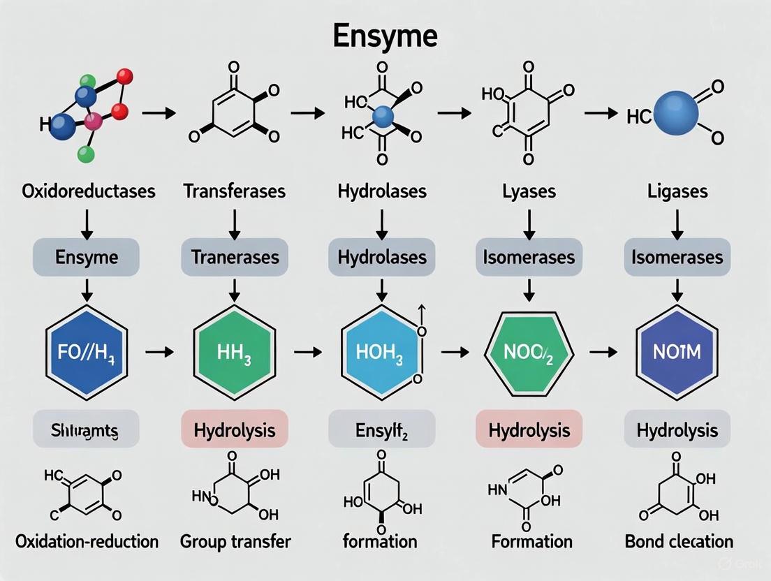
The Six Primary Enzyme Classes: Structure, Function, and Mechanisms
Oxidoreductases (EC 1)
Oxidoreductases catalyze oxidation-reduction reactions where electrons are transferred between molecules. One molecule is oxidized (loses electrons) while another is reduced (gains electrons) [12] [13]. These enzymes typically rely on cofactors such as NAD⁺, NADP⁺, FAD, or metal ions to facilitate electron transfer. Examples include alcohol dehydrogenase, which converts alcohols to aldehydes or ketones during alcohol metabolism, and cytochrome c oxidase, which is essential for cellular respiration [14]. Oxidoreductases are further classified into 23 subcategories based on their specific donors and acceptors, including enzymes acting on CH-OH groups (EC 1.1), aldehyde or oxo groups (EC 1.2), CH-CH groups (EC 1.3), and peroxide as acceptor (EC 1.11) [4].
Transferases (EC 2)
Transferases facilitate the transfer of specific functional groups (e.g., methyl, acetyl, amino, phosphoryl) from one molecule (the donor) to another (the acceptor) [13] [14]. Kinases, a prominent subclass of transferases, catalyze the transfer of phosphate groups from ATP to specific substrates, playing crucial roles in cellular signaling and regulation [14]. Other important examples include aminotransferases (transaminases) that transfer amino groups between amino acids and keto acids, and methyltransferases that mediate methylation processes essential for epigenetic regulation [13]. The transferase class is organized into nine subcategories including one-carbon group transfer (EC 2.1), aldehyde or ketonic group transfer (EC 2.2), and glycosyl transfer (EC 2.4) [4].
Hydrolases (EC 3)
Hydrolases catalyze the cleavage of chemical bonds through the addition of water (hydrolysis) [12]. These enzymes break down larger molecules into smaller units by introducing water across specific bonds. Common examples include lipases that hydrolyze lipids, proteases that cleave peptide bonds in proteins, and amylases that break down starch into sugar molecules [13] [14]. Hydrolases are fundamental to digestive processes and cellular degradation pathways. The hydrolase class encompasses 13 subcategories based on the type of bond hydrolyzed, including ester bonds (EC 3.1), glycosyl bonds (EC 3.2), peptide bonds (EC 3.4), and acid anhydride bonds (EC 3.6) [4].
Lyases (EC 4)
Lyases catalyze the cleavage of C-C, C-O, C-N, and other bonds by means other than hydrolysis or oxidation, often resulting in the formation of double bonds or the addition of groups to double bonds [12] [13]. These enzymes differ from hydrolases in that they do not utilize water in their catalytic mechanism. Notable examples include decarboxylases that remove carbon dioxide from carboxylic acids, and aldolases that catalyze aldol reactions in glycolysis and other metabolic pathways [13]. Lyases are organized into eight subcategories including C-C lyases (EC 4.1), C-O lyases (EC 4.2), and C-N lyases (EC 4.3) [4].
Isomerases (EC 5)
Isomerases catalyze structural rearrangements within a single molecule, converting a substrate from one isomer to another [12]. These enzymes catalyze reactions including racemization, epimerization, cis-trans isomerization, and intramolecular oxidoreductions. Examples include glucose-6-phosphate isomerase (also known as phosphoglucose isomerase) that converts glucose-6-phosphate to fructose-6-phosphate in glycolysis, and racemases that interconvert stereoisomers [13] [14]. The isomerase class comprises six subcategories including racemases and epimerases (EC 5.1), cis-trans isomerases (EC 5.2), and intramolecular oxidoreductases (EC 5.3) [4].
Ligases (EC 6)
Ligases catalyze the joining of two molecules coupled with the hydrolysis of a high-energy phosphate bond in ATP or a similar triphosphate [12] [13]. These enzymes form new C-C, C-S, C-O, and C-N bonds through energy-dependent condensation reactions. DNA ligase, which joins DNA fragments during replication and repair, represents a critically important example [13]. Similarly, aminoacyl-tRNA synthetases attach specific amino acids to their corresponding tRNAs during protein synthesis. Ligases are divided into six subcategories based on the type of bond formed, including C-O bonds (EC 6.1), C-S bonds (EC 6.2), C-N bonds (EC 6.3), and C-C bonds (EC 6.4) [4].
Table 2: The Six Primary Enzyme Classes: Functions, Examples, and Subclasses
| Enzyme Class | Catalytic Function | Representative Example | Example Function | Key Subclasses |
|---|---|---|---|---|
| Oxidoreductases (EC 1) | Catalyze oxidation-reduction reactions | Alcohol dehydrogenase | Alcohol metabolism | EC 1.1 (CH-OH donors), EC 1.2 (aldehyde/oxo donors) |
| Transferases (EC 2) | Transfer functional groups | Alanine aminotransferase | Amino acid metabolism | EC 2.1 (one-carbon groups), EC 2.7 (phosphate groups) |
| Hydrolases (EC 3) | Catalyze bond cleavage with water | Amylase | Starch digestion | EC 3.1 (ester bonds), EC 3.4 (peptide bonds) |
| Lyases (EC 4) | Cleave bonds without hydrolysis/oxidation | Aldolase | Glycolysis | EC 4.1 (C-C bonds), EC 4.2 (C-O bonds) |
| Isomerases (EC 5) | Catalyze molecular rearrangements | Glucose-6-phosphate isomerase | Glycolysis | EC 5.1 (racemases/epimerases), EC 5.3 (intramolecular oxidoreductases) |
| Ligases (EC 6) | Join molecules with ATP hydrolysis | DNA ligase | DNA replication | EC 6.1 (C-O bonds), EC 6.3 (C-N bonds), EC 6.5 (phosphoric ester bonds) |
Experimental Methodologies in Enzyme Research
Enzyme Function Prediction with Machine Learning
Recent advances in machine learning (ML) have revolutionized enzyme function prediction. The SOLVE (Soft-Voting Optimized Learning for Versatile Enzymes) framework exemplifies this approach, utilizing an ensemble learning framework that integrates random forest (RF), light gradient boosting machine (LightGBM), and decision tree (DT) models with an optimized weighted strategy [10]. This method distinguishes enzymes from non-enzymes and predicts EC numbers for mono- and multi-functional enzymes across all four hierarchical levels using only tokenized subsequences from protein primary sequences.
Experimental Protocol: SOLVE Framework Implementation
- Feature Extraction: Protein sequences are converted into numerical features using k-mer tokenization, with systematic analysis identifying 6-mers as optimal for capturing functional patterns while maintaining computational efficiency [10].
- Model Architecture: Implementation of soft-voting ensemble classifier combining predictions from RF, LightGBM, and DT base models with focal loss penalty to address class imbalance.
- Training Protocol: Stratified 5-fold cross-validation to ensure robust performance metrics across different data partitions.
- Validation: Performance evaluation on independent datasets with minimal sequence similarity to training data to assess generalization capability.
- Interpretability Analysis: Application of Shapley (SHAP) analysis to identify functional motifs at catalytic and allosteric sites, providing mechanistic insights into predictions [10].
Enzyme Kinetic Parameter Determination
Accurate prediction of enzyme kinetic parameters is crucial for enzyme exploration and modification. The CataPro model represents a state-of-the-art approach, utilizing deep learning to predict turnover number (kcat), Michaelis constant (Km), and catalytic efficiency (kcat/Km) [15].
Experimental Protocol: CataPro Kinetic Parameter Prediction
- Data Curation: Collection of enzyme-substrate entries from BRENDA and SABIO-RK databases, followed by sequence clustering at 40% similarity threshold to create unbiased evaluation datasets [15].
- Feature Representation:
- Enzyme sequences encoded using ProtT5-XL-UniRef50 embeddings (1024-dimensional vectors)
- Substrate structures represented using MolT5 embeddings (768-dimensional) combined with MACCS keys fingerprints (167-dimensional) [15]
- Model Architecture: Neural network framework processing concatenated enzyme-substrate representations (1959-dimensional vectors) to predict kinetic parameters.
- Validation: Ten-fold cross-validation on unbiased datasets with strict separation between training and test clusters to prevent data leakage and ensure generalization [15].
The following workflow diagram illustrates the integrated experimental and computational pipeline for enzyme function prediction and characterization, from sequence analysis to functional validation.
Table 3: Essential Research Reagents and Databases for Enzyme Classification Studies
| Resource Type | Specific Examples | Research Application | Key Features |
|---|---|---|---|
| Enzyme Databases | BRENDA, SABIO-RK [15] | Kinetic parameter reference | Manually curated experimental data on enzyme kinetics |
| Nomenclature Resources | IUBMB Enzyme Nomenclature [4], ExplorEnz | Standardized classification | Official EC number assignments with reaction mechanisms |
| Sequence Databases | UniProtKB/Swiss-Prot [10], KEGG ENZYME [11] | Sequence-function correlation | Links between sequence data and enzyme nomenclature |
| Machine Learning Tools | SOLVE [10], CataPro [15] | Function prediction | Ensemble models for EC number and kinetic parameter prediction |
| Structural Databases | Protein Data Bank (PDB) [10] | Structure-function analysis | Experimentally determined enzyme structures |
| Research Enzymes | Recombinant enzymes, mutant libraries [16] | Experimental validation | Functionally characterized enzymes for kinetic studies |
Applications in Drug Discovery and Biotechnology
Enzyme classification provides fundamental insights crucial for pharmaceutical development and industrial biotechnology. Understanding the specific reaction mechanisms of enzyme classes enables rational drug design, particularly through the development of targeted inhibitors. The SOLVE framework's capability to identify functional motifs at catalytic and allosteric sites offers significant potential for therapeutic drug design by pinpointing precise intervention sites [10]. In industrial contexts, enzyme engineering leverages classification knowledge to modify or create enzymes with enhanced properties.
The emerging field of synthetic enzymes, or synzymes, represents a promising frontier in modern biocatalysis. These synthetic mimics of natural enzymes are engineered to function under extreme physicochemical conditions unsuitable for natural enzymes, making them suitable for applications in biomedicine, industrial biotechnology, and environmental remediation [17]. Synthetic enzymes have demonstrated remarkable efficacy in neutralizing oxidative stress, a critical factor in many diseases, and have been explored in biosensing, gene editing, and neuroprotection models [17].
Machine learning approaches are increasingly integrated with traditional classification systems to enhance predictive capabilities. The exceptional performance of SOLVE across all EC number levels—from main class (L1) to substrate specificity (L4)—demonstrates how computational methods can complement traditional biochemical approaches to enzyme characterization [10]. Similarly, CataPro's accurate prediction of enzyme kinetic parameters enables more efficient enzyme discovery and engineering, as validated by the identification and optimization of Sphingobium sp. CSO with 19.53-times increased activity compared to the initial enzyme [15].
The systematic classification of enzymes into six primary classes—oxidoreductases, transferases, hydrolases, lyases, isomerases, and ligases—provides an essential conceptual framework for understanding biocatalysis. This classification system, grounded in reaction mechanism specificity rather than sequence similarity, continues to enable critical advances in basic research and applied biotechnology. Contemporary integration of machine learning with traditional enzymology has created powerful synergies, enhancing our ability to predict enzyme function from sequence alone and engineer novel catalysts with tailored properties. As databases expand and computational methods evolve, the fundamental framework of enzyme classification will continue to serve as an indispensable foundation for exploring the vast functional landscape of biocatalysts, accelerating discoveries in therapeutic development, industrial biotechnology, and basic biological research.
Molecular recognition, the specific interplay between an enzyme and its substrate, constitutes the very bedrock of enzymatic catalysis and a pivotal concept in biochemical research. This process governs the remarkable specificity that allows enzymes to selectively bind their cognate substrates from a myriad of cellular molecules, thereby orchestrating the complex metabolic pathways essential for life. The quest to understand the physical and chemical principles underlying this specificity has propelled the development of two seminal conceptual models: Emil Fischer's Lock-and-Key Hypothesis and Daniel Koshland's Induced-Fit Model. These frameworks are not merely historical footnotes; they provide the foundational language and mechanistic intuition that continue to guide contemporary investigations into enzyme function, classification, and catalytic mechanisms.
Fischer's Lock-and-Key model, introduced in 1894, proposed a static and rigid complementarity between enzyme and substrate [18] [19]. In this analogy, the enzyme's active site (the lock) is pre-configured to precisely accommodate the geometry and chemical properties of its specific substrate (the key). This model successfully explained the high degree of specificity observed in enzymatic reactions but fell short of explaining the stabilization of the transition state that enzymes achieve. Decades later, Koshland's Induced-Fit model (1958) addressed this limitation by introducing the concept of flexibility [18] [19]. This model posits that the active site is not static; rather, the binding of the substrate induces a conformational change in the enzyme, reshaping the active site to achieve a optimal fit for catalysis and transition state stabilization.
Understanding the nuances and applications of these models is crucial for modern drug development professionals and researchers. The principles of molecular recognition directly inform rational drug design, where small molecules are engineered to fit into the active sites of pathogenic enzymes or cellular receptors, thereby modulating their activity. Furthermore, in the evolving field of enzyme classification and catalytic mechanism research, these models provide a conceptual scaffold for interpreting structural data and understanding enzyme evolution, enabling scientists to decipher the complex relationship between protein structure, dynamics, and function.
Historical Context and Theoretical Frameworks
The evolution of our understanding of enzyme-substrate interactions mirrors the broader advancements in biochemical and structural sciences. The journey began with Emil Fischer's seminal proposition in 1894, which introduced the Lock-and-Key Model [19]. This model was groundbreaking for its time, providing a intuitive analogy that explained enzyme specificity. It suggested that the enzyme and substrate possess specific complementary geometric shapes that fit exactly into one another, much like a key fits into its matching lock [18] [19]. This implied a rigid, pre-formed active site on the enzyme that was structurally complementary to the substrate. The model successfully highlighted the fact that only the correct-size-and-shape-of-the-substrate-(the-key)-would-fit-into-the-active-site-(the-keyhole) of the enzyme (the lock) [19]. However, a significant limitation of this static model was its inability to satisfactorily explain the stabilization of the transition state that enzymes achieve to catalyze reactions [19].
Building on this foundation, Daniel Koshland proposed a more dynamic theory in 1958: the Induced-Fit Model [18] [19]. This model was developed to account for experimental observations that the Lock-and-Key theory could not reconcile, particularly the stabilization of the transition state and the allosteric behaviors of some enzymes. Koshland's model suggested that the active site of the enzyme is not perfectly complementary to the substrate in its initial state. Instead, the binding of the substrate induces a conformational change in the enzyme's structure [18]. This reshaping aligns catalytic groups, optimizes binding interactions, and ultimately forms a transition state complex that lowers the activation energy of the reaction. The Induced-Fit model thus portrays enzymes as flexible structures whose final shape and charge distribution are determined upon substrate binding [19]. This fundamental shift in perspective-from a rigid to a dynamic interface-reshaped the study of enzymology and provided a more robust explanation for catalytic power and specificity.
Table 1: Core Principles of Lock-and-Key vs. Induced-Fit Models
| Feature | Lock-and-Key Model | Induced-Fit Model |
|---|---|---|
| Proponent & Date | Emil Fischer (1894) [19] | Daniel Koshland (1958) [18] [19] |
| Shape Complementarity | Complementary before binding; shapes fit exactly [18] | Not fully complementary before binding; shapes become complementary after binding [18] |
| Enzyme Active Site | Static and rigid; a single entity [18] | Flexible and dynamic; undergoes conformational change [18] [19] |
| Binding Interaction | Inflexible and very strong [18] | Flexible and not very strong initially [18] |
| Transition State | A transition state does not develop [18] | A transition state develops before reactants undergo changes [18] |
| Catalytic Group | No separate catalytic group; no weakening of substrate bonds [18] | Has a separate catalytic group that weakens substrate bonds [18] |
Quantitative Comparison of Model Attributes and Experimental Evidence
While the historical and conceptual distinctions between the two models are clear, a quantitative and mechanistic comparison is essential for a rigorous scientific understanding. The differences extend beyond simple shape complementarity to encompass the very nature of the binding interaction, the formation of the transition state, and the strategic involvement of catalytic residues.
In the Lock-and-Key model, the binding is characterized as inflexible and very strong, a result of the perfect and immediate steric and chemical complementarity [18]. The enzyme's active site is viewed as a single entity, and crucially, this model does not involve the development of a distinct transition state nor does it propose a separate catalytic group to weaken substrate bonds [18]. The catalytic power, therefore, was thought to arise primarily from the precise orientation of the substrate within the active site. In contrast, the Induced-Fit model describes a more nuanced process. Binding is initially flexible and not very strong, allowing for the necessary conformational adjustments [18]. The active site is composed of multiple components that can move relative to one another [18]. A key tenet of this model is the development of a transition state, which the enzyme actively helps to stabilize. This is often achieved through a separate catalytic group (e.g., a specific amino acid side chain or cofactor) that performs nucleophilic or electrophilic attacks to weaken the critical bonds within the substrate, thereby facilitating the chemical reaction [18].
Modern structural biology provides overwhelming evidence for the induced-fit mechanism. Techniques like cryo-electron microscopy (cryo-EM) and X-ray crystallography have captured enzymes in multiple conformational states—apo (unbound), substrate-bound, and transition-state analog-bound—visually demonstrating the structural shifts that occur upon binding. For instance, studies on enzymes like lysozyme and hexokinase have shown clear differences in the conformation of the active site when comparing the unbound and bound states, with movements of entire domains or loops that serve to enclose the substrate and bring catalytic residues into precise alignment. This experimental evidence solidifies the Induced-Fit model as a more accurate and widespread mechanism, though the Lock-and-Key analogy remains useful for describing systems with very high pre-formed complementarity, such as some antibody-antigen interactions.
Table 2: Mechanistic and Functional Differences
| Aspect | Lock-and-Key Model | Induced-Fit Model |
|---|---|---|
| Binding Nature | Inflexible, very strong [18] | Flexible, optimized after binding [18] |
| Transition State | Not explicitly explained or developed [18] | Explicitly formed and stabilized by the enzyme [18] |
| Catalytic Strategy | No separate catalytic group; relies on proximity and orientation [18] | Involves separate catalytic groups (e.g., for nucleophilic attack) [18] |
| Representation | Single, static complementary surface [18] | Multi-component active site that changes shape [18] |
| Modern View | Seen as a special case of complementarity; less common | Viewed as the predominant mechanism for many enzymes |
Advanced Research Methodologies in Molecular Recognition
The study of molecular recognition has been revolutionized by sophisticated technologies that allow researchers to probe interactions at the single-molecule level and visualize structures with atomic resolution. These methods provide direct, quantitative data that moves beyond theoretical models into experimental observation.
Atomic Force Microscopy (AFM) for Single-Molecule Recognition
Atomic Force Microscopy (AFM) has emerged as a powerful tool for imaging and measuring interaction forces. A specific advanced application is Jumping Force Mode (JM) AFM, which produces simultaneous topography and tip-sample maximum-adhesion images based on force spectroscopy [20]. In this technique, the AFM tip is functionalized with a specific ligand (e.g., biotin), and the sample surface is immobilized with the corresponding receptor (e.g., avidin or streptavidin). When the tip scans the surface, it measures the specific rupture forces of the ligand-receptor complexes at each point, generating qualitative and quantitative molecular recognition maps [20]. This method has been refined to operate in a repulsive regime applying very low forces, which minimizes non-specific tip-sample interactions, ensuring that the adhesion maps reflect only specific binding events [20]. A key experimental protocol involves:
- Probe Functionalization: Covalently attaching ligands (e.g., biotin) to AFM tips via chemical linkers.
- Sample Immobilization: Anchoring protein molecules (e.g., avidin and streptavidin) to a flat substrate like mica using heterobifunctional cross-linkers (e.g., Sulfo-LC-SPDP) to form stable disulfide bonds, ensuring isolated molecules for single-molecule recognition [20].
- Data Acquisition: Scanning the functionalized tip over the protein-decorated surface in JM mode to record force-distance (Fz) curves at defined points, capturing the maximum adhesion force.
- Analysis: Generating adhesion maps where the contrast is based on the measured rupture forces. This allows for the discrimination between even highly similar proteins, as demonstrated by the differentiation of avidin (rupture force 40–80 pN) from streptavidin (rupture force 120–170 pN) [20].
Structural Biology and Computational Approaches
High-resolution structural techniques like cryo-Electron Microscopy (cryo-EM) have been instrumental in visualizing enzyme-substrate complexes and understanding catalytic mechanisms. For example, the recent determination of the human glycogen debranching enzyme (hsGDE) structure at 3.23 Å resolution provided atomic-level insights into its substrate selectivity and the conformational changes associated with its dual catalytic activities [21]. Complementing experimental structures, Molecular Dynamics (MD) simulations allow researchers to model the dynamic process of substrate binding and induced fit. In studies of hsGDE, all-atom MD simulations with substrates like maltopentaose revealed significant dynamics and flexibility within the enzyme's transferase (GT) domain, illustrating the conformational sampling that underpins the induced-fit mechanism [21].
Furthermore, the field is advancing towards "catalysis in silico" [22]. The flood of enzyme data from metagenomic sequencing, coupled with AI-driven protein structure prediction (e.g., AlphaFold), has enabled the accurate computational modeling of enzyme structures. Emerging bioinformatic approaches are now being developed to capture and compare reaction mechanisms computationally. One such novel method involves calculating mechanism similarity based on the bond changes and charge transfers at each catalytic step, using a data entity called an "arrow-environment" (arrow-env) to represent electronic transfers [23]. This allows for the pairwise comparison of enzyme mechanisms from databases like the Mechanism and Catalytic Site Atlas (M-CSA), facilitating the discovery of convergent and divergent evolutionary relationships independent of sequence or structural similarity [23].
Diagram 1: Single-Molecule Recognition via JM-AFM Workflow.
The Scientist's Toolkit: Key Reagents and Methods
Table 3: Essential Research Reagents and Materials for Molecular Recognition Studies
| Reagent / Material | Function / Application | Example Usage |
|---|---|---|
| Heterobifunctional Cross-linkers (e.g., Sulfo-LC-SPDP) | Covalent, site-directed immobilization of proteins to solid supports while preserving functionality [20]. | Immobilizing avidin/streptavidin on mica for AFM studies via amine-to-sulfhydryl chemistry [20]. |
| Functionalized AFM Probes | Serve as sensors for specific molecular recognition in force spectroscopy; the tip is the "key" [20]. | Biotinylated AFM tips for quantifying binding forces with avidin-family proteins [20]. |
| Stable Substrates (e.g., APTES-functionalized Mica) | Provide an atomically flat, chemically modifiable surface for biomolecule attachment [20]. | Creating a rigid, non-conductive surface for anchoring proteins in AFM to minimize background noise. |
| Defined Protein Constructs | Isolated enzymes or receptors for structural, biophysical, and kinetic assays. | Purified human glycogen debranching enzyme (hsGDE) for cryo-EM structure determination [21]. |
| Molecular Dynamics (MD) Software | Simulates the dynamic process of substrate binding and induced fit at atomic resolution. | All-atom MD simulations of hsGDE with maltopentaose to study substrate selectivity and dynamics [21]. |
| Mechanism Databases (e.g., M-CSA) | Curated, machine-readable repositories of enzyme mechanisms for comparative analysis [23]. | Performing pairwise comparisons of enzyme mechanisms to uncover evolutionary relationships [23]. |
Implications for Enzyme Classification and Catalytic Mechanism Research
The evolution from a rigid to a dynamic understanding of molecular recognition has profound implications for the fields of enzyme classification and catalytic mechanism research. Traditional classification systems, such as the Enzyme Commission (EC) numbers, primarily categorize enzymes based on the overall chemical reactions they catalyze. While invaluable, this system does not inherently capture the mechanistic diversity or evolutionary relationships that can be revealed by examining the specific steps of catalysis.
The introduction of quantitative methods for comparing enzyme mechanism similarity, as described in recent research, represents a paradigm shift [23]. This approach moves beyond global sequence and structure similarity to focus on the local chemical transformations—the bond changes and charge transfers—defined as "arrow-environments." This allows for the systematic comparison of mechanisms across different enzyme families, enabling the discovery of convergent evolution, where enzymes with different folds evolve the same catalytic step, and divergent evolution, where related enzymes catalyze different overall reactions using a similar core mechanism [23]. For instance, this method can automatically identify if a phosphoryl transfer step in a kinase is mechanistically analogous to a step in an unrelated nuclease, providing a deeper, more principled layer of functional annotation that complements EC classification.
Furthermore, the detailed structural insights gained from techniques like cryo-EM, as applied to enzymes like hsGDE, directly inform the understanding of disease pathogenesis and the development of targeted therapeutics [21]. By elucidating the precise molecular architecture of the active site and the conformational changes during catalysis, researchers can correlate disease-associated mutations with specific disruptions to substrate binding, transition state stabilization, or protein dynamics. This mechanistic understanding of why a mutation causes a loss of function, as seen in Glycogen Storage Disease Type III, is crucial for designing small molecules or gene therapies that can potentially rescue or bypass the defective enzyme activity [21].
The journey from Fischer's Lock-and-Key Hypothesis to Koshland's Induced-Fit Model illustrates the progressive refinement of our understanding of molecular recognition. This evolution from a static to a dynamic paradigm has been critically supported by advanced experimental and computational methodologies, including single-molecule force spectroscopy, high-resolution structural biology, and novel bioinformatic analyses of mechanism similarity. These models are far from obsolete; they are active frameworks that guide cutting-edge research in enzymology.
For researchers and drug development professionals, these principles are indispensable. The ability to distinguish between rigid and flexible binding interfaces informs the rational design of high-affinity inhibitors and drugs. The emerging capability to quantitatively compare catalytic mechanisms and deconvolute the structural consequences of disease-causing mutations opens new avenues for enzyme engineering, functional prediction, and the development of targeted therapeutic strategies. As the volume of structural and mechanistic data continues to grow, driven by AI and high-throughput methods, the nuanced understanding of molecular recognition provided by both the Lock-and-Key and Induced-Fit models will remain a cornerstone of fundamental biochemical research and its translational applications.
The enzymatic active site represents one of the most sophisticated catalytic environments in nature, where precise spatial arrangement of amino acid residues and helper molecules enables the remarkable rate enhancements characteristic of biological catalysts. Within the context of enzyme classification and catalytic mechanisms research, understanding active site architecture is paramount for elucidating the relationship between protein structure and function. This specialized region, typically comprising only 10-20% of the enzyme's volume, creates a unique chemical microenvironment that facilitates substrate binding, transition state stabilization, and product release [24] [25] [26]. The active site's composition dictates enzyme specificity and catalytic efficiency through a complex interplay between key amino acid residues and essential non-protein components, including metal ions and organic cofactors [24]. Contemporary research continues to reveal surprising aspects of active site dynamics, including the role of conformational flexibility and the emerging understanding of composition-driven activities in certain protein regions [27]. This technical guide examines the structural and functional components of enzyme active sites, providing researchers and drug development professionals with a comprehensive framework for understanding, investigating, and manipulating these fundamental biological catalysts.
Structural Anatomy of the Active Site
Hierarchical Organization and Architecture
The catalytic power of enzymes originates from their precisely organized active sites, which emerge from the hierarchical structure of the protein itself. The primary structure—the linear sequence of amino acids—determines the ultimate three-dimensional configuration of the active site [24]. This sequence folds into localized secondary structures such as α-helices and β-sheets, which further organize into the overall tertiary structure of the protein chain [24]. For multi-subunit enzymes, the arrangement of these subunits constitutes the quaternary structure [24]. The active site itself typically exists as a groove or crevice on the enzyme surface, filled with free water when not binding substrate [24]. This architectural complexity creates a specific chemical environment perfectly suited for stabilizing transition states and facilitating chemical transformations.
The architecture of the active site enables two crucial binding modes: the historic lock-and-key model proposes perfect complementarity between enzyme and substrate, while the more contemporary induced fit model hypothesizes that both enzyme and substrate undergo conformational adjustments upon binding to achieve optimal catalytic alignment [24] [28] [25]. This dynamic binding mechanism maximizes the enzyme's catalytic efficiency by precisely positioning reactive groups and substrates.
Key Amino Acid Residues and Chemical Microenvironments
The specific chemical properties of amino acid residues within the active site create a unique microenvironment essential for catalysis. These residues provide key functional groups that participate directly in catalytic mechanisms through various strategies:
- Covalent catalysis: Transient covalent bond formation between amino acid residues and substrates
- General acid-base catalysis: Proton transfer reactions facilitated by amino acid side chains
- Catalysis by approximation: Spatial orientation of multiple substrates for optimal reaction geometry
- Metal ion catalysis: Enhancement of nucleophilicity and charge stabilization via metal ions [24]
The composition and spatial arrangement of these residues determine substrate specificity and catalytic mechanism, with even single amino acid substitutions dramatically altering enzyme function, as demonstrated in protein engineering studies [29]. Recent perspectives also highlight that some protein activities are driven more by overall amino acid composition than specific sequence, particularly in intrinsically disordered regions and prion-like domains [27].
Table 1: Key Amino Acid Residues and Their Catalytic Roles in Active Sites
| Amino Acid | Chemical Properties | Catalytic Roles | Examples of Participating Mechanisms |
|---|---|---|---|
| Histidine | pKa ~6.5; imidazole ring | Proton shuttle; general acid/base catalysis | Hydrolysis reactions; phosphoryl transfer |
| Cysteine | Thiol group (-SH); nucleophilic | Covalent catalysis; redox reactions | Proteases; redox enzymes |
| Aspartic Acid | Carboxylic acid; anionic at pH 7 | Acid/base catalysis; metal ion binding | Proteases; lyases |
| Glutamic Acid | Carboxylic acid; anionic at pH 7 | Acid/base catalysis; metal ion binding | Proteases; isomerases |
| Serine | Hydroxyl group; nucleophilic | Covalent catalysis; nucleophile | Serine proteases; esterases |
| Lysine | Amino group; cationic at pH 7 | Schiff base formation; electrostatic stabilization | Dehydrogenases; decarboxylases |
| Arginine | Guanidinium group; cationic | Anion binding; charge stabilization | Dehydrogenases; phosphoryl transfer enzymes |
| Tyrosine | Phenolic hydroxyl; amphoteric | Acid/base catalysis; redox reactions | Phosphatases; redox enzymes |
Cofactors and Coenzymes: Essential Non-Protein Components
Classification and Functional Roles
Many enzymes require non-protein components to achieve full catalytic capability. These helper molecules are classified as either cofactors—typically inorganic ions such as Zn²⁺, Mg²⁺, or Fe²⁺—or coenzymes—organic compounds, often derived from dietary vitamins [24] [28]. The protein component alone, without its essential helper molecules, is referred to as an apoenzyme, which is catalytically inactive until it forms a complex with its required cofactor to create the functional holoenzyme [24] [26].
Metal ion cofactors contribute to catalysis through multiple mechanisms: they act as Lewis acids to stabilize negative charges, facilitate oxidation-reduction reactions through reversible changes in oxidation state, mediate substrate orientation through coordinate covalent bonds, and shield negatively charged groups that might otherwise repel substrate binding [24]. The specific properties of the metal ion, including its size, charge density, and preferred coordination geometry, determine its functional role within the active site.
Major Coenzyme Classes and Functions
Coenzymes function as transient carriers of specific functional groups during catalytic cycles. Unlike cosubstrates, which bind and release like substrates, many coenzymes remain tightly associated with their enzymes throughout multiple catalytic turnovers. These organic molecules frequently contain additional chemical functionality not available from the standard amino acid side chains, significantly expanding the catalytic repertoire of enzymes.
Table 2: Essential Coenzymes and Their Catalytic Functions
| Coenzyme | Vitamin Precursor | Chemical Group Transferred | Representative Enzyme Classes |
|---|---|---|---|
| NAD⁺/NADP⁺ | Niacin (B3) | Hydride ion (H⁻) | Dehydrogenases, reductases |
| FAD/FMN | Riboflavin (B2) | Electrons | Oxidoreductases |
| Coenzyme A | Pantothenic acid (B5) | Acyl groups | Transferases, synthases |
| Thiamine pyrophosphate | Thiamine (B1) | Aldehydes | Decarboxylases, transketolases |
| Pyridoxal phosphate | Pyridoxine (B6) | Amino groups | Transaminases, racemases |
| Biocytin | Biotin (B7) | Carbon dioxide | Carboxylases |
| Tetrahydrofolate | Folate (B9) | One-carbon units | Methyltransferases, synthetases |
| Cobalamin | Cobalamin (B12) | Methyl groups; rearrangements | Isomerases, methyltransferases |
Experimental Approaches for Active Site Characterization
Methodologies for Probing Active Site Structure and Function
Understanding active site composition and mechanism requires integrated experimental approaches that provide complementary information about structure, dynamics, and function. The following protocols represent key methodologies employed in contemporary enzymology research.
Protocol 1: Site-Directed Mutagenesis of Active Site Residues
Purpose: To determine the functional contribution of specific amino acid residues to catalytic mechanism and substrate binding.
Procedure:
- Identify candidate residues through sequence alignment with homologous enzymes or structural analysis
- Design mutagenic primers to substitute target codons (e.g., charged to alanine substitutions)
- Perform PCR-based mutagenesis on plasmid DNA encoding the enzyme of interest
- Verify mutations by DNA sequencing
- Express and purify mutant enzyme variants
- Determine kinetic parameters (KM, kcat) and compare to wild-type enzyme
- Assess structural integrity via circular dichroism or thermal denaturation assays
Interpretation: Significant reductions in kcat suggest direct involvement in chemical catalysis, while changes in KM may indicate alterations in substrate binding. Maintenance of structural integrity confirms that observed effects result from specific residue substitution rather than global unfolding [29].
Protocol 2: Substrate-Multiplexed Screening (SUMS) for Active Site Diversification
Purpose: To efficiently explore sequence-function relationships in engineered enzyme variants by simultaneously screening activity against multiple substrates.
Procedure:
- Select substrate panel with diverse electronic and steric properties but similar parent enzyme activity levels
- Construct focused library targeting active site residues via recombination of beneficial mutations
- Use wild-type primer doping during library assembly to enrich for functional multi-site mutants
- Prepare equimolar mixture of selected substrates
- Incubate enzyme variants with substrate mixture and monitor product formation via LC-MS
- Calculate fold-activity change for individual products relative to parent enzyme
- Train logistic regression model on screening data to identify active sequence space
- Validate hits through detailed kinetic analysis of purified variants
Interpretation: SUMS distinguishes variants with impaired activity across all substrates from those with altered specificity profiles, enabling efficient navigation of sequence space while maintaining broad substrate promiscuity [29].
Protocol 3: X-ray Crystallography for Active Site Visualization
Purpose: To determine atomic-level three-dimensional structure of enzyme active sites, including geometry of substrate binding and catalytic residues.
Procedure:
- Purify and concentrate target enzyme to >10 mg/mL
- Screen crystallization conditions using commercial sparse matrix screens
- Optimize initial hits to produce diffraction-quality crystals
- Soak crystals with substrates, inhibitors, or analogues to trap catalytic intermediates
- Flash-cool crystals in liquid nitrogen for cryoprotection
- Collect X-ray diffraction data at synchrotron source
- Solve structure by molecular replacement or experimental phasing
- Model protein, ligands, and solvent molecules into electron density
- Refine structure with restraints for geometric parameters
Interpretation: High-resolution structures (<2.0 Å) reveal precise atomic interactions between enzyme and ligand, conformational changes associated with substrate binding, and the spatial relationships between catalytic residues [22].
Computational and Structural Perspectives on Catalytic Mechanisms
Emerging Tools for Mechanism Analysis and Prediction
Recent advances in computational methodologies have revolutionized our understanding of enzyme catalytic mechanisms. The EzMechanism tool automatically infers mechanistic paths for given three-dimensional active sites and enzyme reactions based on catalytic rules compiled from the Mechanism and Catalytic Site Atlas (M-CSA) database [30]. This knowledge-based approach leverages the rich literature on biological catalysis to generate testable mechanistic hypotheses.
Complementing this approach, novel methods for comparing enzyme mechanisms enable quantitative analysis of catalytic steps across diverse enzyme families. These methods use arrow-environments (arrow-env)—representations of electron movement and associated atoms—as the fundamental unit for mechanism comparison [23]. By calculating similarity based on bond changes and charge transfers at each catalytic step, researchers can systematically explore mechanistic relationships independent of sequence or structural homology, revealing both convergent and divergent evolutionary patterns [23].
The following diagram illustrates the workflow for computational analysis of enzyme mechanisms:
Diagram 1: Computational Workflow for Enzyme Mechanism Analysis
Structural Insights into Catalytic Mechanisms
High-resolution structural studies continue to provide unprecedented insights into the molecular details of enzyme catalysis. Analysis of the M-CSA database, which contains detailed machine-readable descriptions of 734 distinct enzyme mechanisms, reveals remarkable conservation of catalytic strategies across phylogenetically diverse enzymes [23]. Despite the vast diversity of enzyme-catalyzed reactions, the number of unique chemical transformations employed in enzyme active sites is surprisingly limited, with approximately 3,000 arrow-environments sufficient to describe over 19,000 actual catalytic steps [23].
This structural perspective highlights how enzymes employ recurring mechanistic motifs, with proton transfers representing the most common catalytic steps [30]. The most frequent catalytic rule, observed in 61 mechanistic steps across 54 enzymes, involves proton transfer between carboxylic groups and water molecules—a fundamental process for recycling active site states during catalysis [30]. Such analyses underscore the modular nature of enzyme catalysis, where complex mechanisms are constructed from simpler, reusable chemical steps.
The Scientist's Toolkit: Essential Research Reagents and Materials
Table 3: Key Research Reagents for Active Site Studies
| Reagent/Material | Function/Application | Key Characteristics |
|---|---|---|
| Pyridoxal Phosphate (PLP) | Cofactor for amino acid decarboxylases, transaminases, and racemases | Aldehyde group forms Schiff base intermediates with substrates [29] |
| Non-canonical Amino Acids (ncAAs) | Substrate analogs for probing active site specificity and engineering novel activities | Modified side chains test steric and electronic tolerance [29] |
| NAD(P)H/NAD(P)+ | Redox cofactors for dehydrogenases and reductases | Hydride transfer in oxidation-reduction reactions [24] |
| Metal Chelators (EDTA, EGTA) | Selective removal of metal cofactors to study metalloenzyme mechanisms | Differential affinity for specific metal ions [24] |
| Site-Directed Mutagenesis Kits | Systematic alteration of active site residues | Enables alanine scanning and functional group substitution [29] |
| Cross-linking Reagents | Stabilization of enzyme-ligand complexes for structural studies | Captures transient interactions in active site [22] |
| Isotopically Labeled Substrates (²H, ¹³C, ¹⁵N) | Tracing catalytic pathways and intermediate formation | NMR and MS analysis of reaction mechanisms [23] |
| X-ray Crystallography Reagents | Structure determination of enzyme-ligand complexes | Cryoprotectants, heavy atom derivatives for phasing [22] |
The intricate architecture of enzyme active sites represents the culmination of evolutionary optimization for efficient and specific catalysis. Through the precise spatial organization of key amino acid residues and the integration of essential cofactors and coenzymes, enzymes create specialized chemical environments that lower activation barriers and accelerate biological reactions. Contemporary research continues to reveal new dimensions of active site function, from the dynamic nature of substrate binding described by the induced fit model to the emerging understanding of composition-driven activities in certain protein regions. The integration of structural biology, protein engineering, and computational approaches provides researchers with powerful tools to dissect catalytic mechanisms and manipulate enzyme function. For drug development professionals, understanding active site anatomy enables rational design of targeted inhibitors, while protein engineers can leverage this knowledge to create novel catalysts for industrial applications. As research in this field advances, particularly through applications of artificial intelligence and high-throughput screening methodologies, our understanding of the fundamental principles governing enzyme catalysis will continue to deepen, opening new frontiers in biochemistry and biotechnology.
Enzyme catalytic mechanisms form the cornerstone of biological processes, enabling the efficient and specific chemical transformations that sustain life. These remarkable biological catalysts accelerate biochemical reactions by orders of magnitude while operating under mild physiological conditions, achieving rate enhancements ranging from thousands to millions-fold compared to uncatalyzed reactions [31]. The study of enzyme catalysis has yielded profound insights into the dynamic interactions at the active site, with three fundamental strategies emerging as central to understanding enzymatic power: covalent catalysis, acid-base catalysis, and transition state stabilization. These mechanisms provide the physical and chemical basis for the extraordinary efficiency and specificity that enzymes exhibit, distinguishing them from non-biological catalysts [31].
Within the context of enzyme classification and catalytic mechanisms research, understanding these fundamental strategies provides a framework for deciphering the vast universe of possible enzymatic functions. Scientific research has revealed only a minuscule fraction of the enzymes that evolution has generated, and an even tinier fraction of the vast universe of possible biocatalysts [32]. The investigation into how enzymes employ covalent catalysis, acid-base catalysis, and transition state stabilization not only illuminates nature's existing diversity but also offers a route to genetically encoding almost any chemistry through artificial intelligence-driven enzyme discovery and design [32]. This whitepaper provides an in-depth technical examination of these three core catalytic strategies, framed within contemporary research paradigms and highlighting emerging methodologies that are expanding our understanding of enzyme function.
Transition State Stabilization
Fundamental Principles and Mechanisms
Transition state stabilization represents one of the most fundamental and widely accepted mechanisms of enzyme catalysis. This process involves the enzyme's selective stabilization of the transition state (TS), which is the highest-energy, ephemeral intermediate in a reaction pathway, thereby lowering the activation energy barrier and accelerating the reaction rate [31]. The theoretical foundation of this mechanism is rooted in transition state theory, which posits that enzymatic rate acceleration is due to the enzyme's much higher affinity for the transition state relative to its substrates [33]. This concept has been experimentally supported by the high affinities measured for transition-state analogues (TSAs), which have led to the design of TSA as high-affinity enzyme inhibitors [33].
Recent research has revealed a more nuanced understanding of transition state stabilization, demonstrating that enzymes stabilize transition states through enhanced charge densities of catalytic atoms. These atoms experience a reduction in charge density between ground states (GS) and transition states [34]. Importantly, whether enzymes catalyze reactions by TS stabilization or ground state destabilization, they ultimately reduce reaction free energy barriers (ΔG‡) by enhancing the charge densities of catalytic atoms that undergo charge reduction between GS and TS [34]. The key distinction lies in how this enhancement is achieved: in TS stabilization, the charge density of catalytic atoms is enhanced prior to enzyme-substrate binding, whereas in ground state destabilization, this enhancement occurs during enzyme-substrate binding [34].
Emerging Concepts: Transition State Ensembles
Traditional views of transition state stabilization often implied a relatively unique structure at the dividing surface of the free-energy landscape. However, contemporary research has challenged this perspective, revealing that proteins exist as large ensembles of conformations. This understanding has led to the recognition of broad transition-state ensembles (TSE) as a key component for efficient enzyme catalysis [33]. A conformationally delocalized ensemble, including asymmetric transition states, is rooted in the macroscopic nature of the enzyme, and this wide TSE has been computationally predicted and experimentally confirmed to decrease the entropy of activation [33].
Table 1: Key Experimental Evidence Supporting Transition State Stabilization
| Evidence Type | Experimental Approach | Key Findings | Reference System |
|---|---|---|---|
| Transition State Analogues | X-ray crystallography of enzyme-TSA complexes | TSA bind with much higher affinity than substrates | Multiple enzyme systems |
| Computational Simulations | QM/MM calculations | Identification of broad transition state ensembles | Adenylate kinase |
| Kinetic Analysis | Temperature-dependent kinetics | Decreased entropy of activation | Adenylate kinase with Mg²⁺ |
| Charge Density Analysis | Computational charge mapping | Enhanced charge densities at catalytic atoms | Ketosteroid isomerase |
Research on adenylate kinase (Adk), an essential phosphotransferase found in all cells, has provided compelling evidence for the TSE model. Quantum-mechanics/molecular-mechanics (QM/MM) calculations of the phosphoryl-transfer step in Adk revealed a structurally wide set of energetically equivalent configurations along the reaction coordinate, forming a broad transition-state ensemble [33]. This delocalized transition state ensemble boosts a unifying concept for protein folding and conformational transitions underlying protein function, resolving the apparent paradox between unique transition states and the ensemble nature of protein conformations [33].
Experimental and Computational Protocols
QM/MM Protocol for Transition State Analysis
The investigation of transition state ensembles in adenylate kinase exemplifies cutting-edge approaches to studying transition state stabilization [33]:
System Preparation: Start with X-ray structure of enzyme-inhibitor complex (e.g., Adk with Ap5A, PDB: 2RGX). Build substrate coordinates using the inhibitor as a template and replace crystallographic metal ions as needed (e.g., Zn²⁺ to Mg²⁺).
QM/MM Setup: Define the quantum mechanics (QM) region to include the reactive moieties of substrates, catalytic metal ions, and coordinating water molecules. Treat the remainder of the system with molecular mechanics (MM) using appropriate force fields (e.g., AMBER ff99sb). Solvate the system with explicit water models (e.g., TIP3P).
Equilibration: Perform molecular dynamics simulations to equilibrate the starting structure, verifying agreement with relevant experimental structures.
Reaction Sampling: Employ steered molecular dynamics simulations in both forward and reverse directions to sample the reaction coordinate. Use multiple steered molecular dynamics with Jarzynski's Relationship to determine free-energy profiles.
State Analysis: Characterize the transition state ensemble by identifying structurally diverse but energetically equivalent configurations along the reaction coordinate.
Kinetic Validation Protocol
Computational predictions of transition state ensembles require experimental validation [33]:
Temperature-Dependent Kinetics: Measure enzyme activity across a temperature range (e.g., 10-40°C) to determine activation parameters (ΔH‡ and ΔS‡).
pH Profile Analysis: Determine reaction rates across pH values to identify catalytic groups and their protonation states.
Metal Ion Effects: Compare kinetic parameters in presence and absence of catalytic metal ions (e.g., Mg²⁺).
Crystallographic Studies: Solve structures of enzyme complexes with substrates, products, and transition state analogues to correlate structural features with catalytic efficiency.
Figure 1: Transition State Ensemble Model Showing Multiple Pathways Through Energetically Equivalent Transition States
Covalent Catalysis
Mechanisms and Chemical Principles
Covalent catalysis involves the transient formation of a covalent bond between the enzyme and substrate during the catalytic cycle, creating a reaction intermediate with altered chemical properties that facilitate the reaction [31]. This temporary bonding lowers the activation energy required for the reaction by providing an alternative reaction pathway with more favorable energetics. After the reaction is complete, the covalent bond is broken, regenerating the enzyme in its original state [31]. This catalytic strategy is particularly common in enzyme classes such as transferases, hydrolases, and lyases, where it enables challenging chemical transformations that would otherwise require high activation energies.
The mechanism of covalent catalysis typically involves nucleophilic attack by an amino acid side chain from the enzyme on an electrophilic center of the substrate. The most common catalytic residues involved in covalent catalysis include serine (-OH), cysteine (-SH), histidine (imidazole), lysine (-NH₂), and glutamate/aspartate (-COOH), each forming characteristic types of covalent intermediates [31]. For instance, serine proteases form acyl-enzyme intermediates, while many kinases form phosphoryl-enzyme intermediates. The key advantage of this strategy is that it changes a single-step reaction with a high energy barrier into multiple steps, each with lower energy barriers, thereby increasing the overall reaction rate.
Expanding Capabilities Through Protein-Derived Cofactors
Recent research has revealed an expanding repertoire of protein-derived cofactors that significantly extend the capabilities of covalent catalysis [35]. These cofactors, formed through posttranslational modification of amino acids or covalent crosslinking of amino acid side chains, represent a rapidly growing class of catalytic moieties that redefine enzyme functionality. Once considered rare, these cofactors are now recognized across all domains of life, with their repertoire growing from 17 to 38 types in just two decades [35]. Their biosynthesis proceeds via diverse pathways, including oxidation, metal-assisted rearrangements, and enzymatic modifications, yielding intricate motifs that underpin distinctive catalytic strategies.
These protein-derived cofactors span both paramagnetic and non-radical states, including mono-radical and crosslinked radical forms, sometimes accompanied by additional modifications [35]. Beyond traditional roles in redox chemistry and electron transfer, these cofactors confer enzymes with expanded functionalities through covalent catalysis mechanisms. Recent studies have unveiled new paradigms, such as long-range remote catalysis and redox-regulated crosslinks as molecular switches, significantly expanding the chemical landscape available to enzymatic systems [35].
Experimental Approaches for Studying Covalent Catalysis
Identifying Covalent Intermediates
Pre-steady-state Kinetics: Perform rapid-quench or stopped-flow experiments to detect transient covalent intermediates. Look for burst kinetics where product formation shows an initial rapid phase followed by a slower steady-state phase.
Isotope Trapping: Use radiolabeled substrates (e.g., ³²P-ATP, ¹⁴C-acetyl-CoA) to trap covalent intermediates by rapid denaturation, followed by identification of labeled enzyme species.
Mass Spectrometry: Employ high-resolution mass spectrometry to detect covalent enzyme-substrate adducts. Compare intact protein masses before and during reaction, looking for mass changes corresponding to covalently bound substrate fragments.
Structural Studies: Use X-ray crystallography or cryo-EM to visualize covalent intermediates trapped with substrate analogues or under non-reactive conditions.
Characterizing Protein-Derived Cofactors
Spectroscopic Methods: Apply electron paramagnetic resonance (EPR) spectroscopy for radical cofactors, resonance Raman spectroscopy for vibrational characterization, and UV-visible spectroscopy for chromophoric cofactors.
Site-Directed Mutagenesis with Advanced Incorporation: Replace catalytic residues with non-canonical amino acids using expanded genetic code systems to probe cofactor biogenesis and function without disrupting assembly.
Chemical Probing: Use specific chemical modifying agents to test accessibility and reactivity of protein-derived cofactors.
Computational Modeling: Employ QM/MM approaches to model the electronic structure and reactivity of protein-derived cofactors in their enzymatic environments.
Table 2: Major Classes of Covalent Catalysis in Enzymatic Systems
| Catalytic Residue | Intermediate Formed | Representative Enzyme Classes | Key Features |
|---|---|---|---|
| Serine (-OH) | Acyl-enzyme, Phosphoenzyme | Serine proteases, Phosphatases | Nucleophilic attack, Stabilized by charge relay |
| Cysteine (-SH) | Thioester, Disulfide | Thiol proteases, Dehydrogenases | Strong nucleophile, Redox-active |
| Histidine | Phosphohistidine | Kinases, Phosphotransferases | General base, Nucleophile at Nε or Nδ |
| Lysine (-NH₂) | Schiff base | Aldolases, Decarboxylases | Nucleophilic addition to carbonyls |
| Glutamate/Aspartate | Acyl-enzyme, Anhydride | Proteases, Ligases | Nucleophilic attack, Charge stabilization |
| Protein-derived Cofactors | Various radical species | Radical SAM enzymes, Oxidases | Expanded reactivity, Redox versatility |
Acid-Base Catalysis
Fundamental Mechanisms
Acid-base catalysis represents one of the most prevalent strategies in enzyme catalysis, where enzymes act as proton donors (acids) or acceptors (bases) to facilitate the transfer of protons during chemical reactions [31]. By manipulating the effective pH of the microenvironment at the active site, enzymes can dramatically increase the rate of reactions that involve proton transfer, including hydration-dehydration, carbonyl addition, elimination, and many isomerization reactions. This catalytic strategy works by stabilizing developing charges in the transition state, providing low-energy pathways for proton transfer, and enabling the formation of reactive intermediates that would be unstable in bulk solution.
In general acid-base catalysis, the enzyme functional groups donate or accept protons in their ground states, accelerating reactions by factors typically ranging from 10 to 100-fold. However, in specific acid-base catalysis, the catalytic groups exhibit pKa values tuned to optimize proton transfer at physiological pH, often achieving much greater rate enhancements. The most common amino acids involved in acid-base catalysis include histidine (pKa ~6-7), glutamate/aspartate (pKa ~4-5), lysine (pKa ~10), tyrosine (pKa ~10), and cysteine (pKa ~8.5), though their precise pKa values can be significantly perturbed by the enzyme microenvironment to optimize catalytic efficiency.
Molecular Insights from Contemporary Research
Recent research has provided deeper insights into the molecular mechanisms of acid-base catalysis, particularly through the study of model systems like ketosteroid isomerase (KSI). Studies exploring the origin of enzyme catalytic power contributed by enzyme-substrate noncovalent interactions have revealed that acid-base catalysis operates by enhancing the charge densities of catalytic atoms that experience a reduction in charge density between ground states and transition states [34]. This charge enhancement facilitates the proton transfer processes central to acid-base catalysis.
The debate between transition state stabilization versus ground state destabilization mechanisms is particularly relevant to acid-base catalysis. Research has shown that in TS stabilization, the charge density of catalytic atoms is enhanced prior to enzyme-substrate binding, whereas in GS destabilization, the charge density enhancement occurs during enzyme-substrate binding [34]. Despite these differences in timing, both mechanisms ultimately reduce reaction free energy barriers (ΔG‡) through similar physical principles involving charge optimization at catalytic atoms.
Experimental Methodologies
Probing Acid-Base Catalytic Mechanisms
pH-Rate Profiles: Determine enzyme activity across a comprehensive pH range (typically pH 3-10) to identify catalytic groups based on their apparent pKa values. Analyze the resulting bell-shaped or sigmoidal curves to determine the ionization states essential for catalysis.
Solvent Isotope Effects: Measure kinetic parameters in D₂O versus H₂O to identify proton transfer steps in the rate-limiting step. Normal solvent isotope effects (kH₂O/kD₂O = 2-3) suggest proton transfer is partially rate-limiting.
Site-Directed Mutagenesis: Systematically replace putative catalytic residues (e.g., His to Ala, Glu to Gln) and measure the effects on kcat and kcat/KM. Rescue functionality by introducing alternative catalytic groups (e.g, His to Glu mutations in acid-base catalysts).
Structural Analysis with Substrate Analogs: Determine crystal structures with transition state analogs or mechanism-based inhibitors to identify the spatial arrangement of acid-base catalytic residues.
Computational Analysis: Employ molecular dynamics simulations and QM/MM calculations to model proton transfer pathways and quantify energy barriers.
Figure 2: Acid-Base Catalysis Mechanism Showing Concerted Action of General Acid and Base Residues
Integrative Approaches and Research Tools
The Scientist's Toolkit: Advanced Research Reagents and Solutions
Contemporary research on enzyme catalytic mechanisms employs an array of sophisticated reagents and computational tools that enable precise dissection of catalytic strategies:
Table 3: Essential Research Reagents and Tools for Studying Catalytic Mechanisms
| Tool Category | Specific Examples | Application in Catalysis Research | Key Features |
|---|---|---|---|
| Transition State Analogues | Phosphonate esters, Boronic acids | Inhibitor design, Structural studies | Mimic geometry and charge of TS |
| Mechanism-Based Inhibitors | Penicillin, DFPs | Trapping covalent intermediates | Form stable covalent adducts |
| Isotopically Labeled Substrates | ¹⁸O-water, ²H-substrates, ¹⁵N-ATP | Tracing atom fate, Kinetic isotope effects | Pathway elucidation, Rate determination |
| Site-Directed Mutagenesis Kits | QuickChange, Gibson assembly | Testing catalytic residue function | Alters specific amino acids |
| Non-canonical Amino Acids | p-Azido-L-phenylalanine, Selenomethionine | Advanced probe incorporation | Expands chemical functionality |
| Computational Software | QM/MM packages (CHARMM, AMBER) | Modeling reaction pathways | Atomistic insight into mechanisms |
| Spectroscopic Probes | EPR spin labels, Fluorescent dyes | Monitoring conformational changes | Reports on dynamics and distances |
| High-Throughput Screening Assays | Fluorescent substrates, Microfluidics | Directed evolution, Enzyme engineering | Identifies improved variants |
Emerging Computational and AI-Driven Approaches
Artificial intelligence methods are revolutionizing how we understand and compose the language of enzyme catalysis [32]. Machine learning approaches, particularly protein language models (PLMs), are enabling new capabilities in predicting catalytic residues and designing novel enzymes. Tools like Squidly demonstrate how contrastive representation learning with biology-informed pairing schemes can distinguish catalytic from non-catalytic residues using per-token Protein Language Model embeddings [36]. These approaches surpass state-of-the-art ML annotation methods in catalytic residue prediction while remaining sufficiently fast to enable wide-scale screening of databases.
The development of mechanistic similarity measures represents another advance in computational enzymology. Recent work has introduced methods to calculate mechanism similarity based on the bond changes and charge transfers occurring at each catalytic step, with adjustable chemical environment sizes surrounding the atoms directly involved in these transformations [23]. Applying this method to perform pairwise comparison of mechanisms in the Mechanism and Catalytic Site Atlas (M-CSA) database has demonstrated how mechanism similarity serves as a powerful tool to navigate known catalytic space and discover both convergent and divergent evolutionary relationships [23].
Unified Protocol for Comprehensive Catalytic Mechanism Analysis
For researchers investigating novel enzymes or engineering catalytic function, the following integrated protocol provides a comprehensive approach to characterize catalytic strategies:
Sequence and Structural Analysis
- Identify conserved motifs and potential catalytic residues using multiple sequence alignment
- Predict catalytic residues using Squidly or similar PLM-based tools, especially for low-homology sequences
- Analyze structural features and electrostatic landscapes of putative active sites
Mechanistic Hypothesis Generation
- Propose catalytic mechanism based on sequence, structural, and phylogenetic analysis
- Identify potential catalytic strategies (covalent, acid-base, transition state stabilization)
- Design mutagenesis strategy to test key catalytic residues
Kinetic Characterization
- Determine steady-state kinetic parameters (kcat, KM) under varied conditions
- Perform pH-rate profiling to identify catalytic pKa values
- Measure solvent isotope effects and kinetic isotope effects
Intermediate Trapping and Identification
- Use rapid-quench methods to identify transient intermediates
- Employ substrate analogs and mechanism-based inhibitors
- Apply mass spectrometry to detect covalent intermediates
Computational Validation
- Build QM/MM models of proposed catalytic pathways
- Calculate energy barriers and compare with experimental kinetics
- Identify transition state ensembles and analyze electronic features
Mechanistic Classification and Comparison
- Use mechanistic similarity measures to compare with known enzymes
- Classify within existing catalytic frameworks
- Identify unique features and evolutionary relationships
The three fundamental catalytic strategies—covalent catalysis, acid-base catalysis, and transition state stabilization—represent complementary approaches that enzymes employ to achieve extraordinary rate enhancements. While each strategy has distinct characteristics, they frequently operate in concert within natural enzyme systems, creating synergistic effects that maximize catalytic efficiency. Contemporary research has revealed that the traditional boundaries between these mechanisms are increasingly blurred, with systems like ketosteroid isomerase employing both transition state stabilization and ground state destabilization through shared molecular mechanisms [34].
The emerging recognition of broad transition-state ensembles [33], the expanding repertoire of protein-derived cofactors [35], and the development of sophisticated computational tools for mechanistic analysis [23] [36] represent significant advances in our understanding of enzyme catalytic strategies. These developments not only provide deeper insights into natural enzyme function but also create new opportunities for enzyme design and engineering. As artificial intelligence methods continue to revolutionize how we understand and manipulate the language of enzyme catalysis [32], the integration of computational predictions with experimental validation will likely uncover new catalytic strategies and expand the universe of accessible enzymatic functions.
The ongoing research into fundamental catalytic strategies continues to refine our understanding of how enzymes achieve their remarkable catalytic proficiency. By framing this knowledge within the context of enzyme classification and evolutionary relationships, researchers can develop more accurate predictive models of enzyme function, design more effective inhibitors for therapeutic applications, and engineer novel catalysts for sustainable chemical transformations. The integration of traditional kinetic analysis with cutting-edge computational and AI-driven approaches promises to accelerate both our fundamental understanding of enzyme catalysis and its practical applications in biotechnology and medicine.
Harnessing AI and Computational Tools for Enzyme Analysis and Discovery
Enzymes are fundamental biocatalysts in living systems, with their function governed by substrate specificity—the precise ability to recognize and act on particular molecular substrates. The experimental determination of this specificity is a major bottleneck in biochemistry and biotechnology. This whitepaper examines a transformative approach to this challenge: EZSpecificity, a novel computational framework that leverages a cross-attention-empowered, SE(3)-equivariant graph neural network. Trained on a comprehensive database of enzyme-substrate interactions, EZSpecificity achieves a 91.7% accuracy in identifying reactive substrates, significantly outperforming previous state-of-the-art models (58.3%) [37] [38]. This technical guide details the architecture, experimental validation, and practical application of this powerful tool, framing it within the broader context of enzyme classification and catalytic mechanism research.
Enzymes catalyze the vast network of chemical reactions essential for life. A deep understanding of their function is critical for advancing biological research, therapeutic drug design, and the development of industrial biocatalysts [39]. The functional annotation of enzymes is most precisely described by the Enzyme Commission (EC) number, a hierarchical system classifying enzymes from broad reaction types (L1) to specific substrate interactions (L4) [10]. A central, yet often unannotated, component of this functional definition is substrate specificity.
This specificity originates from the complementary three-dimensional structure of the enzyme's active site and its target substrate[s] [37]. However, this relationship is complex. Many enzymes exhibit catalytic promiscuity, acting on multiple substrates beyond their primary evolutionary target[s] [37]. Furthermore, with millions of enzymes cataloged in databases like UniProt, the vast majority lack reliable specificity annotation [37]. Traditional computational methods, which often rely on sequence similarity, struggle with these complexities due to phenomena like convergent evolution, where enzymes with similar functions share low sequence similarity [39].
The convergence of artificial intelligence (AI) and structural biology has created new pathways to overcome these limitations. Early machine learning models for enzyme function prediction utilized algorithms like k-Nearest Neighbors (kNN), Support Vector Machines (SVM), and Random Forests [39]. The advent of deep learning—including Convolutional Neural Networks (CNNs), Recurrent Neural Networks (RNNs), and transformers—enabled models to automatically learn intricate patterns from raw sequence data, leading to significant performance improvements [39]. More recently, graph neural networks (GNNs) have emerged as a powerful method for representing and learning from the inherent graph structure of molecules and proteins [40]. EZSpecificity represents the cutting-edge fusion of these approaches, integrating 3D structural data with a sophisticated cross-attention mechanism to achieve unprecedented accuracy in specificity prediction [37] [38].
Architectural Framework of EZSpecificity
EZSpecificity's architecture is specifically designed to model the physical and biochemical principles of molecular recognition. Its core innovation lies in the seamless integration of SE(3)-equivariance and a cross-attention mechanism to process enzyme and substrate graphs [37] [38].
Graph Representation of Molecular Structures
The model represents both enzymes and substrates as graphs, a natural abstraction for molecular systems.
- Nodes: Represent atoms (for substrates) or residues (for enzymes), each with feature vectors encoding chemical properties.
- Edges: Represent biochemical interactions or bonds, capturing the topological connectivity between nodes [38].
This graph-based representation allows the model to learn directly from the relational structure of the molecules, rather than relying on predefined, hand-crafted features.
SE(3)-Equivariant Graph Neural Networks
A critical requirement for modeling molecular systems is equivariance to the Euclidean group in 3D space (SE(3))—meaning the model's predictions should be invariant to rotations and translations of the input structures. The absolute orientation of a molecule in space is arbitrary, but the relative positions of its atoms determine its chemical behavior [38]. EZSpecificity's SE(3)-equivariant GNN layers ensure that the learned representations respect this symmetry, guaranteeing that predictions are based on the correct geometric relationships regardless of how the molecule is initially oriented [37] [38]. This is a fundamental advancement over models that process 3D structures using non-equivariant methods.
The Cross-Attention Mechanism
The cross-attention module is the cornerstone of EZSpecificity's predictive power, enabling dynamic, context-sensitive communication between the enzyme and substrate representations. It works by:
- Generating Query (Q), Key (K), and Value (V) vectors from the node representations of one molecular graph (e.g., the enzyme).
- Using the node representations from the other graph (e.g., the substrate) to compute attention scores.
- Using these scores to compute a weighted sum of the Value vectors, producing an updated representation for each node that is now "aware" of the most relevant parts of the other molecule [37] [38].
This process mimics the "induced fit" model of enzyme-substrate binding, where both molecules adjust their conformations upon interaction. The cross-attention mechanism allows EZSpecificity to learn these subtle, interdependent adjustments directly from data.
Diagram 1: High-level architecture of EZSpecificity, showing the interaction between enzyme and substrate graphs via cross-attention.
Experimental Validation and Performance Benchmarking
The development of EZSpecificity followed a rigorous methodology, from dataset construction to experimental validation, ensuring its reliability and generalizability.
Training and Datasets
EZSpecificity was trained on a large-scale, tailor-made database of enzyme-substrate interactions that integrated both sequence information and three-dimensional structural data [37] [38]. The comprehensiveness and quality of this dataset were fundamental to the model's success, allowing it to learn the complex patterns underlying substrate selectivity across diverse protein families. To prevent fold bias—where a model learns to associate a general protein fold with a function, missing finer, functionally-determinative structural details—the training and test sets were carefully split to ensure low sequence similarity (e.g., <30%) between partitions [40].
Quantitative Performance Comparison
EZSpecificity was evaluated against existing machine learning models using both an unknown enzyme-substrate database and several well-characterized protein families [37]. The most compelling demonstration of its capabilities came from experimental validation on halogenases, a class of enzymes with significant applications in synthetic chemistry and drug development [37] [38].
Table 1: Benchmarking EZSpecificity against State-of-the-Art Models on Halogenase Experimental Data
| Model / Method | Architecture Overview | Key Input Features | Accuracy on Halogenase Substrate Identification |
|---|---|---|---|
| EZSpecificity | Cross-attention SE(3)-equivariant GNN | Enzyme & substrate 3D structures, sequence data | 91.7% [37] [38] |
| ESP (Previous SOTA) | Not Specified (State-of-the-Art) | Not Specified | 58.3% [37] |
| TopEC | 3D GNN (SchNet, DimeNet++) | Localized 3D enzyme structure descriptors | F-score: 0.72 (EC Classification) [40] |
| SOLVE | Ensemble ML (RF, LightGBM, DT) | Protein primary sequence (K-mer tokenization) | High accuracy (EC L1-L4 prediction) [10] |
The results in Table 1 highlight EZSpecificity's dramatic improvement over the previous state-of-the-art model, ESP. Its 91.7% accuracy in identifying the single reactive substrate from a pool of 78 candidates across eight halogenase variants underscores its potential for high-precision prediction in complex real-world scenarios [37] [38].
Detailed Experimental Protocol: Halogenase Validation
To illustrate a key validation experiment, we outline the protocol used to test EZSpecificity's predictions on halogenases [37].
1. Objective: To experimentally verify EZSpecificity's accuracy in identifying the true reactive substrate for a given halogenase enzyme from a large set of candidate molecules.
2. Materials:
- Enzymes: Eight variant halogenase enzymes.
- Substrates: A diverse set of 78 potential substrate molecules.
- Prediction Model: Pre-trained EZSpecificity model.
3. Methodology:
- Computational Screening: For each of the eight halogenases, all 78 substrates were computationally screened using EZSpecificity. The model generated a prediction score for each enzyme-substrate pair, indicating the likelihood of a catalytic reaction.
- Experimental Assay: The top-ranked substrate predictions, along with negative controls, were subjected to standard experimental enzymatic assays. These assays typically measure the formation of a halogenated product over time using techniques like HPLC or mass spectrometry.
- Analysis: A substrate was confirmed as "reactive" if the assay showed significant product formation above the background level observed in negative controls.
4. Evaluation Metric: Accuracy was calculated as the percentage of enzymes for which EZSpecificity correctly identified the single, experimentally-verified reactive substrate.
Diagram 2: Workflow for the experimental validation of EZSpecificity predictions on halogenase enzymes.
Implementing and utilizing advanced models like EZSpecificity requires a suite of computational and data resources. The following table details key components of the ecosystem for AI-driven enzyme specificity research.
Table 2: Essential Research Reagents and Computational Tools for Enzyme Specificity Prediction
| Resource / Tool | Type | Primary Function in Specificity Research |
|---|---|---|
| EZSpecificity Code [37] | Software | The core model architecture and training code, available on Zenodo, enabling prediction and further development. |
| Protein Data Bank (PDB) [40] | Database | A worldwide repository for 3D structural data of biological macromolecules, providing essential training and testing data. |
| AlphaFold DB [40] | Database | A database of highly accurate predicted protein structures, vastly expanding the universe of enzymes with available 3D models. |
| UniProtKB/Swiss-Prot [10] | Database | A expertly curated protein sequence database with functional information, including EC number annotations. |
| BRENDA / SABIO-RK [41] | Database | Curated repositories of enzyme kinetic parameters, useful for ground-truth data and model validation. |
| RDKit | Software/Chemoinformatics | An open-source toolkit for cheminformatics, used to process substrate molecules from SMILES strings into molecular graphs. |
| SchNet / DimeNet++ [40] | Software/Algorithm | 3D GNN architectures that can be used as building blocks for structure-based function prediction models. |
EZSpecificity demonstrates that deep learning architectures, when grounded in the physical principles of molecular interaction and empowered by mechanisms like cross-attention, can decode the complex language of enzyme specificity with remarkable accuracy. Its success marks a paradigm shift in computational enzymology, transitioning from reliance on sequence homology or static structures to dynamic, physics-aware models of molecular recognition.
The implications for fundamental and applied research are profound. In drug discovery, accurate specificity prediction can accelerate the identification of off-target effects and design of highly specific inhibitors. In synthetic biology and enzyme engineering, models like EZSpecificity enable the in silico screening and design of novel biocatalysts for industrial processes, reducing the time and cost associated with traditional experimental methods [38].
Future advancements in this field will likely involve the integration of even richer dynamic information, such as explicit modeling of enzyme conformational changes over time from molecular dynamics simulations [38]. Furthermore, the development of multi-task models that can jointly predict specificity, turnover rate (kcat) [41], and other kinetic parameters from a unified representation will provide a more holistic view of enzyme function. As these models continue to evolve, they will undeniably become an indispensable tool in the scientist's arsenal, deepening our understanding of biology and empowering the next generation of biocatalytic innovations.
The field of enzymology is undergoing a profound transformation, moving beyond a primary reliance on sequence information to a new paradigm that integrates three-dimensional structural data with advanced computational simulations. While sequence data have long been the foundation for enzyme classification and family annotation, the increasing availability of high-resolution structural information—both experimentally determined and computationally predicted—now provides unprecedented opportunities for understanding catalytic mechanisms at an atomic level [42]. This paradigm shift is particularly crucial for drug discovery, where accurately predicting how small molecules interact with enzyme targets directly impacts the identification and optimization of therapeutic candidates.
Traditional molecular docking approaches, which primarily follow a search-and-score framework, have faced significant challenges in reliably predicting binding poses and affinities due to their simplified treatment of molecular flexibility and their computationally demanding nature [43]. The emergence of deep learning (DL) has begun to transform this landscape, offering docking accuracy that rivals or even surpasses traditional methods while operating at a fraction of the computational cost [43]. This technical guide examines the current state of integrative approaches that combine structural bioinformatics with molecular docking, with a specific focus on their application within enzyme classification and catalytic mechanism research.
The Theoretical Foundation: From Sequence to Structure to Mechanism
The Limitations of Sequence-Centric Approaches
Protein sequences have served as the primary data source for enzyme classification for decades, with resources like Pfam and enzyme commission (EC) numbers providing essential frameworks for functional annotation [23]. However, enzymes with divergent sequences can share highly similar three-dimensional structures and functions, as structure tends to evolve more slowly than sequence [42]. This fundamental limitation of sequence-centric approaches becomes particularly apparent when studying catalytic mechanisms, where spatial arrangement of residues is more critical than linear proximity.
The knowledge gap between sequence and structure has been dramatically reduced in recent years. While UniProt contains hundreds of millions of protein sequences, the Protein Data Bank (PDB) has grown steadily to over 195,000 structures [42]. The revolutionary breakthrough of AlphaFold2 and subsequent structure prediction tools has effectively closed this gap, with the AlphaFold Protein Structure Database now providing over 200 million structural models, covering approximately 76% of the human proteome [42].
Structural Conservation in Enzyme Mechanisms
Enzymes employ a limited repertoire of catalytic residues and cofactors to mediate an extraordinary diversity of chemical transformations. Analysis of the Mechanism and Catalytic Site Atlas (M-CSA) reveals that less than half of the 20 amino acids frequently play direct roles in catalysis, and the number of available cofactors is equally restricted [30]. This conservation at the structural and mechanistic level enables researchers to identify recurring "mechanistic components" across enzyme families, even when their sequences show little similarity.
The most common catalytic rules identified through M-CSA analysis predominantly involve proton transfers, followed by nucleophilic attacks and other fundamental chemical transformations [30]. This conservation pattern underscores the value of structural data for inferring function, particularly for enzymes where sequence-based annotations provide limited mechanistic insight.
Table 1: Most Common Catalytic Rules from M-CSA Analysis
| Catalytic Rule Description | Frequency in Mechanisms | Representative Residues/Cofactors |
|---|---|---|
| Proton transfer between carboxylic acid and water/hydronium | 61 steps across 54 enzymes | Aspartate, Glutamate |
| Proton transfer between amine and carboxylic acid | 44 steps across 40 enzymes | Lysine-Aspartate, Lysine-Glutamate |
| Nucleophilic attack on pyridoxal phosphate | 56 steps across 18 enzymes | Pyridoxal phosphate (PLP) |
| Proton transfer between thiol and imidazole | 37 steps across 33 enzymes | Cysteine-Histidine |
| Hydride transfer between NAD(P) and flavin | 8 steps across multiple enzymes | NAD(P), FAD |
Methodological Approaches: Integrating Structure and Docking
Deep Learning for Molecular Docking
Traditional docking methods primarily rely on search-and-score algorithms that explore possible ligand poses and predict optimal binding conformations based on scoring functions estimating protein-ligand binding strength [43]. These methods are computationally demanding and often sacrifice accuracy for speed by simplifying search algorithms and scoring functions.
Deep learning approaches have emerged as transformative alternatives. Methods such as EquiBind, TankBind, and DiffDock have demonstrated remarkable success in predicting protein-ligand complexes [43]. DiffDock, in particular, introduced diffusion models to molecular docking, progressively adding noise to a ligand's degrees of freedom (translation, rotation, and torsion angles) during training, then using an SE(3)-equivariant graph neural network to learn a denoising score function that iteratively refines the ligand's pose back to a plausible binding configuration [43]. This approach has achieved state-of-the-art accuracy on benchmark datasets while operating at a fraction of the computational cost of traditional methods.
Accounting for Protein Flexibility
A significant limitation of many early docking approaches, both traditional and DL-based, has been their treatment of proteins as rigid bodies. In reality, enzymes are dynamic entities that undergo conformational changes upon ligand binding—a phenomenon known as induced fit [43]. This flexibility presents particular challenges in real-world scenarios such as cross-docking (docking to alternative receptor conformations) and apo-docking (using unbound receptor structures) [43].
Recent advances explicitly address protein flexibility. FlexPose enables end-to-end flexible modeling of protein-ligand complexes irrespective of input protein conformation (apo or holo) [43]. Similarly, DynamicBind uses equivariant geometric diffusion networks to model protein backbone and sidechain flexibility, revealing cryptic pockets—transient binding sites hidden in static structures but revealed through protein dynamics [43]. These approaches mark significant progress toward capturing the dynamic nature of biomolecular interactions.
Structure-Based Interaction Prediction
Beyond docking single ligands, accurately predicting the structures of protein complexes represents another frontier where structural data integration provides substantial benefits. DeepSCFold exemplifies this approach by using sequence-based deep learning models to predict protein-protein structural similarity and interaction probability, providing a foundation for constructing deep paired multiple-sequence alignments for protein complex structure prediction [44].
This method demonstrates that structural complementarity-based information can effectively compensate for the absence of co-evolutionary signals in challenging cases such as antibody-antigen complexes, enhancing the prediction success rate for binding interfaces by 24.7% and 12.4% over AlphaFold-Multimer and AlphaFold3, respectively [44].
Experimental Protocols and Workflows
Protocol 1: Deep Learning-Based Docking with DiffDock
Objective: Predict the binding pose of a small molecule ligand to an enzyme structure using diffusion-based deep learning.
Input Requirements:
- Protein structure in PDB format (experimental or predicted)
- Ligand structure in SDF or MOL2 format
- Defined binding pocket (optional; method can perform blind docking)
Methodology:
- Data Preprocessing: Convert input structures into the appropriate format for the model. For the protein, extract atomic coordinates and element types. For the ligand, generate a graph representation with nodes representing atoms and edges representing bonds.
- Noise Addition: During training, noise is progressively added to the ligand's degrees of freedom (translation, rotation, and torsion angles). At inference time, the process starts from a heavily noised state.
- Pose Refinement: An SE(3)-equivariant graph neural network applies a learned denoising score function to iteratively refine the ligand's pose over multiple steps.
- Pose Selection: Generate multiple candidate poses and rank them based on the model's confidence score.
- Validation: Compare predicted poses with experimental structures when available using metrics like RMSD (Root Mean Square Deviation).
Typical Workflow Duration: 5-30 minutes per ligand depending on complexity.
Applications: Virtual screening, binding mode prediction, structure-based drug design.
Protocol 2: Enzyme Mechanism Proposal with EzMechanism
Objective: Generate potential catalytic mechanisms for a given enzyme active site structure.
Input Requirements:
- Three-dimensional structure of enzyme active site (catalytic residues, substrates, cofactors)
- Information about the reaction catalyzed (reactants and products)
Methodology:
- Active Site Preparation: Identify and extract the catalytic residues, substrates, and cofactors from the enzyme structure.
- Rule Matching: Search the database of catalytic rules compiled from M-CSA to identify transformations that match the chemical groups present in the active site.
- Mechanism Generation: Connect matching rules into potential mechanistic pathways that connect reactants to products.
- Path Ranking: Evaluate and rank proposed mechanisms based on factors such as structural compatibility and rule frequency.
- Hypothesis Generation: Output ranked list of plausible mechanisms for experimental or computational validation.
Typical Workflow Duration: Minutes to a few hours depending on active site complexity.
Applications: Enzyme functional annotation, mechanistic study design, enzyme engineering.
Protocol 3: Flexible Protein-Ligand Docking with FlexPose
Objective: Predict binding poses while accounting for protein flexibility.
Input Requirements:
- Protein structure (apo or holo form)
- Ligand structure
- Optional information about flexible regions
Methodology:
- Flexibility Assessment: Identify potentially flexible regions in the protein, particularly near the binding site.
- Conformational Sampling: Generate alternative protein conformations using molecular dynamics or conformational ensemble methods.
- Ensemble Docking: Perform docking calculations across multiple protein conformations.
- Pose Refinement: Optimize protein sidechain and backbone conformations in response to ligand binding.
- Consensus Selection: Identify consensus poses across multiple conformations and rank based on scoring functions.
Typical Workflow Duration: Several hours to days depending on flexibility extent.
Applications: Cross-docking studies, allosteric inhibitor discovery, cryptic pocket identification.
Visualization of Workflows and Relationships
Integration Workflow for Structural Data and Docking
Enzyme Mechanism Proposal with EzMechanism
Table 2: Key Computational Tools and Databases for Integrated Structure-Docking Research
| Resource Name | Type | Primary Function | Application in Research |
|---|---|---|---|
| AlphaFold Database [42] | Database | Provides protein structure predictions | Source of reliable structural models when experimental structures unavailable |
| M-CSA (Mechanism and Catalytic Site Atlas) [23] [30] | Database | Curated enzyme mechanisms | Training data for mechanism prediction; reference for catalytic rules |
| EzMechanism [30] | Software Tool | Proposes catalytic mechanisms | Generating testable mechanistic hypotheses from active site structures |
| DiffDock [43] | Software Tool | Molecular docking using diffusion models | Predicting ligand binding poses with high accuracy and speed |
| PDB (Protein Data Bank) [42] | Database | Experimentally determined structures | Source of input structures; benchmark for validation |
| DeepSCFold [44] | Software Tool | Protein complex structure prediction | Modeling protein-protein interactions and complexes |
| FlexPose [43] | Software Tool | Flexible protein-ligand docking | Accounting for protein flexibility in docking simulations |
Discussion and Future Perspectives
The integration of structural data with docking simulations represents a paradigm shift in computational enzymology, enabling researchers to move beyond sequence-based predictions to mechanism-aware, structurally grounded models of enzyme function. The quantitative improvements demonstrated by recent methods—such as DeepSCFold's 24.7% enhancement in predicting antibody-antigen binding interfaces over AlphaFold-Multimer [44]—highlight the practical benefits of this integrative approach.
Despite these advances, significant challenges remain. Deep learning models for docking often struggle to generalize beyond their training data and may mispredict key molecular properties like stereochemistry and steric interactions, leading to physically unrealistic predictions [43]. The accurate representation of protein flexibility, particularly in cases involving large conformational changes, continues to pose difficulties for both traditional and DL-based approaches. Furthermore, the validation of predicted mechanisms still requires sophisticated experimental or computational techniques such as QM/MM calculations [30].
Future developments will likely focus on several key areas: improved handling of protein flexibility through more sophisticated geometric deep learning architectures; integration of temporal dynamics to model enzyme catalysis as a process rather than a static snapshot; and the development of multi-scale approaches that combine quantum mechanical accuracy with molecular mechanics efficiency. As these methods mature, they will further accelerate drug discovery, enzyme engineering, and our fundamental understanding of biological catalysis.
The ongoing structural revolution in bioinformatics, powered by both experimental advances and computational breakthroughs like AlphaFold2, ensures that structural data will become increasingly central to enzyme research. By integrating these structural insights with advanced docking simulations, researchers can look forward to unprecedented accuracy in predicting molecular interactions, ultimately advancing both basic science and therapeutic development.
The exponential growth in protein sequence data from genomic and metagenomic sequencing has created a massive annotation gap. While databases like UniProt contain hundreds of millions of protein sequences, less than 1% have experimentally validated functional annotations [45] [46]. This challenge is particularly acute in enzymology, where accurately identifying catalytic functions is essential for advances in drug discovery, metabolic engineering, and synthetic biology [47] [48].
The Enzyme Commission (EC) number system provides a hierarchical framework for classifying enzymes based on the chemical reactions they catalyze [49] [48]. However, traditional experimental methods for EC number assignment are time-consuming and labor-intensive, while conventional computational tools like BLASTp rely on sequence homology and struggle with enzymes that have low similarity to characterized sequences [49] [48].
Deep learning approaches have emerged as powerful solutions for closing this annotation gap. This technical guide explores the integration of two particularly impactful architectures: Long Short-Term Memory (LSTM) networks and protein language models (Prot-BERT) for enzyme functional annotation. These methods can capture complex patterns in protein sequences that elude traditional similarity-based approaches, enabling more accurate function prediction even for distantly related enzymes [49] [48].
Core Methodologies in Deep Learning-Based Enzyme Annotation
Protein Language Models (Prot-BERT, ESM)
Protein language models represent a transformative approach to encoding protein sequences by treating amino acid sequences as textual sentences and applying natural language processing techniques. These models, including Prot-BERT and the Evolutionary Scale Modeling (ESM) series, are pre-trained on massive datasets of protein sequences (e.g., UniProtKB) using self-supervised objectives [46] [49].
The key innovation of PLMs is their ability to learn contextualized representations of amino acids within their sequence environments. Unlike static encoding methods, PLMs generate embeddings that capture evolutionary constraints, structural properties, and functional patterns [46]. The Transformer architecture underlying these models employs self-attention mechanisms to weigh the importance of different sequence regions, enabling the capture of long-range dependencies critical for understanding enzyme function [49].
Prot-BERT specifically adapts the BERT (Bidirectional Encoder Representations from Transformers) architecture to protein sequences, using masked language modeling to learn bidirectional sequence representations [50] [48]. Comparative studies have shown that ESM2 and Prot-BERT outperform traditional one-hot encoding and position-specific scoring matrix (PSSM) methods, with ESM2 particularly excelling for enzymes with low sequence similarity to characterized families [49].
LSTM Networks for Sequence Modeling
Long Short-Term Memory networks belong to the family of recurrent neural networks specifically designed to capture long-range dependencies in sequential data. For protein sequences, BiLSTM architectures process sequences in both forward and backward directions, effectively capturing contextual information from both N-terminal and C-terminal directions [50] [48].
The strength of LSTM networks lies in their gating mechanisms (input, forget, and output gates), which regulate information flow through the sequence and mitigate the vanishing gradient problem. This enables them to learn patterns spanning distant sequence regions that might constitute functional sites or allosteric networks [48]. When combined with Prot-BERT embeddings, BiLSTMs can effectively model the hierarchical relationships between primary sequence and enzyme function.
Integrated Architectures
State-of-the-art approaches increasingly combine PLMs with specialized neural architectures. The ECiLPSE framework exemplifies this trend, integrating Prot-BERT embeddings with a BiLSTM network to classify enzymes across 1,991 EC classes with remarkable accuracy (98.41% on test sets) [48]. Similarly, BBATProt employs a BERT-BiLSTM-Attention-TCN architecture that leverages transfer learning from pre-trained PLMs while capturing both local and global sequence features through its multi-component design [50].
Table 1: Performance Comparison of Deep Learning Models for Enzyme Function Prediction
| Model | Architecture | Key Features | Reported Accuracy | Strengths |
|---|---|---|---|---|
| ECiLPSE [48] | Prot-BERT + BiLSTM | 1,991 EC classes | 98.41% (test) | High accuracy for broad classification |
| BBATProt [50] | BERT-BiLSTM-Attention-TCN | Multi-level feature extraction | 2.96%-41.96% improvement over baselines | Adaptable to various prediction tasks |
| ESM2 with FCNN [49] | ESM2 embeddings + Fully Connected NN | Transformer-based embeddings | Marginal improvement over BLASTp | Excellent for low-homology enzymes |
| EasIFA [45] | PLM + 3D structural encoder | Multi-modal (sequence + structure) | 13.08% higher precision than BLASTp | Active site annotation; 10x faster than BLASTp |
Experimental Protocols and Implementation
Data Preparation and Preprocessing
Successful implementation of deep learning models for enzyme annotation requires rigorous data preprocessing. The standard protocol involves:
Data Sourcing: Extract enzyme sequences and their EC number annotations from curated databases such as UniProtKB/Swiss-Prot. The dataset should include all hierarchical levels of EC classification [49] [48].
Redundancy Reduction: Apply clustering tools (e.g., CD-HIT) at both protein and fragment levels to remove sequence redundancy. Typically, sequences with >40% identity are clustered to prevent overfitting [50] [49].
Dataset Partitioning: Split data into training, validation, and test sets while maintaining the distribution of EC classes across splits. Implement k-fold cross-validation (typically k=10) to ensure robust performance evaluation [50].
Sequence Encoding: For Prot-BERT-based models, tokenize sequences using the appropriate vocabulary (e.g., amino acid tokens + special tokens). Generate embeddings through the pre-trained model, typically extracting the [CLS] token embedding or averaging across sequence positions [48].
Model Training and Optimization
The training protocol for integrated Prot-BERT/LSTM models involves:
Architecture Configuration:
- Prot-BERT component: Use pre-trained weights (freeze or fine-tune based on dataset size)
- BiLSTM component: Typically 1-3 layers with 64-512 units per direction
- Attention mechanism: Add after BiLSTM to weight important sequence regions
- Classification head: Fully connected layers with softmax/output activation [48]
Loss Function Selection: Employ categorical cross-entropy for single-label classification or binary cross-entropy with sigmoid activations for multi-label tasks (accounting for enzyme promiscuity) [49].
Hyperparameter Tuning: Optimize learning rate (typically 1e-4 to 1e-5), batch size (16-64 based on model size), and dropout rate (0.3-0.5) to prevent overfitting [48].
Performance Evaluation
Comprehensive evaluation should include:
- Standard Metrics: Accuracy, precision, recall, F1-score, and Area Under the Receiver Operating Characteristic Curve (AU-ROC) for each EC class level [48]
- Comparative Analysis: Benchmark against traditional methods (BLASTp, DIAMOND) and other deep learning architectures [49]
- Robustness Testing: Evaluate performance on enzymes with low sequence similarity (<25% identity) to training sequences [49]
Table 2: Essential Research Reagents and Computational Resources
| Resource Type | Specific Tools/Databases | Function/Purpose |
|---|---|---|
| Protein Databases | UniProtKB/Swiss-Prot, PDB, M-CSA | Source of annotated sequences, structures, and mechanisms |
| Pre-trained Models | Prot-BERT, ESM-1b, ESM2 | Generate protein sequence embeddings |
| Sequence Processing | CD-HIT, MMseqs2 | Remove redundant sequences and create non-redundant datasets |
| Deep Learning Frameworks | PyTorch, TensorFlow, Keras | Implement and train neural network architectures |
| Evaluation Metrics | scikit-learn, custom scripts | Calculate accuracy, precision, recall, F1-score, AU-ROC |
| Structure Prediction | AlphaFold2, ColabFold | Generate 3D structures for structure-based annotation |
Connection to Enzyme Catalytic Mechanism Research
From Sequence to Catalytic Mechanism
Deep learning models for enzyme annotation provide critical bridges between sequence information and catalytic mechanisms. While EC numbers describe the overall chemical transformation, mechanistic understanding requires identifying the specific bond changes, intermediate states, and catalytic residues involved [47] [23].
Advanced annotation tools like EasIFA demonstrate how multi-modal deep learning can predict active site residues by integrating sequence representations from PLMs with 3D structural information [45]. This enables not just functional classification but mechanistic inference by identifying the key residues involved in catalysis.
Mechanistic Similarity and Enzyme Evolution
Quantitative measures of mechanistic similarity represent an emerging frontier where deep learning can contribute significantly. Traditional classification systems often group enzymes with different mechanisms while separating those with similar catalytic strategies [23]. Methods that compute mechanism similarity based on bond changes and charge transfers at each catalytic step enable the discovery of convergent and divergent evolutionary patterns that are invisible to sequence-based comparisons alone [23].
PLM-based annotation can support these analyses by enabling the identification of distantly related enzymes that share catalytic strategies despite sequence divergence. As mechanistic databases like M-CSA (Mechanism and Catalytic Site Atlas) grow, integrating mechanistic similarity metrics with deep learning predictions will become increasingly feasible [23].
Future Directions and Challenges
Despite significant advances, several challenges remain in deep learning-based enzyme annotation. Model interpretability continues to be a limitation, though attention mechanisms and gradient-based visualization methods are making progress in identifying sequence regions most relevant to functional predictions [50].
The integration of multi-modal data—combining sequence, structure, chemical, and mechanistic information—represents the most promising direction for advancing enzyme annotation. Tools like EasIFA demonstrate the power of combining PLMs with structural encoders [45], while mechanistic similarity metrics offer opportunities for incorporating chemical intelligence into function prediction [23].
Another critical challenge is the development of models that can accurately annotate understudied enzyme families and rare functions where training data is limited. Few-shot learning approaches and transfer learning strategies that leverage large-scale pre-training will be essential for addressing the long-tail distribution of enzyme functions in nature [49].
As deep learning models become more sophisticated and integrated with chemical and mechanistic knowledge, they will increasingly support not just annotation but also enzyme design and engineering, closing the loop between sequence, function, and mechanism to accelerate biological discovery and biotechnological innovation.
Understanding enzyme function and evolution has traditionally relied on comparing protein sequences and three-dimensional structures. However, enzymes with dissimilar sequences and folds can catalyze identical reactions, while closely related enzymes may diverge in function. This paradox highlights a critical gap in our analytical capabilities: the inability to quantitatively compare the detailed chemical mechanisms—the sequential bond changes and charge transfers—that define enzymatic catalysis. Until recently, no standardized methods existed for measuring enzyme mechanism similarity, creating a significant bottleneck in translating mechanistic data into biological insight [23].
The growing volume of enzymatic data, driven by metagenomic sequencing and structural biology advances, has intensified the need for sophisticated comparison tools. UniProt now contains millions of protein sequences annotated with Enzyme Commission (EC) numbers, while databases like the Mechanism and Catalytic Site Atlas (M-CSA) have curated machine-readable representations of hundreds of enzyme mechanisms [22] [23]. These resources provide the foundation for a new paradigm in enzymology—one that moves beyond sequence and structure to examine the chemical logic of catalysis itself.
We address this gap by introducing a novel computational method that enables pairwise comparison of enzyme mechanisms based on their constituent bond changes and electronic rearrangements. This approach, developed through analysis of 734 unique mechanisms in the M-CCSA database, represents the first systematic framework for quantifying mechanistic similarity [23]. By capturing the chemical essence of catalysis, our method enables researchers to discover previously hidden evolutionary relationships, characterize convergent evolution, and navigate the expanding universe of known enzymatic functions.
Methodology: A New Framework for Mechanism Comparison
Data Representation: The Arrow-Environment Concept
The foundation of our similarity method is a novel data entity called an "arrow-environment" (arrow-env), which encapsulates a single curly arrow representing electron movement during catalysis, along with the atoms and bonds directly involved in this transfer [23]. Each arrow-env defines the smallest comparable unit of a mechanism, capturing both the electronic rearrangement and its immediate chemical context.
- Chemical Context Definition: The specificity of an arrow-env is determined by the number of atomic shells included around the reaction centers (atoms directly involved in electron transfer). We tested multiple definitions:
- One-away: Includes only the reaction centers and atoms one bond away.
- Two-away: Extends to atoms two bonds away from reaction centers (our primary definition).
- EzMechanism-like: A specialized definition where second-shell carbon and hydrogen atoms are considered equivalent, as they typically don't differentially impact the reaction [30].
- Graph-Based Step Representation: For each catalytic step, all arrow-envs are assembled into a graph structure where nodes represent individual arrow-envs, and directed edges connect arrows that sequentially follow one another (where an arrow's tail touches atoms contacted by the previous arrow's tip) [23]. This graph representation accommodates non-linear, branched, or cyclic electron pathways within a single concerted step.
- Mechanism Representation: Complete mechanisms are represented as linear sequences of these catalytic step graphs, preserving the temporal progression from substrate to product through all intermediates and transition states.
Table 1: Scale of the M-CSA Database Used for Method Development
| Entity | Count |
|---|---|
| Unique Mechanisms | 734 |
| Catalytic Steps | 3,036 |
| Individual Curly Arrows | 19,311 |
| Unique One-away Arrow-Envs | 3,042 |
| Unique Two-away Arrow-Envs | 5,006 |
| Unique EzMechanism-like Arrow-Envs | 4,591 |
Similarity Calculation Workflow
The calculation of pairwise mechanism similarity follows a structured workflow that transforms raw mechanistic data into a quantitative similarity score. The process is implemented computationally and can be adjusted for different research questions.
The similarity calculation workflow follows these key stages:
- Mechanism Decomposition: Each enzyme mechanism is decomposed into its constituent catalytic steps, which are then further broken down into individual arrow-envs according to the chosen chemical environment definition [23].
- Pairwise Arrow-Env Comparison: Every arrow-env from the first mechanism is compared against all arrow-envs from the second mechanism. The comparison algorithm assesses topological equivalence of the involved atoms and bonds, as well as the direction and type of electron transfer.
- Similarity Scoring: For each arrow-env pair, a similarity score is calculated based on the degree of match. This score reflects both the structural alignment of the chemical environments and the conservation of electronic rearrangement patterns.
- Maximum Score Assignment: Each arrow-env from the first mechanism is assigned the highest similarity score found among its comparisons with arrow-envs from the second mechanism.
- Score Aggregation: The final mechanism similarity score is derived by aggregating the individual arrow-env scores, typically through averaging or weighted averaging that accounts for the chemical significance of different steps.
The method's flexibility allows researchers to adjust the chemical environment size and atom type equivalences, enabling control over the specificity of the comparison. Tighter definitions (including more atomic shells) produce fewer matches and lower overall similarity, while broader definitions increase potential matches [23].
Key Experimental Protocols
Implementing this similarity method requires careful attention to data preparation and computational execution:
- Data Source and Curation: The primary data source is the Mechanism and Catalytic Site Atlas (M-CSA), which contains 734 expert-curated enzyme mechanisms in a machine-readable format [23] [30]. Each entry represents a unique mechanism in terms of catalytic residues, reaction catalyzed, and overall fold, effectively grouping homologous enzymes to avoid redundancy.
- Arrow-Environment Generation Protocol:
- Parse the two-dimensional curly arrow diagrams from M-CSA entries.
- Identify all reaction centers (atoms directly involved in bond formation/cleavage).
- For each curly arrow, extract all atoms within the specified number of bonds (typically two) from the reaction centers.
- Apply atom type equivalence rules (e.g., treating second-shell carbon and hydrogen as equivalent).
- Store the resulting arrow-env as a structured data entity with associated bond change and charge transfer information.
- Similarity Calculation Implementation:
- Represent each arrow-env as a molecular graph with annotated electron pathways.
- Employ graph isomorphism algorithms to identify equivalent substructures.
- Calculate similarity scores based on the maximum common subgraph between arrow-env pairs.
- Normalize scores to account for differences in mechanism complexity and arrow-env count.
- Validation Procedure: The method was validated through pairwise comparison of all mechanisms in the M-CSA database. Results were assessed for biological plausibility by checking whether enzymes with known mechanistic relationships received high similarity scores, while unrelated enzymes received low scores [23].
Results and Applications
Quantitative Analysis of Mechanistic Diversity
Application of our method to the full M-CSA database revealed significant redundancy in enzyme catalysis chemistry, despite the diversity of overall reactions and protein folds. Approximately 3,000-5,000 unique arrow-envs were sufficient to describe nearly 20,000 actual curly arrows across all known mechanisms [23]. This constrained chemical vocabulary reflects the limited set of catalytic residues and cofactors available in enzyme active sites.
Table 2: Analysis of Arrow-Environment Distribution in Enzyme Mechanisms
| Arrow-Env Type | Total Unique | Common (>20 steps) | Rare (Single Occurrence) |
|---|---|---|---|
| One-away | 3,042 | 94 | 1,894 |
| Two-away | 5,006 | 47 | 3,615 |
| EzMechanism-like | 4,591 | 52 | 3,223 |
The distribution of arrow-envs follows a long-tail pattern: a small number of common chemical transformations appear repeatedly across many enzymes, while a large number of rare transformations occur in only single mechanisms [23]. Common arrow-envs typically represent fundamental catalytic actions like proton transfers between specific chemical groups, while rare arrow-envs often reflect specialized chemistry unique to particular enzyme families.
Research Applications and Implications
Our mechanism similarity method enables multiple research applications that extend beyond conventional sequence- and structure-based analyses:
- Evolutionary Relationship Discovery: By clustering enzymes based on mechanism similarity rather than sequence or fold, researchers can identify both divergent evolution (where related enzymes catalyze different reactions through similar mechanisms) and convergent evolution (where unrelated enzymes evolve similar mechanisms independently) [23]. This provides a complementary perspective to phylogenetic analyses based solely on sequence similarity.
- Mechanism Prediction and Validation: The arrow-env concept facilitates the extraction of "catalytic rules" — recurring chemical transformations observed across multiple enzymes [30]. Tools like EzMechanism leverage these rules to propose plausible mechanisms for enzymes with known structures but uncharacterized mechanisms, generating testable hypotheses for experimental validation.
- Enzyme Engineering and Design: When engineering enzymes for novel functions, our similarity method can identify natural mechanisms that serve as optimal starting points. By quantifying mechanistic relationships, protein engineers can select parental enzymes with compatible chemical strategies, potentially increasing the success rate of directed evolution campaigns [23].
- Functional Annotation of Uncharacterized Enzymes: For the vast majority of enzymes in databases that lack experimental characterization, mechanism similarity can suggest molecular functions by identifying well-studied enzymes with analogous catalytic strategies. This approach complements sequence-based annotation methods, particularly for distantly related proteins [23].
Table 3: Key Databases and Software Tools for Enzyme Mechanism Research
| Resource Name | Type | Primary Function | Relevance to Mechanism Similarity |
|---|---|---|---|
| M-CSA (Mechanism and Catalytic Site Atlas) | Database | Repository of expert-curated enzyme mechanisms with machine-readable representations | Provides the foundational data for arrow-environment extraction and similarity calculation [23] [30] |
| EzMechanism | Software Tool | Automated proposal of catalytic mechanisms based on 3D active site structure and reaction | Implements catalytic rules derived from arrow-environment analysis [30] |
| UniProt | Database | Comprehensive protein sequence and functional information | Source of enzyme sequences annotated with EC numbers and reactions [23] |
| Protein Data Bank | Database | Repository of experimentally determined 3D protein structures | Source of active site structures for mechanism prediction and validation [22] |
| SMARTS | Chemical Language | Line notation for specifying substructural patterns in molecules | Used to represent catalytic rules in machine-readable format [30] |
The introduction of a quantitative method for comparing enzyme mechanisms based on bond changes and charge transfers represents a significant advance in computational enzymology. By focusing on the chemical essence of catalysis—the electron movements that transform substrates into products—this approach provides a powerful complement to traditional sequence and structure analyses. The ability to measure mechanism similarity enables researchers to navigate the expanding universe of enzymatic functions, revealing evolutionary relationships that remain invisible to other methods.
Looking forward, several developments will enhance the utility and application of this method. As the number of experimentally characterized mechanisms grows, the coverage and accuracy of similarity calculations will improve accordingly. Integration with AI-based structure prediction tools like AlphaFold will enable mechanism similarity analysis for the vast space of enzymes without experimental structures [22]. Additionally, extending the method to include radical reactions and redox transformations involving metals will expand its applicability to broader enzyme classes.
Perhaps most importantly, this work establishes a foundation for "catalysis in silico"—the computational prediction and design of enzyme function from first principles. By capturing the building blocks of biological catalysis in a quantitative, comparable framework, we move closer to the ultimate goal of understanding, predicting, and designing enzymatic activity based on chemical principles rather than evolutionary relatedness. This has profound implications for biotechnology, medicine, and our fundamental understanding of the chemical basis of life.
The integration of computational methodologies has fundamentally transformed the landscape of drug discovery, enabling the rapid and cost-effective identification and optimization of therapeutic candidates. These workflows are particularly crucial in the context of enzyme-targeted drug development, where understanding catalytic mechanisms at an atomic level is paramount. By leveraging techniques such as virtual screening, molecular docking, and quantum mechanics/molecular mechanics (QM/MM) simulations, researchers can probe enzyme classification and function, design novel inhibitors, and elucidate reaction pathways with exceptional detail. This technical guide outlines the core principles, methodologies, and applications of these integrated computational approaches, providing a framework for their application in modern drug discovery pipelines. The ability to design enzymes with high catalytic efficiency entirely computationally, as demonstrated by recent work on Kemp eliminases, underscores the power of these methodologies [51].
Core Computational Methodologies
Virtual Screening and Molecular Docking
Molecular docking is a foundational computational technique used to predict the preferred orientation and binding affinity of a small molecule (ligand) when bound to a target enzyme. It functions as a virtual screen, rapidly evaluating thousands to millions of compounds from chemical libraries to identify potential drug candidates that favorably interact with the target's active site. The process involves sampling different conformational poses of the ligand within the binding site and ranking them based on a scoring function that estimates the binding free energy. Docking is indispensable for identifying initial hit compounds and understanding structure-activity relationships (SARs) [52] [53].
The performance and limitations of docking are well-illustrated in kinase drug discovery. Kinases represent a major drug target class, but their conserved ATP-binding sites pose a significant challenge for achieving inhibitor selectivity. Docking can rapidly predict plausible binding modes of potential inhibitors, helping to address this selectivity issue virtually before experimental testing [52]. However, a significant limitation of conventional docking is its reliance on static protein structures, which fails to capture the dynamic conformational changes inherent to enzyme function [52].
Molecular Dynamics (MD) Simulations
Molecular Dynamics (MD) simulations address the limitations of static docking by modeling the time-dependent behavior of biological systems. By applying Newton's equations of motion, MD simulations track the movement of every atom in a protein-ligand complex, providing a dynamic view of interactions, conformational changes, and stability over time. This is critical for understanding binding mechanisms, assessing the stability of docked complexes, and capturing phenomena like induced-fit binding that are invisible to docking [52] [53].
A typical MD workflow, as applied in a study of SARS-CoV-2 main protease inhibitors, involves system preparation (placing the protein-ligand complex in a water box and adding ions), energy minimization to remove steric clashes, gradual heating to the target temperature (e.g., 300 K), system equilibration, and finally, a production run that can span from nanoseconds to microseconds [54]. The resulting trajectory is analyzed for stability metrics such as root-mean-square deviation (RMSD) and for specific ligand-residue interactions, offering validation for docking results and deeper mechanistic insights [54].
Hybrid QM/MM Simulations
For studying enzyme catalysis itself, including the detailed mechanism of chemical reactions within the active site, hybrid Quantum Mechanics/Molecular Mechanics (QM/MM) approaches are the gold standard. This method partitions the system: the QM region (e.g., the substrate, catalytic residues, and cofactors) is treated with quantum mechanics to accurately model bond breaking/formation and electronic rearrangements, while the MM region (the rest of the protein and solvent) is handled with molecular mechanics, maintaining computational efficiency [55] [53].
QM/MM was pivotal in elucidating the mechanism of a computationally designed cytochrome P411 enzyme engineered for C–H amination. The study revealed how a mutation from a catalytic cysteine to serine altered the electron density at the iron center, enabling the formation of a reactive iron-nitrenoid species and facilitating the non-native amination reaction. Concurrently, other mutations introduced steric bulk that enhanced enantioselectivity, showcasing how QM/MM can decode the electronic and steric basis of engineered enzyme function [55]. The protocol involves running MD simulations to obtain representative snapshots, then performing QM/MM geometry optimizations and energy calculations on these snapshots to map the reaction pathway [55].
Integrated Workflows and Quantitative Applications
The true power of computational drug design lies in the strategic integration of these methodologies into cohesive workflows. A prominent example is the fully computational design of high-efficiency Kemp eliminase enzymes. This workflow involved: 1) generating thousands of stable TIM-barrel backbones from natural protein fragments; 2) stabilizing these backbones with protein design calculations; 3) using geometric matching to position the catalytic "theozyme" and redesign the active site; and 4) applying fuzzy-logic optimization to select top designs. This pipeline produced enzymes with catalytic efficiencies (kcat/KM up to 12,700 M⁻¹ s⁻¹) that surpassed previous computational designs by two orders of magnitude, all without requiring experimental optimization through mutant-library screening [51].
Another integrated workflow focused on predicting distal hotspots in an artificial enzyme based on the LmrR scaffold. By combining residue network analysis, allosteric pathway mapping, and machine learning-based functional site modeling, the pipeline prioritized distal mutations. Experimental testing of just 20 single-point mutants identified variants that enhanced enzyme activity by 20% and thermostability by 12.5°C, with double mutants yielding even greater improvements [56].
The table below summarizes key quantitative results from recent studies, highlighting the performance achievable with advanced computational design and engineering.
Table 1: Quantitative Outcomes from Computational Enzyme Design and Engineering Studies
| Enzyme / System | Computational Approach | Key Experimental Outcome | Reference |
|---|---|---|---|
| Kemp Eliminase (de novo) | Integrated backbone generation, active site design, & atomistic optimization | Catalytic efficiency (kcat/KM) = 12,700 M⁻¹ s⁻¹; kcat = 2.8 s⁻¹; Tm > 85°C | [51] |
| Kemp Eliminase (optimized) | FuncLib active site redesign without homology restrictions | Catalytic efficiency (kcat/KM) > 10⁵ M⁻¹ s⁻¹; kcat = 30 s⁻¹ | [51] |
| LmrR-based Artificial Enzyme | Distal hotspot prediction via residue networks & machine learning | Activity increase by 50%; Thermostability gain of 22.7°C | [56] |
| SARS-CoV-2 Mpro Inhibitors (Phytochemicals) | Docking, MM-GBSA, MD simulation (200 ns) | Theasinensin B docking score: -8.974 kcal/mol; Stable interactions with ASP153, ARG105 | [54] |
Experimental and Computational Protocols
Protocol for Molecular Docking and Virtual Screening
- Target Preparation: Obtain the 3D structure of the target enzyme from the Protein Data Bank (PDB). Remove water molecules and co-crystallized ligands, add hydrogen atoms, and assign partial charges and protonation states to key residues (e.g., using the LEAP module of AMBER with the ff14SB force field) [55].
- Ligand Library Preparation: Compile a library of small molecules (e.g., from databases like ZINC or IMPPAT). Generate 3D conformers for each compound, optimize their geometries, and assign appropriate charges and atom types (e.g., using GAFF2 force field and RESP charges in AMBER's antechamber) [55] [54].
- Docking Execution: Define the binding site on the enzyme (often the active site). Use docking software (e.g., AutoDock Vina) to perform conformational sampling of each ligand within the defined site. Generate multiple poses per ligand [55] [53].
- Post-docking Analysis: Rank the generated ligand poses based on the docking scoring function. Visually inspect the top-ranking poses for key interactions (hydrogen bonds, hydrophobic contacts, pi-stacking). Select the most promising candidates for further analysis via MD simulations [54] [53].
Protocol for MD Simulations
- System Setup: Solvate the protein-ligand complex in an explicit solvent box (e.g., TIP3P water) with a buffer of at least 10 Å. Add ions (e.g., Na⁺ or Cl⁻) to neutralize the system's total charge [55] [54].
- Energy Minimization: Perform a two-step minimization to remove bad contacts. First, minimize only the solvent and ions while restraining the protein-ligand atoms. Then, minimize the entire system without restraints using algorithms like steepest descent and conjugate gradient [55].
- Heating and Equilibration: Gently heat the system from a low initial temperature (e.g., 10 K) to the target temperature (e.g., 300 K) over 50-100 ps using an NVT ensemble. Then, equilibrate the system for ~1 ns using an NPT ensemble to stabilize the density at 1 atm pressure [55] [54].
- Production MD Run: Run an unrestrained simulation for a timeframe dependent on the biological process of interest (typically 100 ns to 1 µs). Use a multi-trajectory approach with different initial velocities to improve sampling. Employ tools like the SHAKE algorithm to constrain bonds involving hydrogen and the Particle Mesh Ewald (PME) method for long-range electrostatics [55] [54].
- Trajectory Analysis: Analyze the resulting trajectory by calculating RMSD, root-mean-square fluctuation (RMSF), radius of gyration (Rg), and specific hydrogen bonds or interaction distances over time to assess complex stability and interaction persistence [54].
Protocol for QM/MM Calculations
- System Partitioning: From an MD snapshot, define the QM region to include the reacting atoms (e.g., substrate, key catalytic residues, metal ions, and cofactors). The MM region encompasses the remainder of the protein, solvent, and ions. A link atom scheme handles the boundary between QM and MM regions [55].
- QM/MM Geometry Optimization: Optimize the geometry of the entire QM/MM system. Use a hybrid code like Chemshell, combining a QM package (e.g., Turbomole at the UB3LYP/def2-SVP level) for the QM region and an MM package (e.g., DL_POLY with the AMBER force field) for the MM region [55].
- Reaction Pathway Mapping: Identify potential transition states and reaction intermediates along the proposed catalytic pathway using QM/MM methods. Perform frequency calculations to confirm the nature of stationary points (minima vs. first-order saddle points).
- Energy Calculation and Analysis: Perform single-point energy calculations on optimized structures using a higher level of QM theory (e.g., UB3LYP/def2-TZVP). Compute the activation energies and reaction energies for the catalytic steps. Analyze electronic structure changes (e.g., spin densities, orbital interactions) to elucidate the catalytic mechanism [55].
Visualization of Workflows and Pathways
The following diagram illustrates a generalized, integrated computational workflow for structure-based drug design, showcasing the synergy between the methods discussed.
Diagram 1: Integrated Computational Drug Design Workflow.
The QM/MM partitioning scheme is fundamental to understanding how these calculations are structured, as visualized below.
Diagram 2: QM/MM System Partitioning.
Successful execution of the described workflows relies on a suite of specialized software tools and computational resources. The table below details key components of the computational chemist's toolkit.
Table 2: Essential Computational Tools and Resources for Drug Design Workflows
| Tool Category | Representative Software/Packages | Primary Function | Application Example |
|---|---|---|---|
| Molecular Docking | AutoDock Vina, Glide | Predict ligand binding poses and affinity via virtual screening. | Initial hit identification against SARS-CoV-2 Mpro [54]. |
| Molecular Dynamics | AMBER, GROMACS | Simulate atomic-level dynamics of biomolecular systems over time. | Refining docked poses and assessing complex stability [55] [54]. |
| QM/MM Calculations | Chemshell (with Turbomole, AMBER) | Model chemical reactions in enzymes with quantum accuracy. | Elucidating the mechanism of C-H amination in designed P411 [55]. |
| Force Fields | ff14SB, GAFF2 | Define potential energy functions for atoms in MD and MM. | Parameterizing proteins and organic ligands for simulations [55]. |
| System Preparation | MODELLER, LEAP (AMBER) | Add missing residues to protein structures, add H, solvate, add ions. | Preparing protein-ligand systems for MD simulations [55]. |
| Analysis & Visualization | VMD, PyMOL, CPPTRAJ | Visualize structures, trajectories, and analyze simulation data. | Calculating RMSD, RMSF, and interaction analysis from MD [54]. |
| Protein Design | Rosetta, PROSS, FuncLib | Design and stabilize protein sequences for a given fold/function. | De novo design of stable, high-efficiency Kemp eliminases [51]. |
Overcoming Challenges in Enzyme Engineering and Specificity Profiling
Enzyme promiscuity, defined as the ability of enzymes to catalyze reactions beyond their primary physiological functions, has emerged as a pivotal concept in modern enzyme engineering and evolutionary biochemistry [57] [58]. This phenomenon stands in direct contrast to enzyme specificity, where enzymes exhibit precise selectivity for particular substrates and reaction types. While essential for reliable metabolic function, high specificity can constrain biocatalytic applications in industrial and pharmaceutical contexts. Enzyme promiscuity reflects the inherent flexibility of a single active site to catalyze different chemical reactions by stabilizing diverse transition states (catalytic promiscuity) or accommodating structurally varied substrates (substrate promiscuity) [58].
The study of enzyme promiscuity provides crucial insights into enzyme classification systems and catalytic mechanisms. The Enzyme Commission (EC) number system, established to standardize enzyme nomenclature, classifies enzymes based on their primary catalyzed reaction into six main classes: oxidoreductases (EC 1), transferases (EC 2), hydrolases (EC 3), lyases (EC 4), isomerases (EC 5), and ligases (EC 6) [59] [60]. This classification system faces challenges in accommodating promiscuous enzymes, as it primarily catalogs the main physiological reaction while secondary promiscuous activities often remain unclassified [59]. This limitation highlights the complexity of enzyme function that extends beyond traditional classification paradigms.
From an evolutionary perspective, promiscuity serves as a fundamental driver for the emergence of new enzymatic functions. Studies of enzyme superfamilies reveal how divergent evolution optimizes active site properties, allowing closely related enzymes to act on different substrates while conserving mechanistic features [59] [58]. Analysis of functional changes within enzyme superfamilies demonstrates that while approximately 60% of evolutionary transitions occur within the same EC class, the remaining 40% involve shifts between different EC classes, indicating substantial functional diversification throughout evolution [59]. This evolutionary plasticity, mediated by promiscuous intermediates, provides organisms with adaptive advantages in changing environments and serves as the starting point for the evolution of novel metabolic pathways [61].
Molecular Mechanisms Underlying Enzyme Promiscuity
The structural basis of enzyme promiscuity resides in the physicochemical properties and conformational dynamics of enzyme active sites. While enzyme active sites are typically buried pockets that accommodate specific substrates, promiscuous enzymes exhibit particular structural features that enable functional versatility [57] [62]. The active site represents a relatively small portion within the enzyme molecule, formed by amino acid residues that may originate from different regions of the linear amino acid sequence, creating a three-dimensional catalytic entity [62]. In promiscuous enzymes, these active sites often display broader substrate-binding cavities, alternative conformational states, and versatile catalytic residues capable of facilitating multiple reaction types.
The mechanistic basis of catalytic promiscuity primarily involves three fundamental steps: (1) substrate binding and formation of the enzyme-substrate complex, (2) stabilization of high-energy transition states to lower activation energy barriers, and (3) product formation and release [58]. Promiscuous enzymes achieve mechanistic versatility through several molecular strategies. Active site flexibility allows conformational adjustments that accommodate different substrates or transition states, as described by the induced fit hypothesis where enzyme-substrate interaction triggers geometrical changes in the active site [62]. Electrostatic versatility enables catalytic residues to stabilize multiple transition states through alternative bonding patterns or protonation states. Substrate ambiguity occurs when active sites possess sufficient volume and chemical compatibility to bind structurally related substrates, while catalytic ambiguity involves the utilization of different catalytic mechanisms within the same active site [58].
The balance between enzyme specificity and promiscuity is governed by evolutionary constraints and metabolic requirements. Natural selection typically optimizes enzymes for their primary physiological functions, with promiscuous activities representing functional "side effects" that may become biologically relevant under certain conditions [58] [61]. For instance, research on E. coli has demonstrated how promiscuous activity of acetohydroxyacid synthase II (AHAS II) enables a recursive pathway for isoleucine biosynthesis through condensation of glyoxylate with pyruvate, representing a previously unknown metabolic route that arises from enzyme promiscuity [61]. Such examples underscore the physiological significance of promiscuous activities in expanding metabolic capabilities.
Experimental Characterization of Promiscuous Activities
Kinetic Analysis of Enzyme Promiscuity
Comprehensive kinetic characterization forms the foundation for understanding and quantifying enzyme promiscuity. The key kinetic parameters—turnover number (kcat), Michaelis constant (Km), and catalytic efficiency (kcat/Km)—provide crucial metrics for comparing an enzyme's primary and promiscuous activities [63] [64]. Experimental determination of these parameters follows established enzymological protocols, typically measuring initial reaction rates under varying substrate concentrations while maintaining constant enzyme concentration, temperature, and pH conditions.
The standard methodology for kinetic characterization involves several critical steps. First, researchers must select appropriate substrates representing both native and potential promiscuous activities, including natural substrates and synthetic analogs. Reaction conditions must be carefully controlled, with temperature maintained using circulating water baths or Peltier-controlled spectrophotometers, and pH regulated using appropriate buffer systems. Continuous assays are preferred when possible, employing spectrophotometric, fluorometric, or coupled enzyme systems to monitor product formation or substrate depletion in real-time. For discontinuous assays, precise quenching and detection methods must be implemented, followed by data analysis using nonlinear regression to fit the Michaelis-Menten equation or linear transformations for initial parameter estimation [63] [65].
Advanced kinetic analysis further illuminates the structural basis of promiscuity. Comparison of kinetic isotope effects between native and promiscuous reactions can reveal differences in rate-limiting steps or transition state structures. Substrate inhibition studies at high substrate concentrations may indicate non-productive binding modes relevant to promiscuous activities. pH-rate profiles identify catalytic residues with altered pKa values in promiscuous reactions, while temperature dependence studies provide information on activation parameters and transition state stabilization [65].
Structural Biology Approaches
Structural characterization of enzyme-substrate complexes provides atomic-level insights into the molecular mechanisms of promiscuity. X-ray crystallography remains the gold standard for determining high-resolution structures of enzymes bound to substrates, inhibitors, or transition state analogs. Mapping the binding modes of different substrates within the same active site reveals the structural basis of substrate promiscuity, while structures with mechanism-based inhibitors can capture alternative catalytic configurations relevant to catalytic promiscuity [57] [64].
The integration of kinetic and structural data enables the construction of comprehensive structure-activity relationships for promiscuous enzymes. Databases such as SKiD (Structure-oriented Kinetics Dataset) and IntEnzyDB have been developed specifically to bridge this gap, containing thousands of enzyme structure-kinetics pairs that facilitate correlation of structural features with catalytic efficiency across different substrates [63] [64]. These resources employ relational database architectures with flattened data structures to enable rapid mapping between enzyme kinetics and three-dimensional structural features, supporting advanced statistical analysis and machine learning applications in enzyme engineering [63].
Table 1: Key Database Resources for Enzyme Promiscuity Research
| Database Name | Primary Focus | Key Features | Applications in Promiscuity Research |
|---|---|---|---|
| BRENDA | Comprehensive enzyme information | Manually curated kinetic parameters from literature | Identifying documented promiscuous activities across enzyme families |
| SABIO-RK | Enzyme kinetic data | High-quality manually extracted parameters | Comparative kinetics of primary vs. promiscuous reactions |
| IntEnzyDB | Integrated structure-kinetics | Relational database mapping kinetics to PDB structures | Structure-function analysis of promiscuous enzymes |
| SKiD | Structure-kinetics relationships | 13,653 enzyme-substrate complexes with 3D structures | Molecular basis of substrate binding and catalysis |
| FunTree | Enzyme evolution in superfamilies | Phylogenetic trees with functional and mechanistic data | Evolutionary analysis of promiscuity within enzyme families |
Computational Strategies for Analyzing and Predicting Promiscuity
Bioinformatics and Phylogenetic Analysis
Bioinformatic approaches provide powerful tools for identifying potential promiscuous activities based on sequence and structural relationships within enzyme superfamilies. The fundamental premise is that enzymes sharing significant sequence similarity and common structural folds often retain traces of promiscuous activities from their evolutionary ancestors [59]. Phylogenetic analysis of enzyme superfamilies, as implemented in resources like FunTree, enables reconstruction of evolutionary trajectories and identification of conserved structural features that enable functional diversification [59].
Systematic analysis of enzyme superfamilies has revealed several important patterns in the evolution of promiscuity. Mechanistic conservation is frequently observed, where divergent enzymes share common chemical strategies for transition state stabilization despite catalyzing different overall reactions. Active site architecture often shows higher conservation than overall sequence, with key catalytic residues maintained while surrounding regions diversify to accommodate different substrates. Functional transitions between EC classes follow recognizable patterns, with certain transitions (e.g., transferases becoming oxidoreductases, hydrolases, or lyases) occurring more frequently than others [59]. These evolutionary patterns provide valuable guidance for predicting potential promiscuous activities and engineering novel functions.
Computational tools like EC-BLAST enable quantitative comparison of enzyme reactions based on bond change, reaction center, and substructure similarity [59]. By detecting similarities between apparently distinct enzymatic reactions, these tools can identify potential promiscuous activities that might not be evident from sequence or structural comparisons alone. This approach is particularly valuable for predicting catalytic promiscuity, where enzymes catalyze chemically distinct reactions using the same active site machinery.
Molecular Modeling and Machine Learning
Molecular dynamics simulations provide dynamic insights into enzyme flexibility and conformational sampling that underlie promiscuous activities. By simulating atomic-level movements over time, researchers can identify alternative active site configurations, substrate-binding modes, and conformational states that may facilitate promiscuous functions [57]. Advanced sampling techniques allow investigation of rare events and transition paths between different functional states, providing mechanistic understanding of how enzymes balance specificity and promiscuity.
The rapidly advancing field of machine learning offers powerful new approaches for predicting enzyme promiscuity and guiding engineering efforts. Deep learning algorithms trained on comprehensive enzyme databases can identify complex patterns in sequence-structure-function relationships that elude traditional analysis methods [66]. These models show increasing capability for predicting catalytic residues, substrate specificity, and kinetic parameters from sequence and structural data, enabling virtual screening of potential promiscuous activities across enzyme families.
Recent developments in database architecture have focused on improving machine readability to support these artificial intelligence applications. Standardized data formats like EnzymeML facilitate the transfer of enzyme data among experimental platforms, modeling tools, and databases, while implementation of FAIR (Findable, Accessible, Interoperable, Reusable) principles ensures that enzyme data can be effectively utilized by machine learning algorithms [66]. These advances are particularly relevant for studying enzyme promiscuity, where comprehensive datasets spanning multiple substrates and reaction conditions are essential for developing accurate predictive models.
Engineering Strategies for Enhancing Substrate Selectivity
Rational and Semi-Rational Design
Rational design approaches leverage structural and mechanistic knowledge to engineer enhanced substrate selectivity in promiscuous enzymes. These methods employ a targeted strategy focused on specific residues or structural elements identified as crucial for substrate discrimination. The rational design workflow typically begins with structural analysis of enzyme-substrate complexes using crystallographic data or homology models to identify residues involved in substrate binding and catalysis. Computational tools then predict the functional consequences of specific mutations, followed by site-directed mutagenesis to experimentally validate the predictions [57] [58].
Key strategic approaches in rational design include active site reshaping through substitution of residues that directly contact substrates to alter steric constraints and molecular recognition patterns. Electrostatic engineering modifies hydrogen-bonding networks and charge distribution to preferentially stabilize transition states of desired reactions. Substrate channeling designs create structural barriers that restrict access to specific regions of the active site, while allosteric regulation introduces distal mutations that influence active site conformation through long-range effects [57]. These approaches require detailed understanding of structure-function relationships but can yield highly specific enzymes with minimal experimental iteration.
Semi-rational design combines structural knowledge with limited screening to explore targeted regions of sequence space. Methods like focused mutagenesis target specific regions (e.g., substrate-binding loops or catalytic residues) with limited amino acid diversity, while statistical coupling analysis identifies co-evolving residues that functionally couple to the active site. Saturation mutagenesis of predetermined "hotspot" residues allows comprehensive exploration of specific positions with demonstrated functional importance [58]. These approaches balance the precision of rational design with the exploratory power of directed evolution, making them particularly effective for optimizing substrate selectivity.
Directed Evolution and High-Throughput Screening
Directed evolution mimics natural selection in laboratory settings to engineer enzymes with enhanced substrate selectivity. Unlike rational design, directed evolution requires no prior structural knowledge, instead relying on iterative cycles of diversity generation and screening to identify improved variants. The fundamental process involves creating genetic diversity through random mutagenesis or recombination, expressing the variant library in a suitable host system, screening or selecting for desired specificity attributes, and iterating the process with beneficial mutations [58].
Critical to successful directed evolution campaigns is the design of effective screening strategies that accurately report on substrate selectivity. High-throughput assays employ chromogenic or fluorogenic substrates that generate detectable signals upon conversion, enabling rapid screening of variant libraries. Selection systems link enzyme activity to survival or growth advantages, allowing screening of extremely large libraries (>10^6 variants) through simple growth-based assays. Multi-substrate profiling evaluates variants against both target and off-target substrates to directly quantify selectivity improvements, while biosensor-based screening utilizes transcription factors or other sensing elements that respond to specific reaction products [58].
Recent advances in directed evolution methodology have addressed the challenge of optimizing selectivity without compromising catalytic efficiency. Compartmentalized screening techniques like droplet microfluidics enable ultra-high-throughput analysis of enzyme kinetics at the single-cell level. Deep mutational scanning combines comprehensive variant libraries with next-generation sequencing to simultaneously assess the functional consequences of thousands of mutations. Orthogonal replication systems allow continuous evolution campaigns with minimal researcher intervention, while machine learning-guided evolution uses algorithmic analysis of variant libraries to predict beneficial mutations and guide subsequent diversity generation [58] [66].
Table 2: Comparison of Enzyme Engineering Strategies for Enhancing Substrate Selectivity
| Engineering Strategy | Key Principles | Required Resources | Typium Timeframe | Advantages | Limitations |
|---|---|---|---|---|---|
| Rational Design | Structure-based computational prediction | High-resolution structures, molecular modeling software | Weeks to months | Precise, minimal library size, deep mechanistic insight | Requires extensive structural knowledge, risk of dysfunctional designs |
| Semi-Rational Design | Focused mutagenesis of predicted hotspots | Structural data, medium-throughput screening capability | 1-3 months | Balanced efficiency and exploration, manageable library sizes | May miss beneficial mutations outside targeted regions |
| Directed Evolution | Iterative random mutagenesis and screening | High-throughput screening method, library generation tools | 3-12 months | No structural knowledge needed, explores vast sequence space | Resource-intensive screening, potential for epistatic interactions |
| De Novo Design | Computational protein design from first principles | Advanced modeling software, structural validation | Months to years | Ultimate control over enzyme properties, novel functions | Technically challenging, limited success rate for complex reactions |
Research Reagent Solutions for Selectivity Engineering
The experimental workflows for engineering substrate selectivity require specialized reagents and tools designed for precise manipulation and characterization of enzyme function. The following table summarizes essential research reagents and their applications in promiscuity research and selectivity engineering.
Table 3: Essential Research Reagents for Enzyme Selectivity Engineering
| Reagent Category | Specific Examples | Function in Selectivity Engineering | Technical Considerations |
|---|---|---|---|
| Expression Systems | E. coli BL21(DE3), P. pastoris, cell-free systems | Heterologous production of enzyme variants | Codon optimization, fusion tags for purification, solubility enhancement |
| Mutagenesis Kits | Site-directed mutagenesis kits, Golden Gate assembly | Introduction of specific mutations or construction of variant libraries | Mutation efficiency, library diversity, seamless cloning capability |
| Chromogenic/Fluorogenic Substrates | p-Nitrophenyl esters, coumarin derivatives, FRET substrates | High-throughput screening of enzyme activity and selectivity | Signal sensitivity, substrate solubility, kinetic parameters |
| Chromatography Standards | Authentic chemical standards for substrates and products | Quantification of enzyme activity and selectivity ratios | Purity certification, stability, compatibility with detection methods |
| Crystallography Reagents | Cryoprotectants (glycerol, oils), crystal screens | Structure determination of enzyme-substrate complexes | Diffraction quality, ligand occupancy, conformational stability |
| Kinetic Assay Components | Cofactors (NAD/H, ATP), coupling enzymes | Measurement of precise kinetic parameters | Coupling efficiency, background activity, temperature stability |
Visualization of Strategic Framework and Methodologies
The following diagrams illustrate the key conceptual relationships and experimental workflows in enzyme promiscuity research and selectivity engineering.
Diagram 1: Enzyme Promiscuity Classification Framework. This diagram illustrates the hierarchical relationship between different types of enzyme promiscuity and their applications in evolutionary studies and protein engineering.
Diagram 2: Selectivity Engineering Workflow. This diagram outlines the integrated experimental and computational pipeline for converting promiscuous enzymes into selective biocatalysts, highlighting the role of database resources throughout the process.
The strategic enhancement of substrate selectivity in promiscuous enzymes represents a frontier in enzyme engineering with significant implications for industrial biocatalysis, pharmaceutical development, and basic enzymology. As reviewed in this technical guide, contemporary approaches integrate deep mechanistic understanding with advanced engineering methodologies to precisely control enzyme specificity. The field is progressing from simple optimization of existing activities toward true design of novel selectivity profiles that meet specific application requirements.
Future advances in enzyme selectivity engineering will be driven by several converging technological developments. The exponential growth in enzyme structural and kinetic data, coupled with improved database architectures that enhance machine readability, will empower more sophisticated machine learning algorithms for predicting mutational effects and designing variant libraries [66]. Integration of computational design with automated experimental workflows will accelerate the engineering cycle, enabling comprehensive exploration of sequence-function relationships. Additionally, advanced structural biology techniques such as time-resolved crystallography and cryo-electron microscopy will provide dynamic insights into enzyme conformational changes that underlie substrate discrimination.
The systematic study and engineering of enzyme promiscuity not only produces valuable biocatalysts but also deepens our fundamental understanding of enzyme evolution and function. As the field advances, the integration of computational prediction, high-throughput experimentation, and mechanistic analysis will enable increasingly precise control over enzyme selectivity, expanding the toolbox of available biocatalysts for scientific and industrial applications. This progress will further illuminate the intricate relationship between enzyme structure, function, and evolution, enhancing both our theoretical understanding and practical manipulation of biological catalysis.
The Role of Protein Dynamics and Conformational Fluctuations in Catalytic Efficiency
For decades, the "structure determines function" paradigm has dominated enzymology, with the static crystallographic snapshot serving as the primary model for understanding catalytic mechanisms. However, this framework fails to capture the dynamic reality of proteins in their native environments. Emerging research now establishes that protein dynamics—the temporal fluctuations in atomic positions across various timescales—are not merely incidental but fundamental to enzymatic function. Within the broader context of enzyme classification and catalytic mechanism research, this paradigm shift necessitates moving beyond the qualitative descriptions of the Enzyme Commission (EC) system toward a more quantitative, mechanism-based classification that incorporates dynamic information [67] [68].
Proteins in solution exist as dynamic ensembles rather than rigid structures, constantly undergoing conformational changes driven by thermal energy and collisions with solvent molecules. This review synthesizes recent advances in understanding how these conformational fluctuations contribute to catalytic efficiency, examining the evidence from experimental biophysics, computational simulations, and protein engineering. By integrating these perspectives, we provide a comprehensive technical guide for researchers seeking to exploit dynamic principles in enzyme engineering and drug development, ultimately arguing that a full understanding of catalytic mechanism requires characterization of the free energy landscape that defines the populations, rates of interconversion, and structures of all significantly populated states along the reaction pathway [69].
Theoretical Framework: Energy Landscapes and Molecular Motions
The Free Energy Landscape Paradigm
The free energy landscape provides a unifying theoretical framework for describing protein dynamics and their relationship to function. In this model, a protein's conformational space is represented as a topographic map where energy minima correspond to stable states and saddle points represent transition states between them. This landscape is hierarchically organized, with tier 0 representing kinetically distinct states (e.g., inactive vs. active) separated by large barriers and interconverting on slow timescales (milliseconds to seconds). Tier 1 comprises smaller barriers with nanosecond interconversions, while the foundation consists of numerous entropic substates interconverting on picosecond timescales [69].
The functional significance of this hierarchy is profound: catalytic efficiency depends not only on the static arrangement of amino acids in the active site but on the entire dynamic ensemble and the rates of interconversion between conformational substates. The height of the free energy barriers between states determines these rates, with larger barriers corresponding to slower transitions. This landscape is not fixed but responds to environmental conditions, ligand binding, and mutations, providing a physical basis for understanding allosteric regulation and evolutionary optimization of enzyme function [70] [69].
Timescales of Protein Motions and Functional Correlations
Protein dynamics occur across a vast range of timescales, each with distinct functional correlates. The following diagram illustrates this hierarchical organization and the experimental methods used to probe dynamics at each level.
Table 1: Timescales of Protein Motions and Their Functional Correlations
| Timescale | Type of Motion | Functional Role in Catalysis | Experimental Probes |
|---|---|---|---|
| Femtoseconds to Picoseconds | Bond vibrations, Local packing adjustments | Transition state stabilization, Quantum tunneling | FTIR spectroscopy, Ultrafast spectroscopy |
| Nanoseconds to Microseconds | Sidechain rotations, Loop motions, Helix-coil transitions | Substrate binding, Product release, Active site solvation | NMR relaxation, Time-resolved fluorescence |
| Microseconds to Milliseconds | Domain movements, Allosteric transitions, Large loop rearrangements | Conformational selection, Induced fit, Catalytic cycle progression | Stopped-flow, Quenched-flow, Single-molecule FRET |
| Seconds to Hours | Folding/unfolding, Complex formation | Enzyme turnover, Regulation, Cellular localization | Crystallography, Cryo-EM, Activity assays |
Experimental Evidence: Linking Dynamics to Catalytic Function
Crowding Effects and Conformational Stability
Recent investigations into enzyme behavior under crowded conditions have revealed surprising stabilization effects mediated by protein dynamics. Studies of catalase and urease in dense suspensions demonstrate that structural integrity and catalytic activity are preserved for significantly longer durations compared to dilute solutions. In one systematic investigation, enzymes from a 10 µM stock solution retained substantially higher activity after 48 hours compared to those from a 1 nM stock, with the highest reaction rates observed in the most concentrated solutions [71].
The mechanism underlying this stabilization extends beyond simple excluded volume effects. Fluorescence spectroscopy measurements tracking intrinsic fluorescence of aromatic amino acids revealed that conformational fluctuations are suppressed in crowded environments, minimizing structural deviations that lead to inactivation. Circular dichroism spectroscopy further confirmed enhanced secondary structure preservation in dense suspensions, with α-helical content significantly higher in concentrated enzyme solutions. These findings suggest that in crowded cellular environments, transient molecular encounters and weak long-range interactions create a dynamic network that restricts excessive fluctuations and maintains functional conformations [71].
Distal Mutations and Allosteric Networks
Protein engineering studies provide compelling evidence for the functional importance of dynamics, particularly through the characterization of distal mutations—amino acid changes far from the active site that nonetheless enhance catalytic efficiency. In directed evolution campaigns on de novo Kemp eliminases, Shell variants containing only distal mutations demonstrated enhanced catalysis by facilitating substrate binding and product release through tuning structural dynamics to widen the active-site entrance and reorganize surface loops [72].
X-ray crystallography of these engineered enzymes revealed that while active-site (Core) mutations create preorganized catalytic sites optimized for the chemical transformation step, distal mutations modulate conformational landscapes to improve access to these optimized active sites. Molecular dynamics simulations further demonstrated that distal mutations alter collective motions throughout the protein structure, creating allosteric networks that transmit structural changes from surface regions to the active site. This paradigm challenges the traditional focus on active-site optimization alone and emphasizes the need for global dynamic optimization in enzyme engineering [72].
Catalysis-Generated Fluctuations
Perhaps most remarkably, there is growing evidence that catalytic reactions themselves generate mechanical fluctuations that help sustain enzymatic activity. Studies have observed that fluctuations arising from enzyme catalytic reactions play a key role in sustaining enzymatic activity over longer timescales, suggesting a self-reinforcing mechanism where catalytic turnover generates dynamics that in turn maintain catalytic competence [71].
This phenomenon may be particularly important in crowded cellular environments where the dense macromolecular network could facilitate the propagation of these catalysis-generated fluctuations between neighboring enzymes. The energy released during chemical transformations may be partially converted into mechanical motions that help maintain the enzyme in its active conformational ensemble, preventing transition to inactive states [71] [68].
Methodologies for Characterizing Protein Dynamics
Experimental Techniques
A diverse array of biophysical methods enables researchers to probe protein dynamics across the full range of biologically relevant timescales. The following experimental workflow outlines a comprehensive approach for characterizing dynamics and correlating them with catalytic function.
Table 2: Key Experimental Methods for Probing Protein Dynamics
| Method | Timescale Resolution | Spatial Resolution | Key Applications in Catalysis Research | Technical Considerations |
|---|---|---|---|---|
| NMR Relaxation | ps-ns, μs-ms, s | Atomic | Bond vector fluctuations, conformational exchange, chemical shift perturbations | Requires isotopic labeling, limited for large complexes |
| Time-Resolved X-ray Crystallography | ps-ms | Atomic | Light-activated reactions, intermediate trapping | Requires synchrotron access, often limited to photoreactions |
| Hydrogen-Deuterium Exchange Mass Spectrometry (HDX-MS) | ms-min | Peptide level | Solvent accessibility, protein folding/unfolding, allosteric communication | Limited structural resolution, back-exchange artifacts |
| Single-Molecule FRET | μs-s | 2-10 nm distance | Conformational heterogeneity, subpopulation dynamics, folding trajectories | Low throughput, requires site-specific labeling |
| Molecular Dynamics Simulations | fs-μs | Atomic | Atomic-level mechanism, energy landscapes, allosteric pathways | Computational cost, force field accuracy |
| Trajectory Maps | Entire simulation | Backbone residue | Visualizing backbone movements, comparing multiple simulations | Specialized analysis of MD simulations [73] |
Computational Approaches
Molecular dynamics (MD) simulations have become indispensable for characterizing protein dynamics at atomic resolution, complementing experimental approaches. Recent methodological advances have enhanced both the spatial and temporal scope of these simulations, enabling more accurate characterization of catalytic mechanisms. The trajectory maps method represents one such innovation, providing a two-dimensional heatmap visualization of backbone movements throughout simulations that facilitates intuitive interpretation of complex dynamic data [73].
Normal mode analysis offers a complementary approach for identifying collective motions relevant to catalytic function. When integrated with graph-based deep learning, as in the ProDAR method, dynamic correlation information from normal mode analysis significantly enhances the identification of functionally important residues, demonstrating that dynamic fingerprints contain valuable information for functional prediction beyond static structural features [74].
Advanced sampling techniques, including metadynamics and replica-exchange MD, enable more efficient exploration of conformational landscapes, particularly for rare events like transition state passage. These methods facilitate the construction of free energy surfaces along carefully chosen reaction coordinates, providing quantitative insights into the thermodynamic and kinetic parameters governing catalytic efficiency [70].
Research Reagent Solutions Toolkit
Table 3: Essential Research Reagents and Resources for Protein Dynamics Studies
| Reagent/Resource | Function/Application | Specific Examples | Technical Considerations |
|---|---|---|---|
| Macromolecular Crowders | Mimicking cellular environment, studying crowding effects | Glycerol, Ficoll 70, Ficoll 400, Dextran 70, BSA | Size-matched crowders provide most physiologically relevant conditions [71] |
| Isotope-Labeled Compounds | NMR spectroscopy, MS studies | ^2^H-, ^13^C-, ^15^N-labeled amino acids, D~2~O for HDX | Metabolic labeling requires specialized expression systems |
| Transition State Analogs | Trapping catalytic intermediates, structural studies | 6-nitrobenzotriazole (6NBT) for Kemp eliminases | Analog design critical for accurate representation |
| Site-Directed Mutagenesis Kits | Probing functional roles of specific residues | Commercial kits (Q5, QuikChange) | Saturation mutagenesis valuable for exploring dynamic networks |
- Fluorescent Dyes for Spectroscopy: Site-specific labeling for FRET studies (e.g., Cy3/Cy5 pairs), environment-sensitive dyes for conformational changes [69]
- Stopped-Flow Instruments: Rapid mixing techniques for studying kinetics of conformational changes (ms-s timescale) [69]
- Specialized Software: MD analysis packages (GROMACS, AMBER), trajectory visualization tools (TrajMap.py [73]), correlation analysis programs
Implications for Enzyme Engineering and Drug Development
Engineering Strategies Informed by Dynamics
The recognition that intrinsic protein dynamics are fundamental to catalytic efficiency has transformative implications for enzyme engineering. Traditional approaches focused predominantly on optimizing active-site architecture must now expand to include dynamic landscape engineering. This involves targeting residues far from the active site to modulate conformational distributions and allosteric networks [70] [72].
Successful engineering campaigns demonstrate that beneficial substitutions often function by altering dynamic coupling between protein regions, enabling more efficient transmission of conformational changes necessary for catalysis. This explains why seemingly neutral "passenger" mutations distant from the active site can dramatically enhance catalytic efficiency when combined with active-site modifications—they optimize the energetic landscape for functional motions rather than directly participating in chemistry [70].
The emerging paradigm emphasizes global coordination of motions across the protein structure, where distal mutations enhance catalysis by facilitating substrate binding and product release while active-site mutations optimize the chemical transformation step. This division of labor suggests that designing more efficient artificial enzymes will require distinct strategies to balance the structural rigidity essential for precise active-site alignment with the flexibility needed for efficient progression through the catalytic cycle [72].
Dynamic Perspectives in Drug Discovery
The central role of dynamics in enzyme function presents novel opportunities for pharmaceutical intervention. Rather than targeting only the active site, modern drug discovery can exploit allosteric sites that modulate enzyme activity through dynamic networks. This approach offers potential for developing more selective inhibitors with reduced off-target effects [68] [69].
The dynamic energy landscape model also provides insights for combating drug resistance. Many resistance mutations function not by directly altering drug-binding sites but by shifting conformational equilibria to disfavor drug-binding competent states. Understanding these dynamic mechanisms enables rational design of inhibitors that lock enzymes in inactive conformations or that maintain efficacy against dynamically altered variants [69].
The integration of protein dynamics into our understanding of catalytic mechanisms represents a fundamental shift in enzymology. The evidence is clear: enzymes are not static structural scaffolds but dynamic energy converters that harness molecular motions to accelerate chemical transformations. This paradigm necessitates rethinking enzyme classification schemes, moving beyond the static architecture-based EC system toward more quantitative, mechanism-based classifications that incorporate dynamic information [67].
Future research directions will need to address several key challenges: developing experimental methods with improved simultaneous spatial and temporal resolution, creating computational models that accurately simulate catalytic timescales, and establishing theoretical frameworks that quantitatively relate dynamic parameters to catalytic efficiency. The integration of artificial intelligence and machine learning approaches with physical models shows particular promise for predicting dynamic effects from sequence and structure [22] [74].
As these methodologies mature, the ability to rationally engineer dynamic landscapes for desired catalytic properties will transform biotechnology and pharmaceutical development. By embracing proteins as dynamic entities rather than static structures, researchers can unlock new dimensions of functional understanding and manipulation, ultimately leading to more efficient biocatalysts and more precisely targeted therapeutics.
Navigating Limitations in Traditional EC Number Assignment and Automated Annotation Systems
The Enzyme Commission (EC) number system represents a fundamental framework for classifying enzymatic functions based on catalytic activities. While this standardized nomenclature has enabled systematic organization of enzyme knowledge for decades, significant limitations persist in traditional assignment methods and automated annotation systems. This technical guide examines these constraints through the lens of modern biochemical research and computational innovation, highlighting how emerging artificial intelligence approaches are addressing critical gaps in enzyme function prediction. We analyze both computational and experimental methodologies that are advancing the field toward more accurate, efficient, and comprehensive enzyme classification systems, with particular relevance to drug discovery and metabolic engineering applications.
The Enzyme Commission (EC) number system provides a hierarchical classification scheme for enzymes based on the chemical reactions they catalyze rather than their sequence or structural characteristics [2]. Developed in 1961 by the International Union of Biochemistry and Molecular Biology (IUBMB), this system organizes enzymes into seven primary classes: oxidoreductases (EC 1), transferases (EC 2), hydrolases (EC 3), lyases (EC 4), isomerases (EC 5), ligases (EC 6), and translocases (EC 7), with the latter added in 2018 to address previous classification gaps [2]. Each EC number consists of four components (e.g., EC 1.1.1.1) representing progressive levels of functional specificity [2].
Despite its widespread adoption and utility, the EC number system faces substantial challenges in the era of high-throughput sequencing and genomics. The exponential growth of uncharacterized protein sequences has overwhelmed traditional experimental characterization methods, which remain time-consuming and resource-intensive [39] [75]. As of October 2025, only 6,919 active EC numbers have been officially recognized [2], representing merely a fraction of the enzymatic diversity existing in nature. This annotation gap has necessitated the development of automated computational methods, which themselves introduce new limitations including error propagation, database bias, and inadequate handling of enzyme promiscuity [39] [37].
Critical Limitations in Traditional EC Number Assignment
Fundamental Constraints of the Classification Framework
The EC number system itself introduces several inherent limitations that impact both manual and automated annotation approaches. The hierarchical structure assumes discrete enzymatic functions, failing to adequately capture the catalytic promiscuity exhibited by many enzymes [37]. This promiscuity—where enzymes catalyze multiple distinct reactions—creates classification ambiguities that the rigid four-level hierarchy cannot gracefully accommodate [76] [37]. Additionally, the system's requirement for well-defined chemical reactions creates challenges for annotating enzymes with broad substrate specificity or those acting on complex macromolecular structures [2].
The historical development of the classification system has also resulted in inconsistent specificity across different branches of the hierarchy. Some EC numbers provide exquisitely detailed functional information to the substrate level, while others remain broadly defined at higher classification levels [2]. This inconsistency complicates computational prediction, as models must accommodate varying levels of specificity across different enzyme families without clear boundaries between functional classes.
Limitations in Traditional Computational Methods
Traditional bioinformatics approaches for EC number assignment have primarily relied on sequence similarity-based methods, which exhibit significant limitations. The fundamental assumption that sequence similarity implies functional similarity fails consistently due to evolutionary processes such as convergent evolution (where proteins with similar functions show low sequence similarity) and divergent evolution (where proteins with different functions share high sequence similarity) [39]. These phenomena regularly lead to both false positive and false negative annotations in enzyme function prediction.
Table 1: Limitations of Traditional EC Number Annotation Methods
| Method Category | Specific Limitations | Impact on Annotation Accuracy |
|---|---|---|
| Sequence Similarity (BLAST, etc.) | Convergent/divergent evolution issues; database bias; transitive annotation errors | High error propagation; limited novel function discovery |
| Manual Curation | Time-intensive; limited scalability; subjective interpretation | Bottleneck in high-throughput era; inconsistent assignments |
| Early Machine Learning | Limited feature extraction; dependency on manual feature engineering | Poor generalization; inadequate for rare EC classes |
| Structure-Based Methods | Sparse structural data; computational intensity; structure-function mapping complexity | Limited coverage; high resource requirements |
The database bias inherent in similarity-based approaches creates a self-reinforcing cycle where well-characterized enzyme families receive disproportionate research attention, while poorly annotated families remain understudied [39] [77]. This bias particularly impacts the prediction of novel enzymatic functions that diverge from established patterns, limiting discovery in uncharted areas of enzyme function space.
Advanced Computational Approaches Overcoming Traditional Limitations
Deep Learning Architectures for Enzyme Function Prediction
Recent advances in deep learning have revolutionized enzyme function prediction by enabling models to automatically extract relevant features from raw sequence data without manual intervention [39] [75]. Convolutional Neural Networks (CNNs) have demonstrated remarkable capability in capturing local sequence patterns indicative of enzyme function, while Recurrent Neural Networks (RNNs) and Transformer architectures excel at modeling long-range dependencies in protein sequences [39]. The integration of protein language models like ESM-2 has been particularly impactful, leveraging unsupervised learning on millions of protein sequences to generate function-aware representations that capture subtle functional signatures [78] [77].
Graph Neural Networks (GNNs) have emerged as powerful tools for incorporating structural information into function prediction models. These architectures operate on graph representations of protein structures, where nodes correspond to amino acids and edges represent spatial relationships [37]. This approach has proven especially valuable for predicting substrate specificity, as the three-dimensional arrangement of active site residues fundamentally determines enzyme function [37].
Contrastive Learning for Addressing Data Imbalance
A significant breakthrough in computational EC number prediction has been the application of contrastive learning frameworks, which directly address the data scarcity and class imbalance problems that plague traditional methods [78] [7] [77]. These approaches learn embedding spaces where enzymes sharing the same EC number are positioned close together, while those with different functions are pushed apart, effectively leveraging both labeled and unlabeled data [7].
The CLEAN (Contrastive Learning-based Enzyme Annotation) framework exemplifies this approach, demonstrating substantial performance improvements over traditional methods, particularly for understudied EC numbers [77]. Subsequent enhancements integrating structural information (CLEAN-Contact) have further improved prediction accuracy by combining sequence representations from protein language models with structure representations from protein contact maps processed through computer vision models like ResNet50 [77].
Table 2: Performance Comparison of Advanced EC Number Prediction Models
| Model | Architecture | Key Innovations | Reported Accuracy |
|---|---|---|---|
| ProteEC-CLA [78] | Contrastive Learning + Agent Attention | Pre-trained ESM2 embeddings; attention mechanisms | 98.92% (EC4 level, standard dataset) |
| CLEAN-Contact [77] | Contrastive Learning + Structural Inference | Integration of sequence and contact map information | 16.22% improvement in Precision over CLEAN |
| EZSpecificity [37] | SE(3)-equivariant GNN | 3D structure-based substrate specificity prediction | 91.7% accuracy (vs. 58.3% for previous methods) |
| CLAIRE [7] | Contrastive Learning for Reactions | Reaction embedding with data augmentation | 0.861 F1-score on testing set |
Structure-Based and Specificity-Focused Prediction
Beyond general EC number prediction, specialized models have emerged to address the critical challenge of substrate specificity prediction. The EZSpecificity model employs a cross-attention-empowered SE(3)-equivariant graph neural network architecture trained on comprehensive enzyme-substrate interaction data [37]. This approach explicitly models the three-dimensional geometry of enzyme active sites and their interaction with potential substrates, achieving 91.7% accuracy in identifying reactive substrates compared to 58.3% for previous state-of-the-art models [37].
For reaction-centric EC number prediction, models like CLAIRE (Contrastive Learning-based AnnotatIon for Reaction's EC) leverage differential reaction fingerprints (DRFP) and pre-trained reaction language models (rxnfp) to directly predict EC numbers from chemical transformations [7]. This approach has shown particular utility in synthetic biology and metabolic engineering applications, where knowledge of candidate reactions precedes enzyme identification [7].
Experimental Methodologies for Validation and Mechanism Elucidation
Integrated Computational-Experimental Workflows
Robust validation of EC number predictions requires tight integration of computational and experimental approaches. Quantum Mechanical/Molecular Mechanical (QM/MM) simulations provide atomic-level insights into catalytic mechanisms and transition state stabilization, offering powerful validation for computationally predicted functions [76]. These methods partition the system into a QM region containing the active site and reacting substrates, treated with quantum mechanical methods, and an MM region comprising the remaining protein and solvent environment, treated with molecular mechanical force fields [76].
The application of this methodology to human carboxylesterase-1 (CE-1) revealed a novel catalytic mechanism for cocaine metabolism involving a single-step acylation process stabilized by an unconventional hydrogen bonding network involving the substrate's NH group [76]. The computationally predicted free energy barrier of 20.1 kcal/mol showed strong agreement with experimentally derived kinetics (kcat = 0.058 min⁻¹), validating both the methodological approach and the novel mechanistic hypothesis [76].
Computational-Experimental Workflow for EC Function Validation
Essential Research Reagents and Experimental Systems
Table 3: Key Research Reagents for Experimental EC Number Validation
| Reagent/System | Function in Experimental Validation | Example Application |
|---|---|---|
| CE-1 Enzyme [76] | Model system for esterase mechanism studies | Cocaine metabolism pathway elucidation |
| QM/MM Simulation Setup [76] | Atomic-level reaction mechanism modeling | Catalytic pathway prediction for novel enzymes |
| Stochastic Boundary Conditions [76] | Efficient simulation of enzyme active sites | Reduced computational cost for reaction modeling |
| Reaction Coordinate Calculations [76] | Mapping energy landscapes of enzymatic reactions | Transition state identification and energy barrier prediction |
| Kinetic Analysis Platforms [76] | Experimental determination of enzyme catalytic parameters | Validation of computationally predicted reaction rates |
Future Directions and Emerging Solutions
The integration of generative artificial intelligence with bio big data represents the next frontier in enzyme function annotation [39] [75]. These approaches promise not only improved prediction of natural enzyme functions but also the generation of de novo enzymes with customized catalytic properties [39]. Multi-modal learning frameworks that simultaneously incorporate sequence, structure, chemical, and reaction data are showing particular promise in addressing the persistent challenge of enzyme promiscuity and multi-functionality [37] [77].
For the EC number system itself, dynamic, data-driven classification approaches may eventually supplement or replace aspects of the current rigid hierarchy. These systems could accommodate continuous functional landscapes rather than discrete categories, better reflecting the biological reality of enzyme function and evolution [2] [37]. Such advances will be particularly crucial as metagenomic sequencing continues to reveal unprecedented enzymatic diversity with no sequence similarity to characterized enzymes.
Addressing Current Limitations with Emerging Solutions
The limitations in traditional EC number assignment and automated annotation systems are being systematically addressed through integrated computational and experimental approaches. Artificial intelligence, particularly deep learning and contrastive learning frameworks, has demonstrated remarkable progress in overcoming the constraints of similarity-based methods and handling the data imbalance inherent in enzyme function annotation. The combination of these computational advances with rigorous experimental validation through QM/MM simulations and kinetic analyses provides a robust pathway toward more comprehensive and accurate enzyme function prediction. As these methodologies continue to mature, they promise to dramatically accelerate both fundamental understanding of enzymatic mechanisms and practical applications in drug discovery, metabolic engineering, and synthetic biology.
The pursuit of a comprehensive understanding of enzyme classification and catalytic mechanisms is fundamentally constrained by data scarcity. Experimental characterization of enzymes is time-consuming and resource-intensive, creating a significant bottleneck for machine learning (ML) models that require large, high-quality datasets. This scarcity impedes progress in fundamental enzymology and applied drug development. However, two synergistic paradigms offer a path forward: the sophisticated use of phylogenetic inference to extract maximum information from existing data, and the strategic exploitation of massively expanded protein structure and sequence databases. This technical guide details how the integration of these approaches is creating a new paradigm for enzyme research, enabling robust ML even in low-data regimes.
Harnessing Phylogenetic Inference for Data Augmentation
Phylogenetic inference allows researchers to model evolutionary relationships, providing a statistical framework to understand the sequence-function relationships of enzymes beyond simple sequence similarity.
Bayesian Frameworks for Evolutionary Analysis
Bayesian phylogenetic methods are particularly powerful for integrating diverse data types and quantifying uncertainty. The core model involves inferring a phylogenetic tree ℱ from molecular sequence data S [79]. The posterior distribution of the tree given the sequence data is proportional to the product of the likelihood of the sequences given the tree and the prior on the tree topology and branch lengths [79]:
p(ℱ | S) ∝ p(S | ℱ) p(ℱ)
This framework readily accommodates both discrete and continuous data, enabling the integration of genetic sequences with additional covariates such as geographical location, environmental factors, and phenotypic traits [79]. For enzyme research, this means catalytic mechanisms and kinetic parameters can be modeled as evolutionary traits.
Workflow for Phylogeny-Guided Enzyme Analysis
The following diagram illustrates a standardized workflow for applying phylogenetic inference to annotate enzyme function and infer catalytic mechanisms.
Figure 1: Phylogenetic workflow for enzyme functional inference.
Key Experimental Protocol Steps:
- Sequence Collection & Curation: Gather homologous enzyme sequences from databases like UniProt [36] [80]. Filter for quality and remove fragments.
- Multiple Sequence Alignment: Use tools like MAFFT or ClustalOmega to create a residue-by-residue alignment. Manually curate to ensure alignment in active site regions.
- Phylogenetic Tree Inference: Implement a Bayesian analysis using software such as BEAST 2 or MrBayes. Model selection (e.g., using ModelTest) is critical for choosing the appropriate substitution model.
- Trait Mapping & Ancestral Reconstruction: Map experimentally verified catalytic residues or Enzyme Commission (EC) numbers onto the tree tips. Use stochastic character mapping to infer ancestral character states and identify conserved functional motifs across clades.
Leveraging Expanded Structure and Sequence Databases
The explosion of protein structure predictions and metagenomic sequencing has provided an unprecedented resource of protein data, which can be leveraged to overcome traditional data scarcity.
Landscape of Modern Protein Databases
The table below summarizes key databases that provide structural and functional information crucial for training ML models in enzymology.
Table 1: Key Protein Structure and Sequence Databases for Enzyme Research
| Database Name | Content Type | Scale | Primary Application in Enzyme Research |
|---|---|---|---|
| AlphaFold Protein Structure Database (AFDB) [81] [82] | Predicted 3D Structures | ~200 million models [81] | Provides high-quality structural models for functional annotation and active site analysis. |
| ESMAtlas [81] | Predicted 3D Structures & Sequences | >600 million models [81] | Metagenome-focused; expands the known structural diversity of enzymes. |
| AlphaSync [82] | Synchronized Structure Models | 2.6 million models [82] | Offers UniProt-synchronized, up-to-date structural models with residue-level annotations. |
| UniProt Knowledgebase [36] [80] | Annotated Protein Sequences | >2.4 billion sequences [80] | The central hub for sequence and functional annotation, including catalytic residues. |
| CataloDB [36] | Catalytic Residue Annotations | 232 low-identity test sequences [36] | A benchmark dataset designed to rigorously test catalytic residue prediction tools. |
Creating a Unified Functional Landscape
Integrating these databases reveals a shared functional landscape. Studies have projected representative structures from AFDB, ESMAtlas, and the Microbiome Immunity Project (MIP) into a unified low-dimensional space using structural feature extraction tools like Geometricus [81]. This analysis shows that while each database occupies distinct structural regions, they exhibit significant overlap in their functional profiles, with high-level biological functions clustering in specific areas [81]. This means an enzyme of unknown function can be assigned a putative functional role based on its structural proximity to characterized enzymes in this unified space, even in the absence of sequence similarity.
Integrated ML Approaches for Low-Data Regimes
Combining phylogenetic principles with data from expanded databases has inspired novel ML architectures that are robust to data scarcity.
Contrastive Learning with Biological Pairing
Contrastive learning is a powerful technique for learning discriminative representations from unlabeled data. For enzymes, its efficacy is dramatically improved by using a "biology-informed pairing scheme" [36]. Instead of random positive/negative pairs, sequences are paired based on their hierarchical EC classification, creating "hard negatives" (enzymes that are structurally similar but catalyze different reactions) to force the model to learn fine-grained functional distinctions [36].
Experimental Protocol for Contrastive Learning (e.g., Squidly):
- Data Preparation: Obtain enzyme sequences with catalytic residue annotations from M-CSA and UniProt. Split data into training and test sets with low sequence identity (e.g., <30%) to ensure robustness [36].
- Biology-Informed Pairing: For a given enzyme anchor, select a positive pair from the same EC sub-subclass, and a hard negative from a different EC sub-subclass but within the same EC class.
- Model Training: Use a Protein Language Model (PLM) like ESM-2 to generate per-residue embeddings. Train a contrastive learning model to minimize the distance between positive pairs in the latent space while maximizing the distance from negative pairs.
- Catalytic Residue Prediction: The trained model can then predict catalytic residues from sequence alone, achieving high precision and recall even on enzymes with no annotated homologs [36].
The following diagram illustrates the architecture and data flow of this approach.
Figure 2: Contrastive learning with biological pairing for enzyme function.
Cross-Modal Transfer Learning
Another solution involves transferring knowledge from data-rich modalities (sequence) to data-poor modalities (structure). The CrossDesign framework addresses the scarcity of structure-sequence pairs for enzyme design by aligning protein structures with embeddings from Pretrained Protein Language Models (PPLMs) [83].
Methodology Overview:
- A Structure-to-Sequence stream (Str2Seq) processes protein backbone coordinates.
- An auxiliary PPLM stream provides semantic supervision from a model trained on billions of protein sequences.
- An Inter-Modality Alignment (InterMA) loss function forces the structural representations to align with the sequence-based representations from the PPLM [83].
- This allows the model to imbue structural features with rich evolutionary information learned from sequences, enhancing performance on enzyme design tasks with limited structural data.
The Scientist's Toolkit: Essential Research Reagents
The following table details key computational tools and resources that are essential for implementing the strategies discussed in this guide.
Table 2: Research Reagent Solutions for Advanced Enzyme Analysis
| Tool/Resource Name | Type | Function in Enzyme Research |
|---|---|---|
| BEAST 2 / MrBayes | Software Package | Performs Bayesian phylogenetic inference, integrating sequence data with evolutionary models to reconstruct histories. |
| ESM-2 | Protein Language Model | Generates evolutionary-aware embeddings from amino acid sequences, useful for function and structure prediction. |
| Foldseek | Algorithm & Software | Rapidly compares and clusters protein structures, enabling the navigation of the vast structural space of new databases [36] [81]. |
| Squidly | ML Tool | A sequence-only tool that uses contrastive learning to predict catalytic residues with high accuracy, even for distant homologs [36]. |
| DeepFRI | ML Tool | Provides functional annotations (e.g., EC numbers, Gene Ontology terms) for protein structures using graph neural networks [81]. |
| CrossDesign | ML Framework | A domain-adaptive framework for computational protein design, effective for engineering enzymes even with limited task-specific data [83]. |
| AlphaSync Database | Data Resource | Provides up-to-date structural models synchronized with UniProt, including residue-level annotations for variant analysis and design [82]. |
The challenges of data scarcity in enzyme research are being met with sophisticated computational strategies that maximize the informational yield from available data. Phylogenetic inference provides a principled statistical framework for understanding evolutionary constraints on enzyme function, while the deluge of data from structural genomics and metagenomics offers a rich, if under-annotated, substrate for machine learning. By integrating these approaches—through contrastive learning with biological insight, cross-modal knowledge transfer, and the analysis of unified structural landscapes—researchers can build predictive models of enzyme classification and catalytic mechanisms that are both accurate and generalizable. This integrated methodology is paving the way for accelerated enzyme discovery, engineering, and ultimately, drug development.
The intricate three-dimensional structures of enzymes, honed by billions of years of evolution, facilitate the vast majority of life-sustaining chemical reactions with unparalleled efficiency and specificity. These natural catalysts offer powerful and sustainable solutions for chemical manufacturing, pharmaceutical development, and environmental remediation [32]. However, native enzymes are often imperfect for human applications; their catalytic properties, substrate specificity, and stability may not meet the demands of industrial processes or therapeutic contexts. Consequently, the ability to optimize enzyme performance constitutes a critical frontier in biotechnology. Two dominant, complementary paradigms have emerged for this purpose: directed evolution, which mimics natural selection in the laboratory, and rational design, which leverages mechanistic and structural insights for targeted improvements. The former harnesses the power of diversity generation and screening, while the latter relies on deep knowledge of catalytic mechanism and structure-function relationships. Within a broader thesis on enzyme classification and catalytic mechanisms, this review examines how these methodologies, especially when integrated with modern computational tools like artificial intelligence (AI) and machine learning (ML), are accelerating the creation of bespoke biocatalysts for challenging applications.
Foundational Principles of Catalytic Mechanisms
A profound understanding of enzyme catalytic mechanisms is the cornerstone of rational design and provides invaluable guidance for interpreting the outcomes of directed evolution. Enzymes accelerate reactions through precise organization of reactive groups within the active site, stabilizing transition states and facilitating key proton transfers, nucleophilic attacks, and other chemical steps [30].
Systematic analysis of known enzyme mechanisms, as cataloged in databases like the Mechanism and Catalytic Site Atlas (M-CSA), reveals recurring patterns or "rules of enzyme catalysis." These rules codify chemical transformations that occur when specific chemical groups are positioned in the active site. For instance, proton transfers between carboxylic acids (Asp/Glu), imidazole rings (His), and water molecules are among the most common catalytic steps [30]. Tools like EzMechanism now automate the proposal of plausible catalytic mechanisms for a given enzyme active site by applying these curated, machine-readable rules derived from literature knowledge [30]. This ability to computationally generate and evaluate mechanistic hypotheses marks a significant advance, bridging enzyme classification data and functional design.
Table 1: Common Catalytic Rules in Enzyme Mechanisms (Adapted from [30])
| Catalytic Rule Description | Example Residues/Cofactors | Frequency in M-CSA |
|---|---|---|
| Proton transfer between carboxylic acid and water | Asp, Glu | 61 mechanistic steps, 54 enzymes |
| Proton transfer between protonated amine and deprotonated carboxylic acid | Lys, Asp/Glu | Common |
| Amine group attack on pyridoxal 5'-phosphate (PLP) | Lys, PLP cofactor | 56 steps, 18 mechanisms |
| Hydride transfer | NAD(P), flavins | Common, though specific rules vary |
Directed Evolution: Harnessing Evolutionary Principles
Directed evolution is a powerful biomimetic strategy that does not require prior mechanistic knowledge. It involves iterative rounds of: (1) creating genetic diversity in a parent gene, (2) expressing the variant library, and (3) screening or selecting for improved functional properties [84] [85]. This process emulates natural evolution in a controlled, accelerated timeframe with a human-defined fitness goal.
Key Methodological Components
The efficacy of a directed evolution campaign hinges on two factors: the quality of the library and the effectiveness of the screening or selection process.
- Diversity Generation: Libraries can be created through random mutagenesis (e.g., error-prone PCR) or semi-rational approaches. Semi-rational design uses sequence-based analyses—such as multiple sequence alignments to identify evolutionarily variable positions—or structure-based computations to focus mutagenesis on "hotspot" residues, thereby reducing library size and increasing the frequency of improved variants [85].
- Screening vs. Selection: Screening involves assessing the performance of individual clones, often using high-throughput assays based on colorimetry, fluorescence, or chromatography. Selection directly links desired enzyme function to host cell survival or reproduction, enabling the evaluation of vastly larger libraries (millions to billions of variants) [85]. However, developing a robust selection is often challenging, particularly for reactions like hydrocarbon production where the products are insoluble, gaseous, and chemically inert [85].
Workflow and Application
The following diagram illustrates the iterative cycle of a typical directed evolution experiment.
Directed evolution has successfully engineered enzymes for diverse applications. For example, it has been used to optimize a ketoreductase for manufacturing a precursor of the cancer drug ipatasertib and to improve a halogenase for the late-stage functionalization of the macrolide soraphen A [80]. A significant limitation of traditional directed evolution is its tendency to perform a local search in sequence space, potentially missing superior solutions distant from the starting scaffold [32].
Rational Design: A Structure-Guided Approach
In contrast to the exploratory nature of directed evolution, rational design is a knowledge-driven process. It requires a detailed understanding of the enzyme's three-dimensional structure, its catalytic mechanism, and the specific interactions that govern substrate binding and transition state stabilization.
Core Strategies and Computational Tools
Rational design employs a suite of computational tools to predict the effects of mutations before experimental validation.
- Homology Modeling: If an experimental structure is unavailable, computational models of the enzyme structure can be built based on evolutionarily related proteins with known structures. The rise of AI-based structure prediction tools like AlphaFold has dramatically reduced the dependency on experimental crystallography, providing highly accurate structural models on demand [22] [85].
- Molecular Docking and Dynamics (MD): Docking simulations predict how substrates and inhibitors bind within the active site. MD simulations provide insights into the conformational flexibility and dynamic motions of the enzyme, which are often critical for catalysis [84].
- Quantum Mechanics/Molecular Mechanics (QM/MM): These advanced simulations are the gold standard for elucidating reaction mechanisms. They model the electronic changes in the reacting atoms (QM) while treating the surrounding protein environment with a classical force field (MM) [30].
Workflow and Application
The rational design pipeline is a hypothesis-driven cycle of computational analysis and experimental testing.
Rational design was pivotal in engineering chemical- and light-inducible systems for controlling cGAS enzyme activity. Mechanistic studies revealed that the binding strength between cGAS and accessory proteins was the key factor affecting its phase separation and activity. This insight directly guided the rational design of strategies to manipulate immune signaling in living cells [86].
The Integrated Power of Semi-Rational Design and AI
The distinction between directed evolution and rational design is increasingly blurred by semi-rational design, which synergistically combines elements of both. Furthermore, the integration of machine learning is revolutionizing both paradigms.
The Synergy of Semi-Rational Design
Semi-rational approaches leverage computational analysis to create smart, focused libraries. This maximizes the potential of finding improved variants while maintaining a library size that is practical for screening. For instance, stability predictions can be used to exclude deleterious mutations from a library design, thereby accelerating the evolution of a de novo designed Kemp eliminase [80].
The Machine Learning Revolution
Machine learning, particularly protein language models (LLMs) trained on billions of protein sequences, is transforming enzyme engineering [32] [80].
- Navigating Fitness Landscapes: ML models can be trained on experimental screening data to learn the complex sequence-function relationships of a specific enzyme. These models can then predict the fitness of unsampled variants, guiding researchers to beneficial combinations of mutations that would be difficult to discover through traditional directed evolution [32] [80].
- Generative Design: Rather than simply predicting the effect of mutations, generative AI models can create entirely novel protein sequences with desired functions. These models can be conditioned on a particular active site geometry or functional property, potentially enabling the de novo design of enzymes for non-natural chemistries [32] [80].
- Addressing Data Scarcity: A major challenge in applying ML is the scarcity of high-quality, consistently labeled experimental data ("assay-labeled data") [32] [80]. Strategies to overcome this include transfer learning (fine-tuning a general model on a small, task-specific dataset) and the use of zero-shot predictors, which make functional predictions based on general biological principles learned from vast sequence databases without needing specific experimental data for the target enzyme [80].
Table 2: Comparison of Enzyme Optimization Strategies
| Feature | Directed Evolution | Rational Design | AI/ML-Guided Engineering |
|---|---|---|---|
| Required Prior Knowledge | Low (requires assay) | High (structure & mechanism) | Varies (data-driven) |
| Library Size | Very Large (>10⁶) | Small (<10³) | Focused or de novo |
| Exploration of Sequence Space | Local search | Highly targeted | Global or guided search |
| Key Tools | Random mutagenesis, HTS | MD, QM/MM, docking | Protein LLMs, Generative AI |
| Primary Challenge | Throughput, screening assay | Accuracy of predictions, epistasis | Data scarcity & quality |
The Scientist's Toolkit: Essential Research Reagents and Solutions
Successful enzyme engineering relies on a suite of molecular biology, computational, and analytical reagents.
Table 3: Key Research Reagent Solutions for Enzyme Engineering
| Reagent / Tool Category | Specific Examples | Function in Enzyme Optimization |
|---|---|---|
| Structure Prediction | AlphaFold, Rosetta | Generates accurate 3D protein models for rational design without need for crystallography [85]. |
| Mechanism Proposal | EzMechanism, M-CSA | Automates hypothesis generation for catalytic mechanisms based on known "rules" of catalysis [30]. |
| Molecular Simulation | GROMACS (MD), ORCA (QM) | Models atomic-level interactions, dynamics, and reaction energies to inform mutation effects [84]. |
| Machine Learning Models | ESM-2, ProtGPT2 | Predicts protein fitness, stability, and function; generates novel protein sequences [32] [80]. |
| High-Throughput Assay Kits | Fluorescent/Colorimetric Substrates | Enables rapid screening of large variant libraries for activity, specificity, or stability. |
| Cloning & Expression Systems | Gibson Assembly, E. coli strains | Facilitates rapid construction and production of variant libraries for functional testing. |
The fields of directed evolution and rational design, once considered distinct paths, are converging into a unified, powerful discipline for enzyme optimization. This convergence is driven by the exponential growth in biological data and the transformative power of artificial intelligence. While directed evolution effectively mimics nature's exploratory power and rational design provides a targeted, hypothesis-driven approach, their integration—supercharged by machine learning—creates a pipeline capable of navigating the vast universe of possible enzyme sequences with unprecedented efficiency. The continued development of automated tools for mechanism elucidation, high-quality experimental data generation, and generalizable AI models promises to unlock a future where designing highly efficient, bespoke biocatalysts for virtually any chemical transformation becomes a standard practice. This will not only advance a fundamental understanding of enzyme classification and mechanisms but also catalyze innovations across sustainable chemistry, medicine, and environmental science.
Benchmarking Performance: Validating AI Predictions Against Experimental Data
The precise prediction of enzyme-substrate specificity represents a central challenge in molecular biology, with profound implications for drug development, synthetic biology, and fundamental enzymology. Despite advances in computational methods, accurately determining which substrates an enzyme can catalyze remains difficult due to enzyme promiscuity, conformational dynamics, and the vast unexplored diversity of enzymatic sequences [22] [87]. Within this context, the recent development of EZSpecificity, a cross-attention-empowered SE(3)-equivariant graph neural network, marks a significant technological advancement [37] [38]. This case study provides an in-depth technical analysis of the experimental validation of EZSpecificity using eight halogenase enzymes and 78 substrates, an evaluation that demonstrated a remarkable 91.7% accuracy in identifying single potential reactive substrates—significantly outperforming the state-of-the-art model ESP at 58.3% accuracy [37] [88]. The validation framework and results presented here establish EZSpecificity as a transformative tool for enzyme classification and catalytic mechanism research.
Background
The Challenge of Enzyme Specificity Prediction
Enzyme specificity originates from the three-dimensional architecture of enzyme active sites and their complicated transition states [37]. The traditional "lock and key" analogy has evolved to encompass more dynamic "induced fit" models where enzymes undergo conformational changes upon substrate binding [88] [89]. This dynamic nature, combined with the phenomenon of enzyme promiscuity—where enzymes catalyze reactions beyond their primary function—creates substantial challenges for specificity prediction [37] [87]. While millions of enzymes have been sequenced, the vast majority lack reliable substrate specificity annotation, creating a critical knowledge gap in our understanding of biocatalytic diversity [37].
Existing Computational Approaches
Previous computational approaches to enzyme specificity prediction have included:
- Structure-based docking methods: These techniques, such as molecular docking with Glide software, successfully identified cognate substrates within the top 1% of virtual metabolite libraries for glycolytic enzymes but faced limitations in handling enzyme flexibility [90].
- Sequence-based methods: Tools like EFICAz leveraged functionally discriminating residues from multiple sequence alignments but achieved limited accuracy without structural information [91].
- Active site classification: Methods like ASC combined structural and sequence information by extracting active site residues from representative structures, improving interpretability but requiring homologous crystal structures [91].
- Mechanism-based comparisons: Emerging approaches focus on comparing catalytic mechanisms using bond changes and charge transfers, though these methods remain in development [23] [30].
Despite these advances, existing models showed limited accuracy, especially for poorly characterized enzyme families, highlighting the need for more sophisticated approaches [37] [88].
EZSpecificity: Architectural Innovation
Computational Framework
EZSpecificity introduces a novel graph neural network architecture that fundamentally advances enzyme specificity prediction through several key innovations:
SE(3)-equivariant graph neural networks: This framework respects the rotational and translational symmetry of three-dimensional space, ensuring that predictions remain consistent regardless of molecular orientation [37] [38]. This property is crucial for molecular systems where relative positioning—not absolute orientation—determines function.
Cross-attention mechanism: The model enables dynamic, context-sensitive communication between enzyme and substrate representations, effectively mimicking the induced fit phenomenon where both partners adjust their conformations upon interaction [38]. This allows the model to capture subtle binding phenomena observed experimentally.
Dual representation of molecular structures: Both enzymes and substrates are modeled as graphs where atoms represent nodes and biochemical interactions form edges, allowing the model to learn from both sequence information and three-dimensional structural data [37] [38].
Training Database Development
The performance of EZSpecificity stems not only from its architecture but from the comprehensive database on which it was trained. Recognizing limitations in existing datasets, the researchers partnered with computational groups to perform extensive docking studies across different enzyme classes [88]. This effort generated millions of docking calculations that captured atomic-level interactions between enzymes and their substrates, providing the missing data needed to build a highly accurate predictor [89]. The resulting database integrated sequence information, three-dimensional structural data, and interaction landscapes across diverse protein families, enabling the model to learn fundamental principles of substrate selectivity rather than memorizing specific examples [37] [38].
Experimental Validation Methodology
Halogenase Enzyme Selection
The validation study focused on eight halogenase enzymes, a class that catalyzes the introduction of halogen atoms into organic compounds [37] [88]. Halogenases represent an ideal test case for several reasons:
- Practical importance: Halogenation reactions are of great value in pharmaceutical development and synthetic chemistry, as halogen atoms can dramatically alter a compound's bioavailability, stability, and biological activity [37].
- Characterization challenges: Despite their utility, halogenases have not been comprehensively characterized, with many potential substrates remaining unexplored [88].
- Industrial relevance: The ability to accurately predict halogenase specificity would accelerate development of halogenated compounds for medicinal and industrial applications [38].
Substrate Library Composition
The researchers assembled a diverse library of 78 potential substrates, representing a broad chemical space that halogenases might encounter in biological systems or industrial applications [37]. This extensive library enabled rigorous testing of EZSpecificity's ability to discriminate between reactive and non-reactive substrates across multiple chemical classes.
Experimental Protocol
The experimental validation followed a systematic protocol to ensure robust and reproducible results:
Computational prediction: EZSpecificity was used to generate specificity predictions for all possible enzyme-substrate combinations (8 enzymes × 78 substrates = 624 pairs). For each enzyme, the model ranked substrates from most to least likely to react [37].
Experimental verification: The top predictions for each enzyme were experimentally tested using established biochemical assays. These assays directly measured catalytic activity by detecting product formation or substrate depletion through appropriate analytical methods (e.g., mass spectrometry, chromatography) [37] [88].
Comparative analysis: The same enzyme-substrate pairs were evaluated using ESP, the previous state-of-the-art model, enabling direct performance comparison under identical experimental conditions [37].
Accuracy calculation: For each enzyme, researchers assessed whether the top-ranked substrate was indeed reactive, then calculated overall accuracy across all eight halogenases [37] [88].
Results and Performance Analysis
Quantitative Performance Metrics
EZSpecificity demonstrated remarkable performance in identifying reactive substrates, significantly outperforming existing computational methods as detailed in Table 1.
Table 1: Performance Comparison of Specificity Prediction Models
| Model | Architecture | Accuracy | Advantages | Limitations |
|---|---|---|---|---|
| EZSpecificity | Cross-attention SE(3)-equivariant GNN | 91.7% | Integrates structural and sequence data; handles molecular symmetry | Not universally validated for all enzyme classes |
| ESP | Not specified | 58.3% | Previously state-of-the-art | Lower accuracy across multiple enzyme families |
| Structure-Based Docking | Molecular docking with MM-GBSA rescoring | Top 1% of library | Identifies cognate substrates; physical interaction model | Computationally intensive; limited by structure availability |
| ASC | SVM on active site residues | High for benchmark families | Interpretable models; identifies specificity-determining residues | Requires homologous crystal structure |
The stark performance difference—91.7% versus 58.3% accuracy—highlighted EZSpecificity's advanced capability in capturing the fundamental determinants of enzyme specificity [37] [88]. This 33.4 percentage point improvement demonstrates the transformative potential of combining equivariant graph networks with cross-attention mechanisms for molecular recognition problems.
Validation Experimental Workflow
The following diagram illustrates the comprehensive workflow followed during the experimental validation process:
Key Advantages in Specificity Prediction
EZSpecificity demonstrated several critical advantages over previous approaches:
Generalizability: The model maintained high accuracy when predicting substrates for enzymes not present in the training data, indicating that it learned fundamental principles of molecular recognition rather than memorizing specific examples [37] [38].
Handling of enzyme promiscuity: The architecture effectively captured the nuanced specificity patterns of promiscuous enzymes that can act on multiple substrates, a challenge for previous methods [37] [87].
Structural insight incorporation: By leveraging both sequence and structural information, EZSpecificity captured the physical and chemical constraints that govern enzyme-substrate interactions more effectively than sequence-only methods [37] [91].
Research Reagents and Experimental Toolkit
The experimental validation of EZSpecificity relied on several key reagents and computational resources, detailed in Table 2.
Table 2: Essential Research Reagents and Resources
| Reagent/Resource | Specifications | Application in Validation |
|---|---|---|
| Halogenase Enzymes | 8 distinct variants | Primary test subjects for specificity predictions |
| Substrate Library | 78 diverse compounds | Comprehensive coverage of potential reactivity |
| EZSpecificity Model | Cross-attention SE(3)-equivariant GNN | Specificity prediction for enzyme-substrate pairs |
| ESP Model | Previously state-of-the-art | Benchmark comparison for performance evaluation |
| Docking Simulation Data | Millions of calculated interactions | Training database enhancement [88] |
| Biochemical Assays | Product detection methods | Experimental verification of predictions |
Implications for Enzyme Classification and Catalytic Mechanism Research
Advancing Enzyme Classification Systems
The unprecedented accuracy of EZSpecificity has significant implications for enzyme classification and functional annotation:
Beyond sequence homology: Traditional enzyme classification, particularly the Enzyme Commission (EC) system, often relies on sequence similarity and demonstrated biochemical function. EZSpecificity enables function prediction based on structural and physicochemical principles, potentially revealing novel functions for uncharacterized enzymes [37] [91].
Mechanistic insight: By accurately predicting which substrates fit an enzyme's active site, the model provides indirect information about catalytic mechanisms, as the arrangement of catalytic residues must complement the reaction's transition state [23] [30].
Family-independent prediction: Unlike methods that require homology to characterized enzymes, EZSpecificity's structure-based approach can potentially predict functions for enzymes from novel families with unique folds [37] [38].
Integration with Mechanistic Studies
EZSpecificity complements emerging approaches in mechanistic enzymology:
Synergy with EzMechanism: Tools like EzMechanism, which proposes catalytic mechanisms based on active site geometry and curated catalytic rules, can be informed by EZSpecificity's accurate substrate predictions [30]. Knowing the native substrate provides crucial constraints for hypothesizing plausible mechanisms.
Mechanism similarity metrics: Recent methods for quantifying mechanism similarity based on bond changes and charge transfers could be integrated with EZSpecificity to explore relationships between enzyme families that share mechanistic features but differ in overall reaction [23].
Bridging specificity and mechanism: The combination of these approaches moves the field toward a comprehensive understanding of how active site architecture determines both substrate selection and catalytic pathway [23] [30].
Technical Implementation and Accessibility
Computational Requirements and Implementation
Implementing EZSpecificity requires significant computational resources, particularly for training the graph neural network on three-dimensional structural data. The SE(3)-equivariant architecture, while more computationally intensive than traditional models, provides essential robustness to rotational and translational transformations [37] [38]. The model was implemented using modern deep learning frameworks, with source code made publicly available through Zenodo to ensure reproducibility and community access [37].
Web Interface and Accessibility
To maximize utility for the research community, the developers created a user-friendly web interface that allows researchers to input substrate structures and protein sequences, receiving specificity predictions without requiring specialized computational expertise or resources [88] [89]. This accessibility lowers the barrier for experimental researchers to leverage advanced AI tools in designing enzymes and interpreting results.
Future Directions
Model Enhancements
While EZSpecificity represents a significant advance, several directions for improvement remain:
Temporal dynamics: Incorporating molecular dynamics simulations could capture the flexibility and conformational changes that are crucial for substrate binding and catalysis but are not fully represented in static structures [87].
Broader reaction coverage: Expanding the training dataset to include more enzyme classes and reaction types will enhance the model's general applicability across diverse enzymology research [37] [88].
Selectivity prediction: The researchers plan to extend EZSpecificity to predict site selectivity—where enzymes with multiple potential modification sites show preference for specific positions—which is crucial for applications in synthetic chemistry and drug development [88] [89].
Integration with Automated Discovery Platforms
EZSpecificity is positioned to become a core component of automated enzyme engineering and discovery platforms:
Biofoundry integration: The model could be integrated with robotic biofoundries like the NSF iBioFoundry, enabling high-throughput computational screening followed by experimental validation in an automated workflow [88].
Retrobiosynthesis: Combining specificity prediction with pathway design tools could enable comprehensive planning of synthetic routes to valuable compounds, considering both thermodynamic feasibility and enzyme compatibility [37].
The experimental validation of EZSpecificity with halogenase enzymes and 78 substrates demonstrates a quantum leap in computational enzymology, achieving 91.7% accuracy in identifying reactive substrates—far surpassing the previous state-of-the-art at 58.3% [37] [88]. This performance stems from a novel graph neural network architecture that combines SE(3)-equivariance with cross-attention mechanisms, trained on an extensive database of structural and interaction information. The validation methodology employed rigorous experimental testing with direct comparison to existing methods, providing compelling evidence for the model's practical utility.
For researchers in enzyme classification and catalytic mechanisms, EZSpecificity represents a transformative tool that bridges sequence-based annotation and structural mechanistic studies. By accurately predicting substrate specificity from structural and sequence information, the model enables more informed hypotheses about enzyme function, supports directed engineering efforts, and accelerates the characterization of unannotated enzymes in genomic databases. As the field advances, integrating these predictive capabilities with mechanistic studies and automated experimentation platforms promises to dramatically accelerate our understanding and utilization of nature's catalytic diversity.
The accurate prediction of enzyme-substrate specificity is a cornerstone of enzymology and biocatalyst development, with profound implications for drug discovery and synthetic biology. This whitepaper presents a comparative analysis of EZSpecificity, a novel cross-attention-empowered SE(3)-equivariant graph neural network, against established state-of-the-art models. Quantitative evaluation demonstrates that EZSpecificity achieves a remarkable 91.7% accuracy in experimental validation, substantially outperforming previous state-of-the-art models which reached only 58.3% accuracy under identical test conditions [37]. This performance advantage is consistent across multiple protein families and extends to challenging prediction scenarios involving enzymes with minimal sequence homology. The architectural innovations of EZSpecificity, particularly its integration of geometric deep learning with explicit 3D structural reasoning, represent a significant advancement in computational enzymology with transformative potential for enzyme engineering and functional annotation pipelines.
Enzyme substrate specificity—the precise molecular recognition that enables enzymes to selectively catalyze reactions with particular substrates—originates from complex interactions between the enzyme's three-dimensional active site structure and the reaction's transition state [37]. This specificity is fundamental to cellular function and represents a critical parameter for developing enzymes as biocatalysts in pharmaceutical and industrial applications. The experimental characterization of specificity remains resource-intensive, creating a signifcant bottleneck; millions of enzymes in databases lack reliable substrate specificity information, impeding both basic research and practical applications [37].
Computational methods for predicting enzyme function have traditionally relied on sequence homology, operating under the principle that enzymes with similar sequences perform similar functions. Tools like BLASTp have served as the gold standard for transferring functional annotations, including Enzyme Commission (EC) numbers, between homologous enzymes [92]. However, these methods fail for enzymes without close homologs and often miss the nuances of substrate promiscuity. The emergence of machine learning (ML) has introduced more nuanced approaches, with models increasingly leveraging both sequence and structural information to predict enzyme function and specificity [93] [92].
Within this context, EZSpecificity represents a paradigm shift, moving beyond sequence-based and template-based methods to a geometry-aware predictive framework that directly models the physical interactions governing enzyme-substrate recognition. This analysis examines the architectural foundations, performance advantages, and practical implications of EZSpecificity relative to established state-of-the-art models, framing these advances within the broader thesis of evolving computational strategies for enzyme classification and catalytic mechanism research.
Methodological Approaches: From Sequence Alignment to Geometric Deep Learning
Traditional and State-of-the-Art Baseline Methods
The landscape of enzyme function prediction is diverse, encompassing methods with varying theoretical foundations and data requirements:
Sequence Alignment (BLASTp/DIAMOND): These tools identify homologous sequences in reference databases and transfer functional annotations from the best hits. They represent the most widely used approach in mainstream annotation workflows due to their reliability for enzymes with clear homologs. Performance degrades significantly when sequence identity falls below 25-30% [92].
Protein Language Models (ESM2, ESM1b, ProtBERT): These models apply transformer architectures pre-trained on millions of protein sequences to learn evolutionary patterns and structural constraints. They generate embeddings that serve as input for EC number prediction classifiers. Comparative studies indicate ESM2 provides the most accurate predictions among LLMs for difficult annotation tasks [92].
Graph Neural Networks: Various architectures represent proteins or enzyme-substrate complexes as graphs, with nodes corresponding to amino acids or atoms and edges representing interactions. These models capture topological relationships but often lack explicit geometric constraints.
Ensemble and Hybrid Approaches: Many state-of-the-art pipelines combine multiple methods, such as DeepEC's integration of CNNs with DIAMOND similarity searches [92] or ProteInfer's ensemble of deep dilated CNNs with BLASTp predictions [94].
EZSpecificity Architecture and Innovations
EZSpecificity introduces a novel cross-attention-empowered SE(3)-equivariant graph neural network architecture specifically designed for enzyme substrate specificity prediction [37]. Its key innovations include:
SE(3)-Equivariance: The model inherently respects the 3D geometric symmetries of molecular structures (rotation and translation invariance), ensuring predictions are consistent regardless of molecular orientation.
Cross-Attention Mechanisms: These enable the model to dynamically focus on the most relevant interactions between enzyme and substrate atoms, mimicking the molecular recognition process.
Structural-Level Representation: The model was trained on a comprehensive, tailor-made database of enzyme-substrate interactions at both sequence and structural levels, capturing atomic-level interactions critical for specificity.
This architectural foundation allows EZSpecificity to directly reason about spatial relationships and steric constraints in the enzyme active site, moving beyond the pattern recognition approach of previous models to a more physically-grounded predictive framework.
Comparative Performance Analysis
Quantitative Benchmarking
Table 1: Performance comparison of EZSpecificity versus state-of-the-art models
| Model | Architecture | Test Scenario | Accuracy | Key Advantage |
|---|---|---|---|---|
| EZSpecificity | SE(3)-equivariant GNN with cross-attention | Experimental validation with 8 halogenases and 78 substrates | 91.7% | Explicit 3D structural reasoning |
| Previous State-of-the-Art | Not specified (presumably ML-based) | Same experimental validation as EZSpecificity | 58.3% | Established performance baseline |
| BLASTp | Sequence alignment | EC number prediction on standard benchmarks | Marginally better than LLMs overall | Reliability for enzymes with clear homologs |
| ESM2-based Predictor | Protein LLM with fully connected network | EC number prediction, enzymes without homologs | Competitive, excels on difficult annotations | Performance without sequence homologs |
| ProteInfer | Deep dilated CNN | EC number prediction | Compensated by combining with BLASTp | Raw sequence processing |
The performance advantage of EZSpecificity is both substantial and statistically significant, demonstrating a 33.4% absolute improvement in accuracy over the previous state-of-the-art model [37]. This performance gap is particularly notable given the challenging test case involving halogenases—enzymes with potential applications in pharmaceutical synthesis—where precise specificity prediction is essential for practical utility.
Performance Across Enzyme Families and Conditions
Beyond the primary benchmark, EZSpecificity demonstrates consistent advantages across diverse evaluation scenarios:
Family-Specific Performance: The model maintained high accuracy across seven proof-of-concept protein families, indicating robust generalization beyond the halogenase family used for primary validation [37].
Low-Homology Scenarios: For enzymes without close homologs (sequence identity <25% to known enzymes), EZSpecificity and other LLM-based approaches significantly outperform sequence alignment methods, which fail completely in these cases [92].
Complementary Strengths: The analysis reveals that BLASTp and LLMs exhibit complementary strengths—while BLASTp provides marginally better results overall for EC number prediction, LLMs excel for certain EC numbers that prove challenging for alignment-based methods [92].
Experimental Protocols and Validation Frameworks
Model Training and Validation Protocol
Table 2: Key research reagents and computational resources for enzyme specificity prediction
| Resource Category | Specific Item | Function in Research |
|---|---|---|
| Database | Mechanism and Catalytic Site Atlas (M-CSA) | Provides curated enzyme mechanisms in machine-readable format for similarity analysis [23] |
| Database | UniProtKB/SwissProt | Source of manually annotated protein sequences and EC numbers for model training [92] |
| Software | BLASTp | Gold standard for sequence similarity-based function transfer [92] |
| Model Architecture | SE(3)-equivariant GNN | Core innovation enabling geometric reasoning in EZSpecificity [37] |
| Validation Framework | Halogenase experimental assay | In vitro testing with diverse substrates for empirical validation [37] |
The experimental validation of EZSpecificity followed a rigorous protocol to ensure robust performance assessment:
Training Data Curation: The model was trained on a comprehensive, tailor-made database of enzyme-substrate interactions incorporating both sequence and structural information [37]. This dataset likely included enzymes with well-characterized specificities from public databases and literature sources.
Benchmark Construction: Performance was evaluated against an unknown substrate and enzyme database, plus seven proof-of-concept protein families to assess generalization capability [37].
Experimental Validation: The most significant evaluation involved 8 halogenase enzymes and 78 diverse substrates, where computational predictions were empirically tested to determine ground truth reactivity [37]. This direct experimental validation provides high-confidence performance metrics.
Comparative Framework: EZSpecificity's predictions were compared head-to-head with the previous state-of-the-art model using identical test sets and evaluation metrics, ensuring a fair performance comparison [37].
Performance Evaluation Metrics
The primary evaluation employed standard classification metrics including accuracy, with additional assessment using area under the receiver operating characteristic curve (AUC) where appropriate. The 91.7% accuracy reflects the proportion of correct substrate specificity predictions across the entire test set. The consistency of this performance advantage across multiple test scenarios strengthens the evidence for EZSpecificity's superior predictive capability.
Visualization of Workflows and Architectural Relationships
Implications for Enzyme Classification and Catalytic Mechanism Research
The superior performance of EZSpecificity has significant implications for ongoing research in enzyme classification and catalytic mechanisms:
Beyond Sequence-Based Classification: The success of EZSpecificity's structure-aware approach challenges the dominance of sequence-based classification paradigms, suggesting that future enzyme classification systems may increasingly incorporate structural and mechanistic features [23].
Mechanistic Similarity Analysis: Emerging methods for quantifying enzyme mechanism similarity based on bond changes and charge transfers at each catalytic step [23] could be integrated with specificity prediction to create a more comprehensive functional annotation framework.
Precision Enzyme Engineering: The ability to accurately predict substrate specificity enables more targeted enzyme engineering approaches, including the design of synzymes (synthetic enzyme mimics) with tailored catalytic properties [17].
Functional Annotation Completeness: For the millions of enzymes in databases lacking experimental characterization, high-accuracy specificity prediction can dramatically improve functional annotation, supporting efforts in metabolic pathway reconstruction and comparative genomics.
The integration of geometric deep learning with enzymology represents a convergence of computational and experimental approaches that will likely accelerate both our fundamental understanding of enzyme function and our ability to engineer novel biocatalysts for pharmaceutical and industrial applications.
EZSpecificity establishes a new state-of-the-art in enzyme substrate specificity prediction, demonstrating a substantial 33.4% accuracy improvement over previous models. This performance advantage stems from its novel SE(3)-equivariant architecture that explicitly models the 3D geometric constraints of enzyme-substrate interactions. While traditional methods like BLASTp remain valuable for enzymes with clear homologs, and protein language models excel in low-homology scenarios, EZSpecificity's integrated approach provides superior predictive capability across diverse enzyme families.
The implications of this advancement extend throughout enzyme research and engineering, from improving functional annotation pipelines to enabling more precise biocatalyst design. As structural databases expand and geometric deep learning methods mature, the integration of 3D structural reasoning with sequence-based approaches will likely become the standard paradigm for computational enzymology. EZSpecificity represents a significant milestone in this transition, demonstrating the transformative potential of structure-aware machine learning for unraveling the complex relationship between enzyme structure, mechanism, and substrate specificity.
The accurate prediction of protein function and the comparative analysis of enzyme catalytic mechanisms are pivotal challenges in bioinformatics and computational biology. With over 200 million proteins in the UniProt database remaining uncharacterized and the vast majority lacking functional annotations, computational methods have become indispensable for translating sequence and structural data into biological insights [95] [96]. This technical guide examines performance metrics and evaluation methodologies for two complementary domains: enzyme mechanism similarity comparison and protein function prediction. These computational approaches are revolutionizing our understanding of biological processes at the molecular level, with far-reaching implications for drug discovery, therapeutic development, and enzyme engineering [97] [23].
The evaluation landscape for these methods spans multiple dimensions, from residue-level activation scores in function prediction to bond-change similarity metrics for catalytic mechanisms. This review provides researchers with a comprehensive framework for assessing methodological performance, complete with standardized metrics, experimental protocols, and visualization tools to ensure rigorous and reproducible evaluations in enzyme classification and catalytic mechanism research.
Performance Metrics for Enzyme Mechanism Similarity
Enzyme mechanism similarity represents a recently developed paradigm for comparing catalytic processes beyond sequence or structural homology. This approach enables researchers to identify convergent evolution in enzymes with different folds and divergent evolution in related enzymes catalyzing different reactions [23].
Foundational Concepts and Metrics
The Mechanism and Catalytic Site Atlas (M-CSA) database provides the primary curated resource for enzyme mechanism comparisons, containing detailed, machine-readable descriptions of 734 distinct enzyme mechanisms representing homologous families [23]. The fundamental innovation in this field is the "arrow-environment" (arrow-env) similarity metric, which compares the bond changes and electronic transfers at each catalytic step, with adjustable parameters for the chemical environment size surrounding directly involved atoms [23].
Table 1: Core Metrics for Enzyme Mechanism Similarity
| Metric Name | Description | Measurement Range | Data Requirements |
|---|---|---|---|
| Arrow-Environment Similarity | Compares bond changes & electronic transfers | 0 (no similarity) to 1 (identical) | Curated mechanism data from M-CSA |
| One-Away Arrow-Envs | Includes reaction centers plus one shell of atoms | Count of distinct chemical transformations | 19,311 actual curly arrows from M-CSA |
| Two-Away Arrow-Envs | Includes atoms up to two bonds away from reaction centers | Count of distinct chemical transformations | 19,311 actual curly arrows from M-CSA |
| EzMechanism-like Similarity | Inspired by "rules of catalysis" from EzMechanism software | 0 to 1 similarity score | Machine-readable mechanism data |
The chemical diversity within enzyme mechanisms can be quantified by the number of unique arrow-envs required to describe known catalytic transformations. Current analyses indicate approximately 3,000 arrow-envs sufficiently cover 19,311 actual curly arrows documented in the M-CSA database, suggesting substantial redundancy in enzyme catalysis chemistry despite diverse overall reactions [23].
Experimental Protocol for Mechanism Similarity Assessment
Objective: Quantify similarity between two enzyme catalytic mechanisms using arrow-environment analysis.
Input Requirements:
- Curated enzyme mechanisms in machine-readable format (from M-CSA)
- Specification of arrow-environment parameters (one-away, two-away, or EzMechanism-like)
Methodology:
- Mechanism Decomposition: Break each mechanism into constituent catalytic steps and further into individual arrow-envs representing single electron transfers or bond changes [23].
- Arrow-Environment Comparison: Perform pairwise comparison of all arrow-envs between two mechanisms using graph-based representation where each arrow-env is a node and consecutive arrows are linked by directed edges.
- Similarity Scoring: Calculate overall mechanism similarity based on the proportion of matching arrow-envs, with exact matches requiring identical bond changes, atom types, and chemical contexts according to the selected parameters.
- Statistical Validation: Assess significance of similarity scores against random expectation using permutation testing or null distributions.
Output Interpretation:
- Scores approaching 1.0 indicate highly similar catalytic mechanisms
- Scores near 0 suggest fundamentally different chemical strategies
- Intermediate values may indicate shared catalytic steps but different overall mechanisms
Evaluation Metrics for Protein Function Prediction
Protein function prediction employs diverse computational approaches, from sequence-based deep learning to structure-informed methods, each requiring specialized evaluation frameworks.
Performance Metrics and Benchmarks
Table 2: Key Metrics for Protein Function Prediction Performance
| Metric Category | Specific Metrics | Application Context | Interpretation |
|---|---|---|---|
| Residue-Level Accuracy | Activation score, Precision, Recall | Identifying functional sites (e.g., binding pockets, catalytic residues) | Higher scores (>0.5) indicate confident residue-function assignments |
| Protein-Level Annotation | Precision, Recall, F1-score, Accuracy | Assigning Gene Ontology terms or Enzyme Commission numbers | Standard classification metrics applied to function prediction |
| Method-Specific Scores | PhiGnet activation score, Evolutionary coupling significance | Quantifying residue importance for specific functions | Residue-level functional significance on a 0-1 scale |
The PhiGnet method exemplifies modern approaches, utilizing statistics-informed graph networks to predict protein functions from sequence data alone. This method demonstrates approximately 75% accuracy in identifying functionally significant residues at the binding interfaces of diverse proteins including cPLA2α, Ribokinase, and thymidylate kinase [95].
Experimental Protocol for Function Prediction Validation
Objective: Evaluate performance of protein function prediction methods using residue-level and protein-level metrics.
Input Requirements:
- Protein sequences or structures
- Experimentally validated functional annotations (e.g., from BioLip database)
- Positive and negative control datasets
Methodology:
- Dataset Curation: Compile benchmark dataset with known functional annotations, ensuring representation of diverse protein families and functional classes [95] [96].
- Function Prediction: Apply prediction method (e.g., PhiGnet, DeepFRI, etc.) to generate function annotations and residue-level importance scores.
- Performance Calculation:
- For residue-level predictions: Calculate precision, recall, and activation scores comparing predicted versus experimentally determined functional sites
- For protein-level predictions: Compute standard classification metrics (precision, recall, F1-score) for Gene Ontology terms or Enzyme Commission numbers
- Comparative Analysis: Benchmark against established methods using standardized datasets and statistical significance testing.
Output Interpretation:
- Residue-level activation scores ≥0.5 typically indicate high-confidence functional residues
- Protein-level F1-scores provide balanced view of annotation accuracy
- Method performance should be contextualized by protein family and function type
Integrated Workflow for Method Evaluation
The following workflow diagram illustrates the comprehensive evaluation process for computational methods in mechanism similarity and function prediction:
Evaluation Workflow for Protein Function and Mechanism
Essential Research Reagents and Computational Tools
Table 3: Research Reagent Solutions for Evaluation Studies
| Resource Name | Type | Primary Function | Application Context |
|---|---|---|---|
| M-CSA (Mechanism and Catalytic Site Atlas) | Database | Curated repository of enzyme mechanisms | Mechanism similarity studies; contains 734 distinct enzyme mechanisms |
| UniProt Knowledgebase | Database | Comprehensive protein sequence and functional annotation | Reference data for function prediction validation |
| PhiGnet | Software Tool | Statistics-informed graph networks for function prediction | Predicts protein functions from sequence; quantifies residue significance |
| MOAST (Mechanism of Action Similarity Tool) | Software Tool | BLAST-inspired workflow for mechanism similarity | Rapid MOA hypotheses for newly screened compounds |
| BioLip Database | Database | Semi-manually curated ligand-binding sites | Reference data for residue-level function prediction validation |
| ESM-1b Model | Pre-trained Model | Protein language model for sequence embeddings | Provides evolutionary information for function prediction methods |
Future Directions and Implementation Challenges
As the field advances, several challenges persist in evaluating computational methods for mechanism similarity and function prediction. For mechanism similarity, the primary limitation remains the relatively small number of experimentally characterized and curated mechanisms available in databases like M-CSA compared to the vast diversity of known enzymes [23]. For function prediction, the reliance on computational structures of varying confidence scores introduces uncertainty, as these structures may not always reliably support function annotation [95].
Promising research directions include the development of integrated evaluation frameworks that simultaneously consider sequence, structure, reaction, and mechanism data to provide a more comprehensive assessment of methodological performance. Additionally, as deep learning approaches continue to evolve, novel evaluation metrics specifically designed for these methods will be necessary to fully capture their capabilities and limitations in protein function annotation and mechanism comparison [95] [96].
The implementation of standardized evaluation protocols, as outlined in this guide, will enable more direct comparison between methods and accelerate progress in computational enzymology, ultimately enhancing our ability to decipher the functional landscape of proteins and their catalytic mechanisms.
The field of enzymology is being transformed by artificial intelligence, with deep learning models now capable of predicting enzyme functions from amino acid sequences with increasing accuracy [39]. However, these computational predictions, no matter how sophisticated, remain hypotheses until they are empirically confirmed. Experimental validation is the critical bridge between in silico predictions and biological reality, providing the tangible proof required for scientific credibility, particularly in drug development and biotechnology applications. Among the various biophysical techniques available, Isothermal Titration Calorimetry (ITC) and Surface Plasmon Resonance (SPR) have emerged as powerful, label-free methods that provide comprehensive characterization of molecular interactions [98] [99]. This technical guide examines the fundamental principles, methodological applications, and complementary nature of ITC and SPR in validating enzyme function predictions and characterizing catalytic mechanisms.
Fundamental Principles of ITC and SPR
Isothermal Titration Calorimetry (ITC): Direct Measurement of Energetics
ITC is a biophysical technique that directly measures the heat changes that occur during molecular interactions at constant temperature [100] [101]. The fundamental components of an ITC instrument include a reference cell filled with solvent and a sample cell containing the molecule of interest (typically the enzyme), with an injection syringe for titrating the binding partner [99]. As the titrant is injected into the sample cell, the instrument measures the power required to maintain a constant temperature difference (typically 0°C) between the sample and reference cells [100] [101].
The key thermodynamic parameters obtained from an ITC experiment include:
- Binding affinity (Kd): The dissociation constant, reflecting interaction strength
- Stoichiometry (n): The binding ratio between interacting molecules
- Enthalpy change (ΔH): The heat released or absorbed during binding
- Entropy change (ΔS): The change in system disorder during binding
- Gibbs free energy (ΔG): The overall energy driving the interaction, calculated from ΔG = ΔH - TΔS [100] [98]
A significant advantage of ITC is its label-free nature, requiring no modification of the interacting partners, which eliminates potential artifacts introduced by tags or fluorophores [100]. The technique provides a complete thermodynamic profile in a single experiment, offering profound insights into the nature of binding forces, whether driven by hydrogen bonding (enthalpy-driven) or hydrophobic interactions (entropy-driven) [100].
Surface Plasmon Resonance (SPR): Real-Time Kinetic Analysis
SPR is an optical technique that exploits the unique properties of surface plasmons—collective oscillations of free electrons at a metal-dielectric interface [102]. In SPR biosensors, a thin gold film serves as the sensor surface. When polarized light hits this film under conditions of total internal reflection, an evanescent wave excites surface plasmons, resulting in a characteristic dip in reflected light intensity at a specific resonance angle [102] [103].
The core principle of SPR sensing relies on detecting changes in the local refractive index near the gold surface. When biomolecules bind to immobilized capture molecules on the sensor chip, the increased mass changes the refractive index, leading to a shift in the resonance angle that is measured in resonance units (RU) [102]. This shift is monitored in real-time, generating a sensorgram that tracks the entire binding event from association to dissociation phases [98].
Key kinetic parameters obtained from SPR include:
- Association rate constant (kₒₙ): How quickly the complex forms
- Dissociation rate constant (kₒff): How quickly the complex dissociates
- Binding affinity (Kd): Calculated as kₒff/kₒₙ
- Stoichiometry: Binding ratio between partners [98]
Localized Surface Plasmon Resonance (LSPR) employs metal nanoparticles instead of continuous gold films, generating a strong resonance absorbance peak sensitive to local refractive index changes [102] [99]. LSPR instruments are typically more compact, affordable, and robust against environmental disturbances than conventional SPR systems [99].
Methodologies and Experimental Protocols
ITC Experimental Design for Enzyme Studies
Instrument Preparation:
- Degassing: Prior to experiment, thoroughly degas all solutions to eliminate microbubbles that could create artifacts during titration [101].
- Temperature equilibration: Set the instrument to the desired temperature (typically 25-37°C for enzyme studies) and allow sufficient time for temperature stabilization.
- Cell loading: Precisely load the enzyme solution into the sample cell (typically 200-400 µL for modern instruments) and the ligand/binding partner into the syringe [101].
Titration Protocol:
- Baseline establishment: Begin with a stable thermal baseline before initiating injections.
- Injection sequence: Program a series of injections (typically 1-20 µL each) with sufficient intervals between injections (120-300 seconds) to allow the signal to return to baseline [98].
- Control experiments: Perform control titrations of ligand into buffer alone to account for dilution heats.
- Concentration optimization: Use an enzyme concentration in the cell that yields a critical parameter value (c = n[E]×Kₐ) between 10 and 100 for optimal binding isotherm shape, with the ligand in the syringe at 10-20 times higher concentration [98].
Data Analysis:
- Integration: Integrate the peak areas from the thermogram to obtain heat per injection.
- Normalization: Normalize the heat values by mole of injectant.
- Curve fitting: Fit the binding isotherm to an appropriate model (e.g., single-site, multiple-site, or sequential binding) to extract thermodynamic parameters [100] [98].
For enzyme kinetics studies, ITC can monitor the heat flow from catalytic reactions in real-time, enabling determination of Michaelis-Menten parameters (Kₘ and kcₐₜ) without requiring modified substrates or coupled assays [101].
SPR Experimental Design for Enzyme-Ligand Interactions
Sensor Chip Preparation:
- Surface selection: Choose an appropriate sensor chip (e.g., CM5 for carboxylated dextran matrix, NTA for His-tagged proteins, or SA for biotinylated capture).
- Immobilization: Immobilize the enzyme or binding partner on the sensor surface using covalent coupling (amine, thiol), affinity capture (His-tag, biotin-streptavidin), or hydrophobic adsorption [98].
- Surface blocking: After immobilization, block any remaining active groups to minimize non-specific binding.
- Reference surface: Prepare a reference flow cell without immobilized enzyme for background subtraction.
Binding Experiment:
- Buffer conditions: Use running buffer compatible with both the SPR measurement and biological activity of the enzyme.
- Flow rate optimization: Set appropriate flow rate (typically 10-100 µL/min) to balance mass transport limitations with sample consumption.
- Association phase: Inject analyte over the sensor surface for sufficient time to observe binding.
- Dissociation phase: Monitor dissociation in running buffer to determine kₒff.
- Regeneration: If reusing the sensor surface, apply regeneration solution to completely remove bound analyte without damaging the immobilized enzyme [98].
Kinetic Analysis:
- Reference subtraction: Subtract the reference cell signal and blank injections from the binding data.
- Model selection: Fit the sensorgram to appropriate binding models (1:1 Langmuir, conformational change, mass transport-limited).
- Global fitting: Simultaneously fit multiple analyte concentrations to obtain robust kinetic parameters [98].
Diagram: Complementary nature of ITC and SPR for enzyme characterization.
Comparative Analysis: ITC vs. SPR and Other Biophysical Techniques
Direct Comparison of ITC and SPR
Table 1: Comparative analysis of ITC and SPR for biomolecular interaction studies
| Parameter | Isothermal Titration Calorimetry (ITC) | Surface Plasmon Resonance (SPR) |
|---|---|---|
| Information Obtained | Full thermodynamics (Kd, n, ΔH, ΔS, ΔG) | Kinetics (kₒₙ, kₒff), affinity (Kd), stoichiometry |
| Affinity Range | nM - μM [98] | pM - mM [98] |
| Sample Consumption | High (typically 50-400 μg protein per experiment) [99] | Low (typically 5-50 μg for immobilization) [98] |
| Throughput | Low (0.25-2 hours per experiment) [99] | High (multiple flow cells, automation) [98] |
| Immobilization | No immobilization required [100] | One binding partner must be immobilized [98] |
| Labeling | Label-free, no modification needed [100] | Label-free, but immobilization required [98] |
| Solvent Compatibility | Narrow (sensitive to buffer mismatch) [98] | Broad (various buffers and additives) [98] |
| Kinetic Information | Limited (recent developments in kinITC) [98] | Comprehensive real-time kinetics [98] |
Comparison with Other Biophysical Techniques
Table 2: Overview of key biophysical techniques for studying enzyme interactions
| Technique | Information Obtained | Advantages | Limitations |
|---|---|---|---|
| Microscale Thermophoresis (MST) | Binding affinity | Small sample size, measures in complex mixtures | Requires fluorescence, no kinetic data [99] [104] |
| Biolayer Interferometry (BLI) | Kinetic parameters, affinity | Label-free, fluidic-free system, crude samples | Lower sensitivity than SPR, immobilization required [99] |
| Differential Scanning Fluorimetry (DSF) | Thermal stability, binding yes/no | High throughput, low sample consumption | Many false positives/negatives [104] |
| Native Mass Spectrometry | Binding affinity, stoichiometry | Label-free, high sensitivity | Limited to certain biological systems [104] |
| Fluorescence Anisotropy | Binding affinity | Low sample consumption, high throughput | Requires fluorescent labeling [104] |
Applications in Enzyme Research and Validation
Validating Enzyme-Ligand Interactions
ITC provides direct evidence for binding events through heat measurement, distinguishing between enthalpy-driven and entropy-driven interactions. This information is crucial for validating computational predictions of enzyme-ligand binding. For example, when AI models predict a specific enzyme-inhibitor interaction, ITC can confirm this interaction and provide insight into the molecular forces governing binding—whether driven by hydrogen bonds, van der Waals forces, or hydrophobic effects [100]. This thermodynamic signature serves as a unique fingerprint for the interaction.
SPR complements this by revealing the kinetic mechanism of inhibition. For instance, a slowly dissociating inhibitor (low kₒff) identified by SPR suggests tight-binding behavior with potential for prolonged therapeutic effects [98]. The combination of thermodynamic data from ITC and kinetic data from SPR provides a comprehensive validation of both the existence and mechanism of enzyme-ligand interactions predicted by computational methods.
Characterizing Enzyme Kinetics and Mechanisms
ITC can directly measure enzyme kinetics by monitoring the heat flow from catalytic reactions in real-time [101]. This approach enables determination of Michaelis-Menten parameters (Kₘ and kcₐₜ) using native substrates without requiring chemical modification or coupled assays. The method is particularly valuable for studying:
- Allosteric regulation: Identifying and characterizing allosteric modulators that bind outside the active site to modulate activity [101]
- Complex substrate systems: Investigating reactions with insoluble or heterogeneous substrates like crystalline chitosan [101]
- Inhibition mechanisms: Distinguishing between competitive, uncompetitive, and mixed inhibition patterns through thermodynamic signatures [101]
SPR can monitor conformational changes associated with enzyme activity in real-time, providing insights into structural dynamics during catalysis. When combined with ITC data, this offers a multidimensional view of enzyme mechanism that powerfully validates structural predictions from computational models.
Applications in Drug Discovery and Development
In pharmaceutical research, the combination of ITC and SPR provides critical data for lead optimization [98] [99]. ITC identifies compounds with optimal thermodynamic profiles, while SPR screens for desirable kinetic properties (slow dissociation for sustained effect). This approach is particularly valuable for kinetic selectivity—ensuring lead compounds have prolonged binding to the target enzyme while rapidly dissociating from off-target enzymes to minimize side effects [98].
SPR's high throughput capability enables screening of compound libraries against immobilized enzyme targets, rapidly identifying hits for further characterization [103]. ITC then provides detailed thermodynamic profiling of the most promising candidates, guiding medicinal chemistry efforts to optimize binding interactions.
Diagram: Workflow for validating AI predictions of enzyme function using ITC and SPR.
Research Reagent Solutions for ITC and SPR Experiments
Table 3: Essential reagents and materials for ITC and SPR experiments
| Reagent/Material | Function | Application Notes |
|---|---|---|
| High-Purity Buffers | Maintain pH and ionic strength | Avoid volatile buffers for ITC; ensure matching buffer composition in ITC [98] |
| SPR Sensor Chips | Provide immobilization surface | Choice depends on immobilization strategy: CM5 (carboxylated), NTA (His-tag), SA (biotin) [98] |
| ITC Cleaning Solution | Maintain instrument cleanliness | Regular cleaning prevents contamination and baseline drift [101] |
| Amine Coupling Kit | Covalent immobilization | For immobilizing enzymes via primary amines on SPR chips [98] |
| Regeneration Solutions | Remove bound analyte | pH extremes, high salt, or mild detergents to regenerate SPR surfaces [98] |
| Reference Proteins | System calibration | Known binding systems for validating instrument performance |
| Degassing Station | Remove dissolved gases | Essential for ITC to prevent bubble formation during titration [101] |
In the evolving landscape of enzyme research, where computational predictions are becoming increasingly sophisticated, the role of experimental validation remains irreplaceable. ITC and SPR offer complementary approaches that bridge the gap between in silico predictions and biological reality. ITC provides a complete thermodynamic profile of molecular interactions, revealing the energetic drivers of binding, while SPR delivers detailed kinetic parameters that describe the temporal dynamics of these interactions. Together, these techniques form a powerful validation toolkit that confirms the existence of predicted interactions and provides deep mechanistic insights that can guide further research and development. As enzyme studies continue to advance, the integration of computational predictions with rigorous experimental validation using ITC, SPR, and other biophysical techniques will remain fundamental to progress in biochemistry, biotechnology, and drug discovery.
The field of enzyme research is undergoing a paradigm shift, moving from traditional, labor-intensive methods to a new era of autonomous, data-driven science. The integration of high-throughput experimental data with artificial intelligence (AI) model refinement is creating a powerful feedback loop that dramatically accelerates the discovery, classification, and engineering of biocatalysts. This synergy is not only enhancing the precision of enzyme function prediction but is also enabling the de novo design of enzymes with tailored properties for applications in drug development, sustainable chemistry, and biotechnology. This technical guide explores the core principles, methodologies, and transformative impact of this integrated approach, framed within the context of advanced enzyme classification and catalytic mechanism research.
The Core Integration Framework: DBTL and Data Refinement
At the heart of modern enzyme engineering lies the Design-Build-Test-Learn (DBTL) cycle. The integration of high-throughput experimentation with AI has transformed this from a sequential process into a rapid, iterative, and autonomous loop [105].
- AI-Powered Design: Machine learning (ML) models, particularly protein Language Models (pLMs) like ESM-2 and evolutionary scale models, are used to propose initial enzyme variants. These models predict the fitness of mutations without requiring prior experimental data for the specific enzyme, a capability known as zero-shot prediction [105] [80].
- Automated Build and Test (High-Throughput Validation): Robotic biofoundries, such as the Illinois Biological Foundry for Advanced Biomanufacturing (iBioFAB), automate the construction of genetic variants and the execution of high-throughput assays. This automation generates large, consistent, and quantifiable fitness data (e.g., enzyme activity, stability, specificity) with minimal human intervention [105] [106].
- Learned Model Refinement: The experimental data generated is used to retrain and fine-tune the initial AI models. This step closes the loop, as the refined models make more accurate predictions for the subsequent DBTL cycle. Specialized low-N machine learning models are particularly valuable for extracting meaningful patterns from relatively small initial datasets, allowing for efficient navigation of the protein fitness landscape [105] [80].
This framework effectively creates a virtuous cycle of validation, where each experimental round enhances the predictive power of the AI, which in turn designs more informative experiments.
AI Models for Enzyme Classification and Functional Prediction
Accurate enzyme classification is a critical first step for understanding catalytic mechanisms. AI tools have become indispensable for predicting Enzyme Commission (EC) numbers and specific functions from sequence and structural data. The evolution of these models is marked by a shift from manual feature extraction to automated, holistic learning [107] [10].
Table 1: Key AI Models for Enzyme Function Prediction and Classification
| Model/Tool | Type | Primary Function | Key Advantage | Experimental Validation/Application |
|---|---|---|---|---|
| SOLVE [10] | Ensemble ML (RF, LightGBM, DT) | Enzyme vs. non-enzyme classification & EC number prediction | High interpretability; identifies functional motifs; handles class imbalance | Accurately identifies catalytic/allosteric sites; predicts up to EC L4 level |
| CLEAN [88] | Machine Learning | Enzyme function prediction from sequence | Highly complementary to specificity tools | Used for functional annotation prior to engineering |
| EZSpecificity [88] | Machine Learning | Predicts enzyme-substrate specificity | 91.7% top-pairing accuracy in validation studies | Experimentally validated on halogenase enzymes |
| ESM-2 [105] | Protein Language Model (pLM) | Variant fitness prediction & library design | Zero-shot prediction without initial experimental data | Used in autonomous platform to design initial diverse libraries |
| AlphaFold2/3 [106] [107] | Deep Learning (Structure Prediction) | Protein structure & protein-ligand interaction prediction | Elucidates 3D structure and dynamic interactions | Accelerates discovery by modeling enzymes of unknown structure |
These models address the critical challenge of data scarcity and bias. As noted by experts, "Experimental datasets are typically small and can be inconsistent, hindering ML models from learning meaningful patterns" [80]. Techniques like transfer learning—where a model pre-trained on a vast dataset of protein sequences is fine-tuned with a smaller, task-specific dataset—are crucial for overcoming this limitation and improving model generalizability [105] [80].
High-Throughput Experimental Methodologies for Data Generation
The quality and throughput of experimental data are fundamental for effective AI refinement. Below are detailed protocols for key high-throughput methodologies cited in recent literature.
Protocol: Automated Library Construction and Screening on a Biofoundry
This protocol, derived from a generalized autonomous enzyme engineering platform, outlines an end-to-end automated workflow [105].
- 1. Library Design: An initial library of enzyme variants (e.g., 180 variants) is designed using a combination of unsupervised models (e.g., ESM-2 pLM and EVmutation epistasis model) to maximize diversity and quality.
- 2. HiFi-Assembly Mutagenesis: This method eliminates the need for intermediate sequencing verification, enabling a continuous workflow.
- Procedure: Mutagenesis PCR is performed with high-fidelity DNA polymerases. The template plasmid is subsequently digested with DpnI. The assembly reaction is then transformed into microbial hosts plated on 8-well omnitray LB plates via a 96-well transformation protocol.
- Validation: Random sequencing of mutants confirms >95% accuracy in targeted mutations [105].
- 3. Protein Expression and Crude Lysate Preparation: Single colonies are picked robotically, cultured in deep-well plates, and induced for protein expression. Cells are then lysed, and the crude lysate is removed automatically for functional assays.
- 4. High-Throughput Enzyme Assay: The assay is tailored to the desired fitness function (e.g., methyltransferase activity, phytase activity at neutral pH). This is performed directly in microtiter plates, with robotic systems handling liquid transfers and kinetic measurements.
- 5. Data Integration: The quantitative assay data (e.g., absorbance, fluorescence) for each variant is automatically logged and formatted for the ML learning phase.
Protocol: High-Throughput Screening for Enzyme Specificity and Selectivity
This protocol supports the validation of AI tools like EZSpecificity and is central to identifying optimal enzyme-substrate pairs [88] [106].
- 1. Docking Simulations: To compensate for limited experimental data on enzyme-substrate interactions, extensive molecular docking studies are performed for various enzyme classes. This generates a large database of atomic-level interaction information, which is used to train the initial specificity model [88].
- 2. Experimental Validation of Specificity:
- Enzyme Panel: A set of enzymes (e.g., eight halogenases) is selected for experimental characterization.
- Substrate Panel: A diverse array of potential substrates (e.g., 78 compounds) is assembled.
- Reaction Setup: Reactions are set up in a high-throughput microplate format, incubating each enzyme with each substrate under optimized conditions.
- Activity Measurement: Product formation is quantified using high-throughput analytics such as UV-Vis spectroscopy, fluorescence, or mass spectrometry.
- Data Analysis: The experimentally determined activity profiles are compared against the AI model's predictions to calculate accuracy (e.g., 91.7% for top pairings) [88].
Quantitative Performance of Integrated AI-Experimental Systems
The success of the integration between AI and high-throughput validation is demonstrated by dramatic improvements in engineering efficiency and outcomes.
Table 2: Performance Metrics of Autonomous AI-Driven Enzyme Engineering
| Enzyme / AI Tool | Engineering Goal | Experimental Throughput & Duration | Key Quantitative Results | AI Model & Validation Data Used |
|---|---|---|---|---|
| AtHMT (Arabidopsis thaliana halide methyltransferase) [105] | Improve ethyltransferase activity & substrate preference | 4 rounds in 4 weeks; <500 variants constructed & characterized | 90-fold improvement in substrate preference; 16-fold improvement in ethyltransferase activity | ESM-2 & EVmutation for design; low-N ML trained on assay data |
| YmPhytase (Yersinia mollaretii phytase) [105] | Improve activity at neutral pH | 4 rounds in 4 weeks; <500 variants constructed & characterized | 26-fold improvement in activity at neutral pH | ESM-2 & EVmutation for design; low-N ML trained on assay data |
| EZSpecificity [88] | Predict optimal enzyme-substrate pairs | Validation against 8 enzymes and 78 substrates | 91.7% accuracy in top pairing predictions | ML model trained on docking data & experimental results |
| SOLVE [10] | Predict EC number from sequence (L1 to L4) | Trained on datasets from UniProtKB/Swiss-Prot | Outperforms existing tools across all evaluation metrics on independent datasets | Ensemble ML (RF, LightGBM, DT) using 6-mer tokenized sequences |
Visualization of Workflows
The following diagrams illustrate the core logical relationships and workflows described in this guide.
Diagram 1: Autonomous Enzyme Engineering DBTL Cycle
Diagram 2: Multimodal AI for Enzyme Function Prediction
The Scientist's Toolkit: Essential Research Reagents and Platforms
Table 3: Key Research Reagent Solutions for Integrated AI-Experimental Workflows
| Tool / Reagent / Platform | Function / Description | Application in Workflow |
|---|---|---|
| iBioFAB / Biofoundry [105] | Fully integrated robotic platform for biological automation | Executes the "Build" and "Test" phases of the DBTL cycle without human intervention. |
| Protein Language Models (pLMs) [105] [107] | AI models (e.g., ESM-2, Ankh) trained on global protein sequences | Used for zero-shot fitness prediction and generating diverse initial variant libraries in the "Design" phase. |
| High-Fidelity (HiFi) Assembly [105] | A DNA assembly method with high accuracy (>95%) | Enables continuous, automated library construction in the "Build" phase without needing sequence verification. |
| EnzymeMiner [106] | A computational tool for automated mining of soluble enzymes | Filters and selects promising, expressible enzyme candidates from databases prior to experimental validation. |
| SOLVE [10] | An interpretable ensemble ML model for enzyme function prediction | Classifies novel sequences as enzymes/non-enzymes and predicts EC numbers, providing functional hypotheses. |
| EZSpecificity [88] | A machine learning model for predicting enzyme-substrate specificity | Identifies the best substrate for a given enzyme sequence, guiding assay design and enzyme selection. |
| FireProtDB & SoluProtMutDB [80] | Databases of mutational effects on protein stability and solubility | Provides curated data for training AI models to predict deleterious mutations and guide protein engineering. |
Future Directions and Converging Trends
The future of validation in enzyme research will be shaped by several converging trends identified in the literature:
- Shift from Single-Modal to Multimodal AI: The next generation of AI tools will integrate multiple data types—sequence, structure, dynamics, and experimental fitness—into unified models for a more holistic understanding of enzyme function [107].
- Rise of Intelligent Autonomous Agents: Systems like Coscientist [105] demonstrate the potential for AI agents that not only predict but also reason and plan entire experimental campaigns, further reducing the need for human expertise.
- Beyond Static Structure to Dynamic Simulation: While AlphaFold provides static snapshots, future AI will increasingly focus on predicting molecular dynamics and conformational changes, which are critical for understanding catalytic mechanisms and allosteric regulation [107] [108].
- Generative AI for De Novo Enzyme Design: Generative models are moving beyond optimizing existing enzymes to creating entirely novel protein scaffolds with custom active sites, as demonstrated by the design of serine hydrolases not found in nature [94] [108].
The integration of high-throughput experimental data with AI model refinement is fundamentally reshaping the landscape of enzyme research and validation. This synergistic approach creates a powerful, autonomous cycle of discovery that is rapidly replacing traditional, linear methods. By leveraging automated biofoundries for validation and sophisticated AI for design and learning, researchers can now navigate the vast complexity of enzyme sequence space with unprecedented speed and precision. As these technologies continue to mature and converge, they promise to unlock new frontiers in drug development, the creation of novel biocatalysts for a sustainable economy, and a deeper, mechanistic understanding of the molecules that power life.
Conclusion
The field of enzymology is undergoing a profound transformation, moving from a static, structure-centric view to a dynamic, data-driven discipline. The integration of foundational biochemical principles with advanced computational methodologies, particularly AI and machine learning, is revolutionizing our ability to classify enzymes, decipher their catalytic mechanisms, and predict their function with remarkable accuracy. Tools like EZSpecificity demonstrate the tangible impact of these advances, offering unprecedented precision in matching enzymes to substrates. The emerging capability to quantitatively compare enzyme mechanisms opens new avenues for understanding evolutionary relationships and functional convergence. For biomedical and clinical research, these developments promise to accelerate drug discovery by enabling more precise targeting of disease-relevant enzymes, facilitate the design of novel biocatalysts for synthetic biology, and deepen our understanding of metabolic networks in health and disease. The future lies in the continued synergy between high-quality experimental data, sophisticated computational models, and a biophysical understanding that embraces enzyme dynamics as a core component of function.
References
- 1. Enzyme Commission number [https://en.wikipedia.org/wiki/Enzyme_Commission_number]
- 2. Enzyme Commission number [https://grokipedia.com/page/Enzyme_Commission_number]
- 3. Classification and Nomenclature of Enzymes ... [https://www.enzyme-database.org/rules.php]
- 4. Enzyme Nomenclature [https://iubmb.qmul.ac.uk/enzyme/]
- 5. Scaling laws in enzyme function reveal a new kind of ... [https://pmc.ncbi.nlm.nih.gov/articles/PMC8892295/]
- 6. TopEC: prediction of Enzyme Commission classes by 3D ... [https://www.nature.com/articles/s41467-025-57324-5]
- 7. CLAIRE: a contrastive learning-based predictor for EC number ... [https://jcheminf.biomedcentral.com/articles/10.1186/s13321-024-00944-8]
- 8. EC2Vec: A Machine Learning Method to Embed Enzyme ... [https://pmc.ncbi.nlm.nih.gov/articles/PMC11898066/]
- 9. tutorials:workshop [http://wiki.cathdb.info/wiki-beta/doku.php?id=tutorials:workshop]
- 10. Prediction of enzyme function using an interpretable optimized ... [https://pubs.rsc.org/en/content/articlehtml/2025/sc/d5sc04513d]
- 11. KEGG ENZYME Database [https://www.genome.jp/kegg/annotation/enzyme.html]
- 12. Enzyme Classification By Reaction Type [https://jackwestin.com/resources/mcat-content/enzyme-structure-and-function/enzyme-classification-by-reaction-type]
- 13. What Are Enzymes? Classification and Evolution of Enzymes [https://ultrezenzymes.com/classification-types-of-enzymes-structure-functions/]
- 14. Enzyme Classification and Its Implications for Biotechnology [https://www.creative-enzymes.com/blog/harnessing-the-power-of-enzymes-enzyme-classification-and-its-implications-for-biotechnology/]
- 15. Robust enzyme discovery and engineering with deep ... [https://www.nature.com/articles/s41467-025-58038-4]
- 16. Enzyme Definition and Classification [https://www.creative-enzymes.com/resource/enzyme-definition-and-classification_18.html]
- 17. Synzymes: The Future of Modern Enzyme Engineering [https://link.springer.com/article/10.1007/s12010-025-05305-1]
- 18. What are the differences between induced fit & lock and ... [https://www.aatbio.com/resources/faq-frequently-asked-questions/what-are-the-differences-between-induced-fit-lock-and-key-model]
- 19. Lock-and-key model Definition and Examples [https://www.biologyonline.com/dictionary/lock-and-key-model]
- 20. Molecular Recognition of Proteins through Quantitative ... [https://pmc.ncbi.nlm.nih.gov/articles/PMC9024611/]
- 21. Molecular architecture and catalytic mechanism of human ... [https://www.nature.com/articles/s41467-025-61077-6]
- 22. A structural perspective on enzymes and their catalytic ... [https://www.sciencedirect.com/science/article/pii/S0959440X25000582]
- 23. a new approach to study enzyme function and evolution - PMC [https://pmc.ncbi.nlm.nih.gov/articles/PMC12366284/]
- 24. Biochemistry, Proteins Enzymes - StatPearls - NCBI Bookshelf [https://www.ncbi.nlm.nih.gov/books/NBK554481/]
- 25. 6.6: Enzymes [https://bio.libretexts.org/Courses/Reedley_College/Biology_for_Science_Majors_I/06%3A_Metabolism/6.06%3A_Enzymes]
- 26. 7.9: Enzymes [https://chem.libretexts.org/Bookshelves/Introductory_Chemistry/Introduction_to_Organic_and_Biochemistry_(Malik)/07%3A_Proteins/7.09%3A_Enzymes]
- 27. Protein activities driven by amino acid composition [https://www.sciencedirect.com/science/article/pii/S0021925825024925]
- 28. Mechanism - Enzymes - MCAT Content [https://jackwestin.com/resources/mcat-content/enzymes/mechanism]
- 29. Active site diversification of a non‐canonical amino acid ... [https://pmc.ncbi.nlm.nih.gov/articles/PMC12541894/]
- 30. EzMechanism: an automated tool to propose catalytic ... [https://www.nature.com/articles/s41592-023-02006-7]
- 31. Journal of Biochemistry and Cell Biology Open Access [https://www.omicsonline.org/open-access/enzyme-catalytic-mechanisms-unlocking-the-secrets-of-biological-catalysts-126121.html]
- 32. Illuminating the universe of enzyme catalysis in the era ... [https://www.sciencedirect.com/science/article/abs/pii/S2405471225002054]
- 33. Wide Transition-State Ensemble as Key Component for ... [https://elifesciences.org/reviewed-preprints/93099]
- 34. Key difference between transition state stabilization and ... [https://pmc.ncbi.nlm.nih.gov/articles/PMC9278421/]
- 35. Protein-derived cofactors: chemical innovations expanding ... [https://pubs.rsc.org/en/content/articlelanding/2025/cs/d4cs00981a]
- 36. Squidly: Enzyme Catalytic Residue Prediction Harnessing ... [https://elifesciences.org/reviewed-preprints/108186]
- 37. Enzyme specificity prediction using cross-attention graph neural networks | Nature [https://www.nature.com/articles/s41586-025-09697-2]
- 38. Predicting Enzyme Specificity with Graph Neural Networks [https://bioengineer.org/predicting-enzyme-specificity-with-graph-neural-networks/]
- 39. Enzyme functional classification using artificial intelligence [https://www.sciencedirect.com/science/article/abs/pii/S0167779925000885]
- 40. TopEC: prediction of Enzyme Commission classes by 3D ... [https://pmc.ncbi.nlm.nih.gov/articles/PMC11923149/]
- 41. Multimodal Regression for Enzyme Turnover Rates ... [https://arxiv.org/html/2509.11782v1]
- 42. Beyond sequence: Structure-based machine learning - PMC [https://pmc.ncbi.nlm.nih.gov/articles/PMC9826903/]
- 43. Beyond rigid docking: deep learning approaches for fully ... [https://pmc.ncbi.nlm.nih.gov/articles/PMC12406700/]
- 44. High-accuracy protein complex structure modeling based ... [https://www.nature.com/articles/s41467-025-65090-7]
- 45. Multi-modal deep learning enables efficient and accurate ... [https://www.nature.com/articles/s41467-024-51511-6]
- 46. Evaluating the advancements in protein language models ... [https://www.frontiersin.org/journals/bioengineering-and-biotechnology/articles/10.3389/fbioe.2025.1506508/full]
- 47. Toward mechanistic classification of enzyme functions [https://www.sciencedirect.com/science/article/abs/pii/S1367593111000585]
- 48. Enzyme classification integrating LSTM and Prot-BERT ... [https://www.sciencedirect.com/science/article/abs/pii/S1568494625010877]
- 49. Comparative Assessment of Protein Large Language ... [https://pmc.ncbi.nlm.nih.gov/articles/PMC11866580/]
- 50. BBATProt: a framework predicting biological function with ... [https://pmc.ncbi.nlm.nih.gov/articles/PMC12599320/]
- 51. Complete computational design of high-efficiency Kemp ... [https://www.nature.com/articles/s41586-025-09136-2]
- 52. Molecular docking and dynamics in protein serine/threonine ... [https://pmc.ncbi.nlm.nih.gov/articles/PMC12598042/]
- 53. Enzyme Inhibitors & Molecular Modeling in Drug Discovery [https://www.bocsci.com/resources/enzyme-inhibitors-and-the-role-of-molecular-modeling-in-drug-discovery.html?srsltid=AfmBOopatOVg4y_SOJuxakHclpr1FSn0L7qs0oYkaN5aYnpo4l0P0nPS]
- 54. Structure-Based Screening of Antiviral Phytocompounds ... [https://www.sciencedirect.com/science/article/pii/S3050787125001015]
- 55. MD simulations and QM/MM calculations reveal the key ... [https://pmc.ncbi.nlm.nih.gov/articles/PMC8580044/]
- 56. A computational pipeline for predicting distal hotspots in an ... [https://www.sciencedirect.com/science/article/pii/S0141813025068345]
- 57. New insights into exploring new functional enzymes ... [https://pubmed.ncbi.nlm.nih.gov/39904435/]
- 58. New insights into exploring new functional enzymes ... [https://www.sciencedirect.com/science/article/abs/pii/S0141813025011250]
- 59. The Classification and Evolution of Enzyme Function - PMC [https://pmc.ncbi.nlm.nih.gov/articles/PMC4576142/]
- 60. Enzyme | Definition, Mechanisms, & Nomenclature [https://www.britannica.com/science/enzyme]
- 61. A recursive pathway for isoleucine biosynthesis arises from ... [https://elifesciences.org/reviewed-preprints/108864]
- 62. Enzymes: Structure, Types, Mechanism, Functions [https://microbenotes.com/enzymes/]
- 63. IntEnzyDB: an Integrated Structure–Kinetics Enzymology ... [https://pmc.ncbi.nlm.nih.gov/articles/PMC10335264/]
- 64. A structure-oriented kinetics dataset of enzyme-substrate ... [https://www.nature.com/articles/s41597-025-05829-5]
- 65. Enzyme Substrate - an overview [https://www.sciencedirect.com/topics/agricultural-and-biological-sciences/enzyme-substrate]
- 66. Enzyme Databases in the Era of Omics and Artificial ... [https://www.mdpi.com/1422-0067/24/23/16918]
- 67. Towards mechanistic classification of enzyme functions [https://pmc.ncbi.nlm.nih.gov/articles/PMC3551611/]
- 68. Protein Catalysis Through Structural Dynamics [https://www.mdpi.com/1424-8247/18/7/951]
- 69. Visualizing Protein Dynamics [https://www.ibiology.org/biophysics/protein-dynamics/]
- 70. Capturing intrinsic protein dynamics for explaining ... [https://www.sciencedirect.com/science/article/abs/pii/S0734975025001466]
- 71. Preserving enzyme conformation and catalytic efficiency in ... [https://pmc.ncbi.nlm.nih.gov/articles/PMC12394905/]
- 72. Distal mutations enhance catalysis in designed enzymes ... [https://www.nature.com/articles/s41467-025-63802-7]
- 73. Trajectory maps: molecular dynamics visualization and ... [https://pmc.ncbi.nlm.nih.gov/articles/PMC10789246/]
- 74. Article Encoding protein dynamic information in graph ... [https://www.sciencedirect.com/science/article/pii/S2666386422002612]
- 75. Enzyme functional classification using artificial intelligence [https://pubmed.ncbi.nlm.nih.gov/40155269/]
- 76. Catalytic reaction mechanism for drug metabolism in ... [https://pmc.ncbi.nlm.nih.gov/articles/PMC7737239/]
- 77. Improved enzyme functional annotation prediction using ... [https://www.nature.com/articles/s42003-024-07359-z]
- 78. Enhancing Enzyme Commission Number Prediction With ... [https://pubmed.ncbi.nlm.nih.gov/40171777/]
- 79. Data integration in Bayesian phylogenetics - PMC [https://pmc.ncbi.nlm.nih.gov/articles/PMC11108065/]
- 80. Machine learning applied to biocatalysis research [https://www.nature.com/articles/s41467-025-64510-y]
- 81. Large protein databases reveal structural complementarity ... [https://www.nature.com/articles/s41467-025-63250-3]
- 82. AlphaSync is an enhanced AlphaFold structure database ... [https://pubmed.ncbi.nlm.nih.gov/41219581/]
- 83. Pan-protein Design Learning Enables Task-adaptive ... [https://arxiv.org/html/2411.17795v1]
- 84. Advances in the Directed Evolution of Computer-aided ... [https://pubmed.ncbi.nlm.nih.gov/40454510/]
- 85. Directed evolution of hydrocarbon-producing enzymes [https://biotechnologyforbiofuels.biomedcentral.com/articles/10.1186/s13068-025-02689-4]
- 86. and light-inducible cGAS activation based on mechanistic ... [https://pubmed.ncbi.nlm.nih.gov/40175538/]
- 87. Controlling the Substrate Specificity of an Enzyme through ... [https://www.mdpi.com/1420-3049/26/18/5693]
- 88. New AI tool helps match enzymes to substrates [https://las.illinois.edu/news/2025-11-03/new-ai-tool-helps-match-enzymes-substrates]
- 89. Revolutionary AI Tool Streamlines Enzyme-Substrate ... [https://bioengineer.org/revolutionary-ai-tool-streamlines-enzyme-substrate-matching/]
- 90. Studying enzyme-substrate specificity in silico: A case ... [https://pmc.ncbi.nlm.nih.gov/articles/PMC2877507/]
- 91. Combining Structure and Sequence Information Allows ... [https://journals.plos.org/ploscompbiol/article?id=10.1371/journal.pcbi.1000636]
- 92. Comparative Assessment of Protein Large Language Models ... [https://bmcbioinformatics.biomedcentral.com/articles/10.1186/s12859-025-06081-9]
- 93. Machine learning in predictive biocatalysis: A comparative ... [https://www.sciencedirect.com/science/article/pii/S0734975025001843]
- 94. Generative artificial intelligence for enzyme design [https://www.sciencedirect.com/science/article/pii/S2452223625000148]
- 95. Accurate prediction of protein function using statistics ... [https://www.nature.com/articles/s41467-024-50955-0]
- 96. Computational prediction of protein functional annotations [https://pubmed.ncbi.nlm.nih.gov/40728605/]
- 97. Editorial: Computational protein function prediction based ... [https://www.frontiersin.org/journals/bioinformatics/articles/10.3389/fbinf.2025.1705252/full]
- 98. Isothermal titration calorimetry and surface plasmon ... [https://www.sciencedirect.com/science/article/abs/pii/S1046202324000707]
- 99. SPR, ITC, MST & BLI: What's the optimal interaction ... [https://nicoyalife.com/blog/spr-vs-itc-vs-mst-vs-bli/]
- 100. Applications of Isothermal Titration Calorimetry in Studying ... [https://pmc.ncbi.nlm.nih.gov/articles/PMC12564194/]
- 101. Enzyme Kinetics by Isothermal Titration Calorimetry [https://pmc.ncbi.nlm.nih.gov/articles/PMC7604385/]
- 102. Advancements in surface plasmon resonance sensors for real ... [https://pubs.rsc.org/en/content/articlehtml/2025/tc/d4tc04890c]
- 103. Surface plasmon resonance biosensors and their medical ... [https://www.sciencedirect.com/science/article/pii/S0956566325001824]
- 104. Differential scanning fluorimetry followed by microscale ... [https://pmc.ncbi.nlm.nih.gov/articles/PMC11885780/]
- 105. A generalized platform for artificial intelligence-powered ... [https://www.nature.com/articles/s41467-025-61209-y]
- 106. Accelerating enzyme discovery and engineering with high ... [https://pubs.rsc.org/en/content/articlehtml/2025/np/d4np00031e]
- 107. From Machine Learning to Multimodal Models: The AI ... [https://www.sciencedirect.com/science/article/pii/S2693125725000457]
- 108. Generating new enzymes with complex active sites [https://www.bakerlab.org/2025/02/13/ai-enzymes-with-complex-active-sites/]