Harnessing Electric Fields for Next-Generation Enzyme Design: From Electrostatic Principles to Biomedical Applications
This article explores the critical role of optimizing intrinsic electric fields for the design of efficient artificial enzymes.
Harnessing Electric Fields for Next-Generation Enzyme Design: From Electrostatic Principles to Biomedical Applications
Abstract
This article explores the critical role of optimizing intrinsic electric fields for the design of efficient artificial enzymes. Tailored for researchers, scientists, and drug development professionals, it provides a comprehensive examination of how electrostatic preorganization, a key strategy used by natural enzymes, can be leveraged to overcome the catalytic limitations of current designed enzymes. We cover the foundational theory, advanced computational and experimental methodologies for field analysis and design, troubleshooting of common pitfalls, and validation through case studies and comparative performance metrics. The synthesis of these areas highlights a paradigm shift from random exploration to rational design, offering a roadmap to create highly active and specific biocatalysts with significant potential for biomedical innovation.
The Principles of Electrostatic Preorganization: How Natural Enzymes Harness Electric Fields for Catalysis
FAQ: Core Principles of Electrostatic Preorganization
What is electrostatic preorganization and why is it crucial for enzyme catalysis?
Electrostatic preorganization is a fundamental concept explaining enzymes' immense catalytic power. Pioneered by Warshel, it proposes that enzyme active sites are preorganized with an optimal electric field that permanently favors the reaction's transition state over the reactants [1]. Unlike in solution, where solvent molecules must reorganize at a significant energetic cost to stabilize charge redistribution during reactions, the enzyme's scaffold—with its precisely oriented permanent dipoles and charges—is already preorganized to provide this stabilization without major rearrangement [1] [2]. This preorganization lowers both the enthalpy and entropy components of the free energy barrier, leading to dramatic rate accelerations [1] [3].
Is catalysis due to stronger enzyme-transition state interactions or preorganization?
A common misunderstanding is that enzymes catalyze reactions solely through stronger interaction energy with the transition state. The preorganization concept clarifies that the interaction energy between the environment and the transition state can be similar in enzymes and in solution [2]. The key difference is the reorganization energy. In water, solvent molecules pay a large reorganization free energy to reorient and stabilize the transition state. In the preorganized enzyme active site, the catalytic groups are already optimally oriented, minimizing this reorganization penalty [2]. Thus, catalysis arises not from stronger interactions per se, but from the enzyme's preorganized architecture that provides those interactions without the energetic cost of reorganization.
How does electrostatic preorganization differ from "substrate preorganization" or strain concepts?
Electrostatic preorganization is a distinct concept from traditional ideas like substrate strain or substrate preorganization into a "near-attack conformation." Electrostatic preorganization specifically refers to the preorganization of the enzyme's own electric field, created by its polar groups and dipoles throughout the protein scaffold, to stabilize the charge redistribution occurring during the chemical reaction step [1] [2]. Proposals that attribute catalytic power primarily to the preorganization of the substrate itself have been challenged by studies showing that without the preorganized protein environment, achieving significant catalysis is extremely difficult [2].
Troubleshooting Guide: Computational Analysis of Electrostatic Preorganization
Problem 1: Inaccurate Electrostatic Models in Simulations
- Problem Description: Molecular dynamics (MD) simulations using standard, non-polarizable force fields (e.g., Amber ff14SB, CHARMM C36m) fail to reproduce the quantum mechanical picture of electric fields inside enzyme active sites [1]. This leads to incorrect predictions of catalytic efficiency and the effects of mutations.
- Recommended Protocols:
- Utilize Polarizable Force Fields: Employ advanced force fields like AMOEBA, which incorporate terms to account for asymmetric electronic distribution around nuclei, providing a more accurate description of electrostatics [1].
- Implement Titratable Residues: Use software that allows protonation states of residues to change during simulation, as fixed protonation states can provide an inaccurate representation of the electrostatic environment, especially for residues with pKa's near physiological pH [1].
- Proper Long-Range Electrostatics: Ensure long-range electrostatic interactions are correctly handled in simulations, for example, using Particle Mesh Ewald summation methods with appropriate cutoffs [1].
Problem 2: Difficulty Quantifying and Comparing Preorganization
- Problem Description: The electric field within an enzyme is a complex, heterogeneous 3D vector field. Simply projecting it onto a single reaction axis or analyzing it at a few discrete points may not capture its full catalytic effect, leading to an incomplete understanding [1] [4].
- Recommended Protocols:
- Analyze Electron Density Topology: Use the geometry and topology of the electron charge density in the active site as a quantitative descriptor. Features like the electrostatic potential and electron density at bond critical points converge with increasing protein model size and correlate with reaction barriers [4].
- Compare Global Field Line Distributions: Instead of comparing fields at points, compare the global topology and distribution of electric field lines around the relevant reacting bonds. Topologically similar electric fields have been shown to correspond to similar reaction barriers in studies of ketosteroid isomerase and Diels-Alder reactions [1].
Problem 3: Poor Catalytic Efficiency in Computationally Designed Enzymes
- Problem Description: De novo computationally designed enzymes often have catalytic efficiencies (
kcat/KM) orders of magnitude lower than natural enzymes. A major factor is the failure of current design protocols to adequately incorporate long-range electrostatic preorganization [1] [3]. - Recommended Protocols:
- Inverse Design of Electric Fields: Tackle the "inverse design problem" by first determining the optimal electric field for accelerating your target reaction. Subsequently, search the vast sequence space for a protein scaffold that can generate this field [1] [3].
- Incorporate Preorganization Metrics in Screening: Use ground-state charge density and electric field topology descriptors as screening tools during the design process. These continuous, information-rich descriptors can help predict the effects of mutations on reactivity without costly transition-state calculations and are suitable for machine learning approaches [1].
Experimental Data and Metrics
Table 1: Key Thermodynamic and Kinetic Parameters from Preorganization Studies
| System / Parameter | Value | Interpretation | Source |
|---|---|---|---|
| HG3 Kemp Eliminase (Computational Design) | kcat/KM ≈ 430 M-1s-1 |
Low efficiency, missing preorganization [1] | [1] |
| HG317 (After Directed Evolution) | kcat/KM ≈ 230,000 M-1s-1 |
Evolution likely optimized preorganization [1] | [1] |
| Natural Enzyme Efficiency | kcat/KM ~ 105 M-1s-1 |
Benchmark for efficient catalysis [1] | [1] |
| UDP-glucuronic acid 4-epimerase | -TΔS‡ = 20 kJ/mol (298 K) |
Significant entropy loss, implies configurational restriction to reach reactive state [5] | [5] |
Table 2: Research Reagent Solutions for Electrostatic Analysis
| Reagent / Tool Category | Specific Example | Function in Analysis |
|---|---|---|
| Polarizable Force Fields | AMOEBA force field [1] | Provides a more accurate quantum-mechanically informed description of electrostatics in molecular dynamics simulations compared to standard fixed-charge force fields. |
| MD Software with Titration | pi-DMD software [1] | Allows protonation states of residues to change during dynamics, critical for modeling the true electrostatic environment, particularly for catalytic residues. |
| Electron Density Analysis | QM/MM Charge Density Topology [4] | Uses the geometry of the electron charge density in the active site (e.g., at bond critical points) as a rigorous metric to quantify electrostatic preorganization effects. |
| Modeling Ions & Modifications | Explicit ion/post-translational modification modeling | Accounts for the influence of solution ions and covalent protein modifications on the active site's electric field, effects often overlooked. |
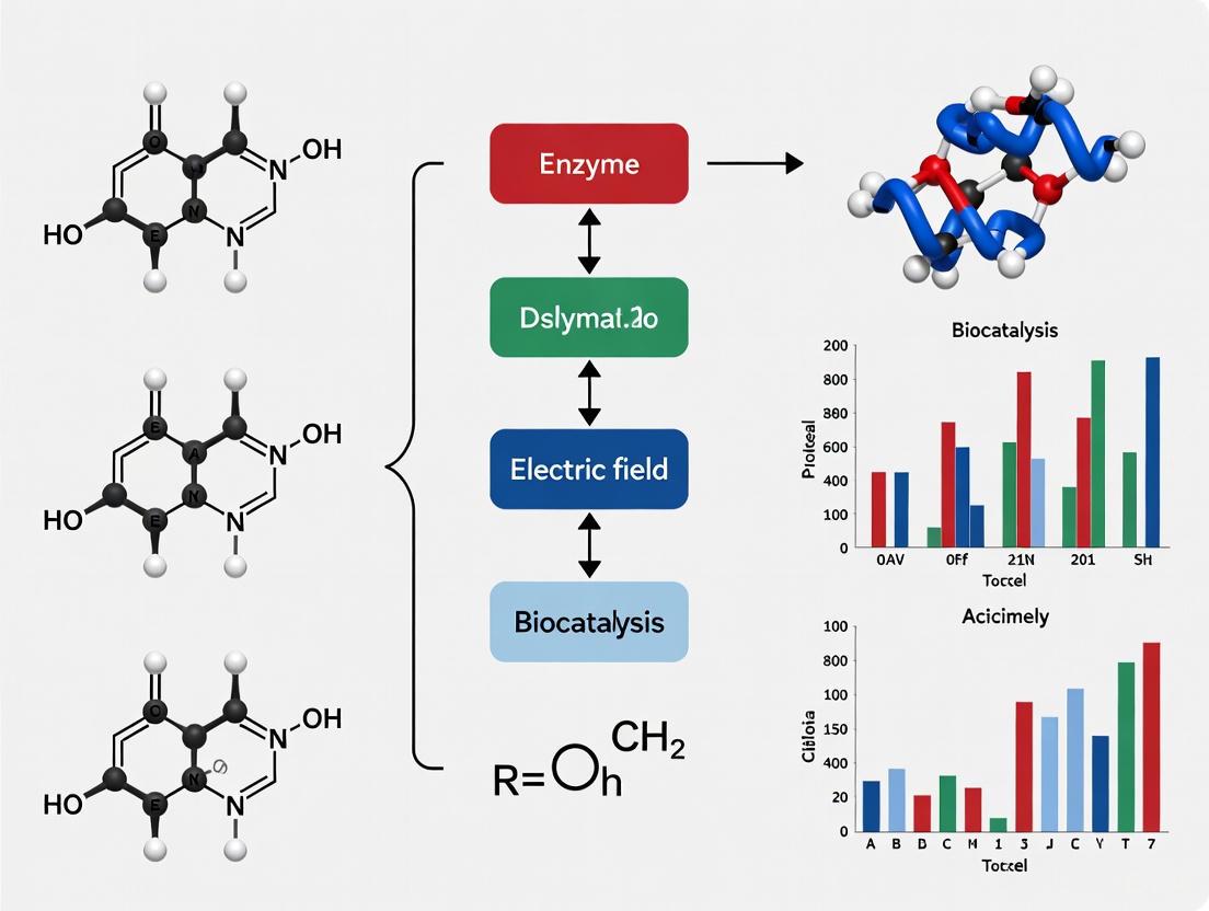
Conceptual Workflow: From Theory to Design
The following diagram outlines the logical relationship between the core theory of electrostatic preorganization and the modern approaches for its analysis and application in enzyme design.
KSI Troubleshooting FAQs
What are the common issues when studying KSI catalysis and their solutions?
Problem: Inconsistent or lower-than-expected reaction rates.
- Possible Cause & Solution:
- Active site residue protonation state: The catalytic efficiency of KSI is highly dependent on the precise protonation states of Asp-38 (general base) and Tyr-14/Tyr-16 (part of the oxyanion hole). Ensure reaction buffer pH is optimized for the specific KSI homolog (typically around pH 7) [6] [7].
- Disruption of the oxyanion hole: Mutations or conditions that disrupt the hydrogen-bonding network of the oxyanion hole (e.g., Asp-99/103, Tyr-14/16) drastically reduce activity. Check enzyme construct and purity [8] [6].
- Incorrect intermediate stabilization: The reaction proceeds through a dienolate intermediate. Using analogues like 4-fluorophenol can help probe and validate the intermediate stabilization capability of your enzyme preparation [6].
Problem: Discrepancies in determining the catalytic mechanism (dienol vs. dienolate intermediate).
- Possible Cause & Solution:
- Indirect measurement methods: Use direct spectroscopic methods to probe the ionization state of the intermediate. FTIR spectroscopy with specifically designed inhibitor probes (e.g., 4-fluorophenol) can report directly and quantitatively on the ionization state of the ligand bound in the active site [6].
- Interpretation of mutational data: The energetic contributions of catalytic residues can be additive rather than synergistic. Perform detailed thermodynamic cycle analysis to discriminate between concerted and stepwise proton transfer mechanisms [6].
Problem: Difficulty quantifying the contribution of electric fields to catalysis.
- Possible Cause & Solution:
- Complex native system: The native enzyme's complexity makes it difficult to isolate the effect of electric fields from other catalytic strategies. Use supramolecular enzyme mimics with strategically placed charged groups to create and measure local, oriented electric fields. Stark spectroscopy can be employed to quantify these electric fields [6] [9].
How can I optimize experimental conditions for enzyme inhibition studies like those relevant to drug development?
Problem: Inaccurate or imprecise estimation of inhibition constants (Kic and Kiu).
- Possible Cause & Solution:
- Inefficient experimental design: Traditional designs using multiple substrate and inhibitor concentrations can introduce bias and are inefficient. Adopt the 50-BOA (IC50-Based Optimal Approach), which requires initial velocity data obtained using a single inhibitor concentration greater than the IC50. This method incorporates the harmonic mean relationship between IC50 and the inhibition constants into the fitting process, reducing the number of experiments by over 75% while improving precision [10].
- Uncertain inhibition type: Using the mixed inhibition model (which applies to all types) without prior knowledge can lead to false reporting. The 50-BOA approach allows for precise estimation without prior knowledge of the inhibition type [10].
Problem: Inconsistencies between in vitro and predicted in vivo enzyme inhibition.
- Possible Cause & Solution:
- Non-optimized in vitro conditions: Use well-characterized experimental systems (e.g., hepatocytes or microsomes) and correct kinetic parameters for nonspecific binding. For Ki or IC50 determinations, use initial product formation rates with less than 20% substrate depletion to avoid artefacts like product inhibition [11].
- Ignoring time-dependent inhibition: Routinely screen for time-dependent (irreversible) inhibition during drug development, as this is a major clinical concern that can lead to drug withdrawal [11].
Experimental Protocols
Protocol 1: Estimating Inhibition Constants Using the 50-BOA Method
This protocol enables precise and accurate estimation of enzyme inhibition constants with a minimal experimental dataset [10].
Determine IC50:
- Perform a preliminary experiment by measuring the initial reaction velocity (V₀) over a range of inhibitor concentrations ([I]) at a single substrate concentration, typically [S] = Kₘ.
- Fit the % control activity data to a log(inhibitor) vs. response model to calculate the IC₅₀ value.
Set Up the Optimal Experiment:
- Measure initial velocities using a single inhibitor concentration [I] > IC₅₀.
- Vary the substrate concentration across at least three values (e.g., 0.2Kₘ, Kₘ, and 5Kₘ) to adequately define the enzyme kinetics.
Data Fitting and Analysis:
- Fit the collected initial velocity data to the mixed inhibition model using a software package that implements the 50-BOA (available for MATLAB and R).
- The model equation is: V₀ = (Vₘₐₓ * [S]) / ( Kₘ * (1 + [I]/Kᵢ𝒸) + [S] * (1 + [I]/Kᵢᵤ) )
- The fitting algorithm will incorporate the relationship between IC₅₀, Kᵢ𝒸, and Kᵢᵤ, providing precise estimates for both inhibition constants and identifying the inhibition type.
Protocol 2: Probing the Ionization State of an Intermediate in KSI using FTIR
This protocol uses IR spectroscopy to directly determine whether a reaction intermediate is neutral or charged, a key question in KSI catalysis [6].
Sample Preparation:
- Use a catalytically compromised mutant of KSI (e.g., Asp40Asn in the P. putida homolog) to trap the intermediate state.
- Prepare a solution of the mutant enzyme in an appropriate buffer (e.g., 50 mM phosphate, pD 7.0). Use D₂O-based buffers to avoid the strong IR absorption of H₂O.
- Titrate the enzyme with a intermediate analog, such as 4-fluorophenol (pKₐ = 10.0, matching the proposed dienol intermediate), which contains an intrinsic IR probe (C-F stretch).
FTIR Spectroscopy:
- Record IR spectra of the free enzyme, free ligand, and the enzyme-ligand complex.
- Focus on the spectral region corresponding to the C-F stretching vibration (around 1100-1300 cm⁻¹). The exact frequency is sensitive to the phenol's ionization state.
Data Analysis:
- Compare the C-F stretch frequency of the bound ligand to that of the neutral and ionized forms of the free ligand in solution.
- A frequency shift towards that of the ionized (phenolate) form indicates the ligand is deprotonated when bound to the active site.
- Quantitatively analyze the spectra to calculate the fraction of bound ligand that is ionized. This fractional ionization provides a direct measure of the free energy difference, informing the thermodynamic advantage of a concerted mechanism.
Key Data for Ketosteroid Isomerase (KSI)
Table 1: Wild-Type KSI Reaction Kinetics on 5-Androstenedione [8]
| Kinetic Parameter | Value |
|---|---|
| kcat (s⁻¹) | 3.0 x 10⁴ |
| Km (μM) | 123 |
| kcat/Km (M⁻¹s⁻¹) | 2.4 x 10⁸ |
Table 2: Key Catalytic Residues in KSI Homologs [8]
| Residue Role | Comamonas testosteroni | Pseudomonas putida |
|---|---|---|
| General Acid/Base | Asp-38 | Asp-40 |
| Oxyanion H-Bond Donor | Asp-99, Tyr-14 | Asp-103, Tyr-16 |
Visualizing Mechanisms and Workflows
KSI Catalytic Mechanism and Electric Field
50-BOA Workflow for Efficient Inhibition Analysis
The Scientist's Toolkit: Research Reagent Solutions
Table 3: Essential Reagents for KSI and Enzyme Inhibition Studies
| Reagent / Material | Function / Application | Example / Note |
|---|---|---|
| KSI Homologs | Model enzyme for studying proton transfer & electrostatic catalysis. | Comamonas testosteroni (TI), Pseudomonas putida (PI) [8]. |
| Intermediate Analogs | Probe the ionization state and binding in the active site. | 4-Fluorophenol (pKₐ 10.0), Equilenin, 19-nortestosterone [6] [7]. |
| Site-Directed Mutagenesis Kits | Generate catalytic mutants to dissect residue contributions. | Used to create D38N, Y14F, D99A/N mutants for mechanistic studies [6]. |
| IC50-Based Optimal Approach (50-BOA) | Software/Tool for precise inhibition constant estimation with minimal data. | User-friendly MATLAB and R packages are available [10]. |
| Methylation-Free E. coli Strains | Propagate plasmids for digestion when restriction sites are susceptible to methylation. | Use dam-/dcm- strains (e.g., E. coli GM2163) if methylation blocks cleavage [12] [13]. |
Frequently Asked Questions (FAQs)
Q1: What are the primary catalytic strategies enzymes use to accelerate chemical reactions? Enzymes primarily utilize transition state stabilization (TSS) and the management of entropic advantages to achieve remarkable rate enhancements. TSS involves the preferential stabilization of the high-energy transition state structure through precise electrostatic interactions and other bonding interactions within the active site. The entropic advantage, or the "Circe effect," involves reducing the unfavorable entropy change required to reach the transition state by preorganizing substrates into reactive conformations and proximity [14].
Q2: How do electric fields contribute to transition state stabilization? The precise orientation of electric fields within an enzyme's active site creates a preorganized electrostatic environment that stabilizes the charge distribution of the transition state. This significantly lowers the activation energy required for the reaction. Recent studies using vibrational Stark effect spectroscopy have directly measured these fields, confirming their critical role in catalysis. The magnitude and direction of these fields differ considerably from those in common solvents, highlighting enzymatic optimization [14] [15].
Q3: What is the difference between ground state destabilization and transition state stabilization? Ground state destabilization (GSD) proposes that enzymes distort substrate bonds toward the transition state geometry, while TSS involves stronger binding to the transition state than to the ground state. The Circe effect is a more thermodynamically plausible form of GSD, where the enzyme selectively destabilizes the substrate's reactive region while maintaining favorable binding interactions with distal parts of the substrate [14].
Q4: Can external electric fields be used to mimic enzymatic catalysis in synthetic systems? Yes, emerging research demonstrates that oriented external electric fields (OEEFs) can catalyze chemical reactions in synthetic systems. For example, carbon nanotubes in microfluidic reactors can apply strong electric fields that influence reaction mechanisms, change rate-limiting steps, and even enable reactions that do not proceed without a field, offering a promising path for sustainable synthesis [16].
Q5: How are electric fields measured and mapped within enzyme active sites? Researchers use vibrational Stark effect (VSE) spectroscopy, which measures shifts in the vibrational frequencies of probe molecules bound to the active site. These shifts reveal the strength and orientation of the local electric field. Novel probes, like modified N-cyclohexylformamide, allow measurement of electric field magnitude and direction, providing a more complete picture of the active site electrostatic environment [15].
Troubleshooting Guide: Electric Field Analysis in Enzyme Studies
Common Experimental Challenges and Solutions
Table 1: Troubleshooting Electric Field and Catalysis Experiments
| Problem | Possible Cause | Recommended Solution |
|---|---|---|
| Inconclusive VSE data | Poor probe binding or orientation; inability to detect key vibrational modes [15]. | Use deuterium isotope exchange (e.g., C-H to C-D bonds) to access measurable vibrational frequencies; employ computational simulations to validate probe placement [15]. |
| Low catalytic activity in enzyme designs | Poorly preorganized electric field in the active site; suboptimal field orientation [14] [15]. | Use two-directional VSE probes to map field orientation; redesign active site residues to optimize the electrostatic environment for transition state stabilization [15]. |
| Difficulty quantifying electrostatic contributions | Overreliance on structural data; inability to separate electrostatic effects from other catalytic factors [14]. | Combine VSE experiments with Quantum Mechanical/Molecular Mechanical (QM/MM) calculations and conceptual Density Functional Theory (CDFT) analysis to correlate field strength with reactivity [17]. |
| External field experiments not yielding results | Incorrect field alignment with the reaction axis; insufficient field strength [16]. | Ensure the substrate is fixed and oriented relative to the field; use high-voltage sources and polarized nanotube surfaces to enhance field strength and control [16]. |
Experimental Protocols
Protocol 1: Mapping Electric Field Orientation with a Two-Directional Probe
This protocol is adapted from research to visualize the electric field in the active site of liver alcohol dehydrogenase [15].
1. Principle A probe molecule (N-cyclohexylformamide) is engineered with two chemical bonds approximately 120 degrees apart. The vibrational Stark effect on these bonds is measured to reconstruct both the magnitude and the orientation of the electric field within the active site.
2. Materials
- Purified enzyme (e.g., Liver Alcohol Dehydrogenase)
- N-cyclohexylformamide probe, synthesized with deuterium substitution at the critical C-H bond
- Appropriate buffer solutions
- Infrared spectrometer
- Computational resources for molecular simulations and quantum mechanical calculations
3. Procedure
- Step 1: Probe Binding. Incubate the deuterium-modified N-cyclohexylformamide probe with the enzyme, allowing it to bind tightly to the active site as an inhibitor.
- Step 2: IR Spectroscopy. Perform infrared spectroscopy on the enzyme-probe complex. Measure the vibrational frequency shifts of the carbon-deuterium (C-D) bond and another key bond (e.g., C=O) in the probe.
- Step 3: Data Analysis. The observed Stark shifts (in cm⁻¹) are proportional to the projection of the electric field onto the respective bond axes. Use the formula: Δν = -Δμ · E, where Δν is the frequency shift, Δμ is the difference in dipole moment, and E is the electric field.
- Step 4: Field Reconstruction. Using the two directional measurements, calculate the vector components of the electric field to determine its overall orientation within the active site.
- Step 5: Computational Validation. Compare experimental results with quantum mechanical/molecular mechanical (QM/MM) simulations of the enzyme-probe system to validate the findings.
Protocol 2: Analyzing Electrostatic Contributions in a Diels-Alderase
This protocol uses conceptual DFT and electric field analysis to unravel the electrostatic basis of catalysis in enzymes like AbyU [17].
1. Principle The reactivity of bound substrates is predicted by calculating atom-condensed Fukui functions, which describe regional susceptibility to electrophilic attack. This reactivity is then correlated with the electric field exerted by the enzyme on key reactive moieties.
2. Materials
- Enzyme-substrate structural data (from crystallography or docking)
- Computational software for QM/MM and conceptual DFT calculations
- Electric field analysis software
3. Procedure
- Step 1: Pose Generation. Generate multiple enzyme-substrate binding poses using molecular docking.
- Step 2: Fukui Function Calculation. For each pose, perform quantum mechanical calculations on the reactant to compute the Fukui function (ƒ⁻) for the key carbon atoms (e.g., the diene carbons in a Diels-Alder reaction).
- Step 3: Electric Field Calculation. For each pose, use QM/MM simulations to calculate the electric field vector projected along the critical reaction axis (e.g., the diene moiety).
- Step 4: Correlation Analysis. Correlate the calculated Fukui function values (reactivity descriptors) with the strength and alignment of the enzyme's electric field.
- Step 5: Mechanism Insight. Identify which poses have electric fields that best align to stabilize the transition state, explaining the enzyme's catalytic power and selectivity.
The Scientist's Toolkit: Key Research Reagents & Materials
Table 2: Essential Reagents for Electric Field and Enzyme Catalysis Research
| Item | Function/Application |
|---|---|
| Vibrational Stark Probe (e.g., N-cyclohexylformamide) | A small molecule inhibitor that binds the active site; its chemically engineered bonds serve as sensors for local electric fields via IR spectroscopy [15]. |
| Isotopically Labeled Compounds (e.g., Deuterated Bonds) | Used to modify probe molecules, making specific chemical bonds (like C-D) spectroscopically accessible for measurement in a protein environment [15]. |
| Polarized Nanotube Surfaces | Provide a platform in microfluidic reactors to apply strong, oriented external electric fields to chemical reactions, mimicking enzyme active sites [16]. |
| Conceptual DFT Descriptors (e.g., Fukui Functions) | Computational tools that predict the intrinsic reactivity of different atoms in a molecule based on electron density, helping to explain enzyme regioselectivity [17]. |
| QM/MM Software | Enables hybrid quantum mechanical and molecular mechanical simulations to model enzyme catalysis and calculate internal electric fields with atomic detail [17] [15]. |
FAQs: Understanding the Protein Scaffold and Long-Range Interactions
Q1: What is the functional role of the protein scaffold beyond providing a structural framework for the active site? The protein scaffold is not a passive structural element but plays an active role in catalysis. It facilitates the formation of conformational ensembles—numerous protein substates in rapid equilibrium—that are essential for function [18]. Through long-range interactions, the scaffold establishes thermally activated dynamical networks that connect the active site to the protein-water interface, acting as conduits for energy transfer and communication [18]. This allows the scaffold to influence the active site remotely.
Q2: How can remote mutations, far from the active site, significantly impact enzyme catalysis? Mutations in the protein scaffold can alter the distribution of conformational substates, shifting the population toward catalytically competent conformations [18]. This is often achieved through rigidification of the active site via improved packing, effectively pre-organizing the site for catalysis [18]. Furthermore, scaffold mutations can fine-tune intramolecular interactions that stabilize remote functional loops, which are critical for complex biological functions like accessing cellular targets [19].
Q3: What is the evidence that electric fields from the protein scaffold contribute to catalysis? Experimental studies using the vibrational Stark effect have provided direct measurements of the strong electric fields present within enzyme active sites [14]. These fields, generated by the precise three-dimensional arrangement of the protein scaffold, can stabilize the transition state of a reaction and are a major contributor to catalytic rate enhancement [14]. Computational designs like the AI.zymes platform successfully improve activity by iteratively selecting variants with stronger catalytic electric fields, demonstrating their importance [20].
Q4: How does the acquisition of remote loops during evolution lead to new enzyme functions? The acquisition of remote loops can grant enzymes access to new biological functions without disrupting the original catalytic activity [19]. For example, in GH19 chitinases, the acquisition of a specific remote loop (Loop II) was necessary for the emergence of antifungal activity [19]. This loop directly accesses the fungal cell wall, but its function depends on long-range interactions with the protein scaffold that restrict its mobility and stabilize a defined structure [19].
Troubleshooting Guide: Experimental Challenges in Analyzing Scaffold Function
| Problem | Possible Cause | Recommended Solution |
|---|---|---|
| Incomplete or No Digestion | Catalytic activity blocked by DNA methylation. | Check the enzyme's sensitivity to Dam/Dcm/CpG methylation; propagate plasmid in a dam-/dcm- E. coli strain [21] [22]. |
| Unexpected Cleavage Patterns (Star Activity) | Altered enzyme specificity due to non-optimal conditions (e.g., high glycerol concentration, long incubation). | Ensure glycerol concentration is <5%; use the recommended reaction buffer; decrease incubation time and enzyme units; use High-Fidelity (HF) engineered enzymes [21] [22]. |
| Low Catalytic Efficiency in Designed Enzyme | Suboptimal conformational sampling; inactive substates are overly populated. | Use directed evolution to select for mutations that shift the conformational ensemble toward catalytically active populations, often by rigidifying the active site through improved packing [18]. |
| Difficulty in Resolving Small/ Flexible Protein Structures | Proteins smaller than ~40 kDa are difficult to visualize at high resolution with cryo-EM. | Utilize a double-shell protein scaffold technology that sandwiches the target protein to increase particle size and enable high-resolution structure determination [23]. |
Key Experimental Protocols
Protocol 1: Ancestral Sequence Reconstruction to Study Remote Loop Evolution
Objective: To identify key structural acquisitions and understand the evolutionary path by which a protein scaffold gains new functions.
Methodology:
- Sequence Collection and Phylogeny: Collect a comprehensive set of modern sequences for the enzyme family of interest. Perform multiple sequence alignment and infer a maximum-likelihood phylogenetic tree [19].
- Ancestral Sequence Reconstruction: Use statistical models to infer the most probable amino acid sequences at ancestral nodes of the phylogenetic tree [19].
- Gene Synthesis and Protein Expression: Synthesize genes coding for the reconstructed ancestral proteins, clone them into an expression vector, and express and purify the proteins [19].
- Functional Characterization: Measure both the core catalytic activity and the newly evolved function (e.g., antifungal activity) of the ancestral proteins and their engineered loop variants (e.g., loop insertions/deletions) [19].
- Structural and Dynamical Analysis: Solve high-resolution structures (e.g., via X-ray crystallography) and perform Molecular Dynamics (MD) simulations to analyze structural differences and loop mobility, correlating them with the acquired function [19].
Protocol 2: Utilizing the Vibrational Stark Effect to Measure Active-Site Electric Fields
Objective: To experimentally measure the magnitude and direction of the intrinsic electric field within an enzyme's active site.
Methodology:
- Probe Incorporation: Introduce a covalent vibrational probe (e.g., a nitrile group) into the enzyme's active site, typically by chemically modifying a bound substrate or inhibitor [14].
- Infrared Spectroscopy: Obtain the infrared (IR) absorption spectrum of the vibrational probe when bound inside the enzyme.
- External Electric Field Application: Place the enzyme-probe complex in an external electric field and record the Stark spectrum, which shows the shift in the IR absorption band in response to the field [14].
- Calibration: Calibrate the vibrational probe's sensitivity to electric fields (its Stark tuning rate) in a known environment.
- Field Calculation: Use the measured Stark effect to calculate the electric field the enzyme exerts on the probe along the relevant reaction coordinate [14].
Research Reagent Solutions
| Reagent / Tool | Function in Research |
|---|---|
| Directed Evolution Platforms | A semi-rational approach to optimize enzyme properties, including those mediated by the scaffold, such as electric fields and conformational stability [20]. |
| Molecular Dynamics (MD) Simulation Software | Used to visualize protein dynamics in real time and analyze the mobility and interactions of remote loops and dynamical networks [18] [19]. |
| Room-Temperature X-ray Crystallography | Allows for the detection of alternate protein side chain conformations and the inference of dynamical networks, providing a more dynamic view of the scaffold than traditional cryo-crystallography [18]. |
| Ancestral Sequence Reconstruction Algorithms | Computational tools to infer ancient protein sequences, enabling the experimental study of evolutionary trajectories and the functional impact of historical scaffold changes [19]. |
| Double-Shell Protein Scaffold | A technology using fusion proteins (e.g., apoferritin and MBP) to cage small, flexible proteins, enabling high-resolution structure determination via single-particle cryo-EM [23]. |
Conceptual Diagrams
Diagram 1: Enzyme Function via Scaffold Dynamics and Remote Loops
Diagram Title: Enzyme Function via Scaffold Dynamics and Remote Loops
Diagram 2: Experimental Workflow for Evolutionary Analysis
Diagram Title: Workflow for Evolutionary Analysis of Scaffolds
Computational and Experimental Tools for Measuring and Designing Catalytic Electric Fields
Troubleshooting Guides
1. Unphysical Energies or Catastrophic Drift in QM/MM Dynamics
- Problem: During a QM/MM molecular dynamics simulation, the total energy behaves erratically or the structure drifts and becomes unphysical.
- Causes:
- Incorrect treatment of long-range electrostatics: Using a simple cutoff method for the QM-MM electrostatic interactions can introduce significant errors, as particles beyond 10-20 Å can still have non-negligible contributions to the energy and forces [24].
- Inadequate convergence of polarizable models: The shell model or Drude oscillator iterations may not have reached convergence, leading to unstable forces [25].
- Parameter mismatch: Van der Waals parameters at the QM/MM boundary, especially for custom-defined atoms, may be too repulsive or attractive [26].
- Solutions:
- Implement a long-range electrostatic correction (LREC) method. The LREC approach uses a smoothing function to scale electrostatic interactions, smoothly reducing them to zero at a finite cutoff. Studies show that energies and forces converge to within 0.2% of Particle Mesh Ewald (PME) results with a cutoff of 20–25 Å [24].
- For polarizable simulations, tighten the convergence criteria for the microiterations. In the
mm_polcosmethod, adjustpolcos_maxdx,polcos_rmsdx, andpolcos_toler_energy[25]. - Check and refine parameters for any user-defined MM atom types in the QM region using the
$force_field_paramssection, paying close attention to Lennard-Jones parameters [26].
2. Failure to Converge in Polarizable QM/MM SCF Calculations
- Problem: The self-consistent field (SCF) procedure for the QM region fails to converge when polarizable MM force fields are active.
- Causes:
- Strong electric field from polarized MM atoms: The induced dipoles or shells in the MM region create a strong, fluctuating electric field that the QM Hamiltonian cannot easily adapt to [25] [27].
- Overlapping atoms or poor initial geometry: This can cause excessively high initial forces from the MM region.
- Solutions:
- Use a two-step relaxation process: First, allow the MM polarizable sites (shells or Drude oscillators) to relax in the field of the initial QM electron density. Then, run the full mutual polarization cycle until self-consistency is achieved [25].
- Ensure the initial structure is well-minimized using a pure MM force field before starting the QM/MM calculation. Verify that no atoms are unnaturally close to each other.
3. Inaccurate Reaction Barriers in Enzyme Design
- Problem: Computed reaction barriers for a designed enzyme are inaccurate compared to experimental results, hindering the rational optimization of electric fields for catalysis [27] [28].
- Causes:
- Lack of electrostatic preorganization: The designed protein scaffold fails to generate the strong, oriented internal electric field necessary to stabilize the transition state [27].
- Neglect of long-range electrostatics: Truncating electrostatic interactions prevents an accurate description of the total electric field experienced by the substrate in the active site [24].
- Use of non-polarizable force fields: Standard mechanical embedding (e.g., ONIOM) does not allow the QM region's electron density to be polarized by the MM environment, missing a key catalytic effect [26].
- Solutions:
- Employ an electronic embedding scheme (e.g., the Janus model in Q-Chem) that includes the MM point charges directly in the QM Hamiltonian [26].
- Always use a robust long-range electrostatic treatment (like LREC or PME) in production calculations to ensure the electric field is properly modeled [24].
- Analyze the electric field in the active site using vibrational Stark effect proxies or by analyzing the QM electron density response to the protein environment [27].
Frequently Asked Questions (FAQs)
Q1: What is the fundamental difference between mechanical and electronic embedding in QM/MM?
- A: In mechanical embedding (e.g., ONIOM), the QM region is not polarized by the MM environment. The total energy is a combination of MM energies for the whole system and the QM region, and a QM energy for the QM region. Interactions between the subsystems are described only at the MM level [26]. In electronic embedding (e.g., the Janus model), the electrostatic potential from the MM point charges is included directly in the QM Hamiltonian. This allows the QM electron density to be polarized by the MM environment, providing a more physically accurate description, which is critical for modeling enzyme catalysis and optimizing electric fields [26].
Q2: When should I use a polarizable MM force field instead of a fixed-charge force field?
- A: You should consider polarizable force fields when:
- Your research focuses on accurately modeling electrostatic preorganization, a key strategy in natural enzyme catalysis [27].
- The system involves significant charge transfer or polarization effects, such as ions in channels or molecules with large dipole moments responding to a protein environment [25].
- You are studying interfaces between regions of high dielectric contrast, like a protein active site and a bulk solvent [24]. Fixed-charge force fields are computationally cheaper and may be sufficient for systems where polarization effects are less critical.
Q3: My simulation is computationally expensive. What is the most efficient way to handle long-range electrostatics in large QM/MM systems?
- A: The QM(LREC)/MM(PME) approach is an excellent compromise between simplicity, speed, and accuracy [24]. In this method:
- QM Region with LREC: The QM-MM electrostatic interactions are handled with the Long-Range Electrostatic Correction method, which converges with a cutoff of 20-25 Å and does not require modifications to the SCF routine [24].
- MM Region with PME: The much larger MM-MM interactions are treated with the highly efficient Particle Mesh Ewald method [24]. This combination avoids the computational cost of a full Ewald treatment for the QM region while maintaining accuracy.
Q4: How do I handle a covalent bond between the QM and MM regions?
- A: This is done using a "link atom" or a "capping atom." Most software offers automated solutions:
- Link Atoms: A hydrogen atom (link atom) is introduced to cap the valency of the QM atom at the boundary. The QM calculation is performed with the link atom, but its interactions are carefully handled to avoid double-counting [26].
- YinYang Atoms: In Q-Chem's Janus model, a single atom acts as a hydrogen cap in the QM calculation but retains its full MM identity for interactions within the MM subsystem. This maintains charge neutrality and improves performance [26].
Experimental Protocols & Data
Table 1: Convergence Parameters for Polarizable QM/MM (polcos)
| Parameter | Description | Recommended Value | Purpose |
|---|---|---|---|
polcos_maxcycle |
Max outer QM/MM iterations [25] | 20 | Controls the number of mutual polarization cycles. |
polcos_inmaxcycle |
Max inner MM SCF iterations [25] | 1000 | Ensures Drude oscillators/shells converge for a fixed QM density. |
polcos_toler_energy |
QM energy change tolerance [25] | 1.0e-8 | Sets convergence based on energy change between outer cycles. |
polcos_maxdx |
Max change in massless charge position [25] | 2.0e-5 a.u. | Sets a force-based convergence criterion for the polarizable particles. |
Table 2: Comparison of Long-Range Electrostatic Methods
| Method | Principle | Advantages | Limitations |
|---|---|---|---|
| Simple Cutoff | Truncates interactions beyond a fixed distance. | Very fast and simple to implement. | Can introduce severe artifacts in energy and forces; not recommended for production runs [24]. |
| Ewald/PME | Sums interactions in both real and reciprocal space for periodic systems. | Highly accurate; standard for periodic MM. | Requires modifications to the SCF routine; can be complex to implement for QM/MM [24]. |
| LREC | Uses a smoothing function to scale interactions to zero at a cutoff. | Simple implementation; no SCF modifications; accurate with 20-25 Å cutoff [24]. | Less common than PME; requires parameterization of the cutoff distance. |
Detailed Protocol: Setting Up a Polarizable QM/MM Simulation in ChemShell
This protocol outlines the steps for a QM/MM calculation with a Drude polarizable force field, based on the mm_polcos method [25].
System Preparation:
- Obtain the initial protein structure (e.g., from a PDB file).
- Prepare the topology and parameter files for the CHARMM or GROMOS polarizable force field.
- Define the QM region (e.g., substrate and key active site residues) and the MM region.
Input File Configuration:
- In the ChemShell input script, specify the
theory=hybridblock. - Set
coupling=shift(or another electrostatic embedding scheme). - Activate polarizability with
mm_polcos=yesand provide a list of control arguments. - The
polcos_atom_polcosqlist must contain the atom ID, polarizability (in a.u.), and the charge (in a.u.) for each polarizable MM atom.
- In the ChemShell input script, specify the
Execution and Monitoring:
- Run the simulation and closely monitor the output log.
- Check for messages indicating convergence of both the QM SCF procedure and the
polcosmicroiterations. - Verify that the changes in energy and Drude oscillator positions (
polcos_maxdx,polcos_rmsdx) are below the specified thresholds.
The Scientist's Toolkit: Research Reagent Solutions
Table 3: Essential Software and Force Fields for Electrostatic-Focused QM/MM
| Item | Function in Research | Relevance to Electric Field Optimization |
|---|---|---|
| ChemShell | A QM/MM integration environment. | Supports advanced polarizable force fields (shell, Drude) and provides the mm_polcos method for mutual polarization, key for modeling environmental response [25]. |
| Q-Chem | A comprehensive quantum chemistry program. | Its stand-alone Janus model enables electronic embedding QM/MM, allowing the MM charge distribution to directly polarize the QM active site [26]. |
| LICHEM | A package for QM/MM simulations. | Implements the LREC method for accurate and efficient treatment of long-range electrostatics in multipolar/polarizable simulations [24]. |
| CHARMM Drude FF | A polarizable force field based on Drude oscillators. | Allows the MM environment to respond to the charge distribution of the QM region, creating a more realistic and responsive internal electric field [25]. |
| AMOEBA FF | A polarizable force field based on atomic multipoles. | Provides a more accurate description of the electrostatic potential around MM atoms, which is critical for calculating precise electric fields in an enzyme active site [24]. |
Workflow Visualization
Diagram Title: QM/MM Setup for Electric Field Optimization
Diagram Title: Polarizable QM/MM Self-Consistent Cycle
The Vibrational Stark Effect (VSE) describes the perturbation of a molecular vibrational frequency by an external electric field, forming the basis for Vibrational Stark Spectroscopy (VSS). This technique has become an indispensable tool for measuring and analyzing in situ electric field strength in diverse chemical environments, including the binding pockets of enzymes. The fundamental relationship is given by the Stark equation:
ν = ν₀ - Δμ⃗ · F⃗ + ½ F⃗ · Δα · F⃗
where ν and ν₀ are the vibrational frequencies with and without the electric field F⃗, respectively, Δμ⃗ is the difference dipole moment (Stark tuning rate), and Δα is the difference polarizability [29].
For the relatively weak electric fields typically encountered (below 100 MV/cm), the quadratic term can often be neglected, resulting in a linear relationship between the vibrational frequency shift and the electric field: Δν = ν - ν₀ ∝ Δμ⃗ · F⃗ [29]. This linear correlation provides the foundation for using VSE as a molecular ruler for electric fields in complex environments like proteins.
Key Assumptions and Theoretical Framework
The application of VSE rests on four critical assumptions that must be validated for reliable experimental results [29]:
- Bond Localization: The normal stretching vibration of the probe bond must be largely decoupled from the rest of the molecule.
- Electric Field Attribution: Frequency shifts from environmental changes can be fully attributed to the external electric field.
- Linearity: The difference dipole moment Δμ⃗ remains unaffected by the external electric field F⃗.
- Field Strength Extrapolation: The linear relationship observed at weak field strengths also holds for the much stronger fields present in enzyme active sites.
The most crucial of these is the first assumption regarding bond localization. Normal vibrational modes are typically delocalized due to mass coupling, meaning the target vibration can mix with other internal coordinates. If this occurs, the measured frequency shift no longer purely reports on the electric field at the target bond, compromising interpretation [29].
Evaluating Probe Bond Localization The Local Vibrational Mode Theory, specifically the Characterization of Normal Modes (CNM) procedure, quantitatively assesses how much a target normal vibration consists of pure bond stretching character. This method determines the degree to which the local stretching mode of the probe bond is decoupled from other local vibrational modes, providing a quantitative score to evaluate potential VSE probes [29].
Experimental Protocols and Methodologies
Selecting and Validating a VSE Probe
The initial and most critical step is selecting an appropriate probe molecule. An ideal VSE probe exhibits a highly localized target bond vibration.
- Recommended Probe Molecules: Based on comprehensive local mode analysis of 68 candidates, 31 polyatomic molecules with localized target bonds are recommended as ideal VSE probes. The table below summarizes key validated probe types [29].
- Probe Validation: Before deployment in complex systems, validate the localization of your chosen probe's target vibration using computational chemistry methods. Perform a local mode analysis (e.g., using the CNM method) to calculate a localization score. Probes with low scores should be avoided as their frequency shifts will be difficult to interpret unambiguously [29].
Basic VSE Measurement Workflow
The following workflow outlines the core steps for a typical VSE experiment in a biochemical context.
Step-by-Step Protocol:
- Probe Selection & Validation: Choose a probe from the recommended list (see Table 1). Confirm bond localization computationally if it is a novel candidate [29].
- Probe Incorporation: Introduce the probe into the protein system. This can be achieved by:
- Site-specific labeling of a native amino acid (e.g., using a cyanobenzothiazole probe to conjugate with a cysteine residue).
- Using a small molecule inhibitor or substrate analog that contains the probe bond (e.g., a carbonyl group).
- Incorporating a non-canonical amino acid bearing the probe bond directly into the protein sequence.
- Reference Spectrum Acquisition: Place the protein-probe system in its native environment (e.g., buffer, crystal, or membrane). Using FTIR or other IR spectroscopy methods, acquire a high-quality IR absorption spectrum of the target bond (e.g., C=O, C≡N) without any externally applied electric field. This provides the reference frequency, ν₀.
- Application of Electric Field: Apply a known, uniform external electric field to the sample. This is typically done using a custom-built electrochemical cell or a capacitor-like setup with transparent electrodes.
- Perturbed Spectrum Acquisition: Acquire a second IR spectrum under the exact same conditions but with the external electric field applied.
- Frequency Shift Measurement: Analyze the two spectra. Precisely measure the shift in the absorption peak of the target bond, Δν = ν - ν₀.
- Electric Field Calculation: Using the previously calibrated Stark tuning rate (Δμ⃗) for the probe, calculate the magnitude of the electric field projected along the bond's axis using the linear Stark equation: |F| ≈ |Δν| / |Δμ⃗|.
Calibrating the Stark Tuning Rate (Δμ⃗)
The Stark tuning rate (Δμ⃗) is a probe-specific constant that must be determined experimentally before the probe can be used as a quantitative ruler.
- Method: The probe molecule is placed in a series of inert solvents with known dielectric properties (or in a frozen glass) where it experiences different internal electric fields. The vibrational frequency of the probe bond is measured in each solvent. The slope of a plot of frequency (ν) versus the known electric field (F) yields the Stark tuning rate, Δμ⃗ [29].
Troubleshooting Common Experimental Issues
FAQ 1: My measured vibrational frequency shift is non-linear with the applied field. What could be wrong?
- Potential Cause 1: Probe Delocalization. The normal mode of your target bond may not be fully localized, mixing with other vibrations.
- Solution: Re-evaluate your probe choice. Consult literature for probes with high localization scores [29]. computationally validate a new probe before use.
- Potential Cause 2: Strong Field Effects. The applied field might be too strong, making the quadratic term in the Stark equation significant.
- Solution: Reduce the applied field strength and confirm the linear response region for your specific probe.
- Potential Cause 3: Environmental Artifacts. Sample heating, electrode polarization, or molecular reorientation could be causing non-linear artifacts.
- Solution: Ensure temperature control, use short field pulses, and verify electrode stability.
FAQ 2: I observe an "anomalous" (negative) Stark shift in my system. How should I interpret this?
- Explanation: A negative Stark shift, where the frequency decreases with increasing field, was historically considered anomalous but has been observed in systems like CO on Pt(111) electrodes [30].
- Investigation Steps:
- Check for Phase Coexistence: High-resolution IR measurements may reveal that a single absorption peak is actually a doublet, indicating two slightly different molecular environments or adsorbate phases. The apparent negative shift can arise from fitting a single peak to a doublet feature where the two components have different intensities that change with potential or field [30].
- High-Resolution Scan: Perform a high-resolution spectral scan in the problematic region. Use peak-fitting software to deconvolute overlapping peaks.
- Control Experiment: If possible, run a control experiment under conditions known to produce a single, homogeneous phase.
FAQ 3: The signal-to-noise ratio for my VSE measurement is poor. How can I improve it?
- Solution A: Increase Probe Concentration. If feasible, increase the concentration of your labeled protein or probe molecule in the sample path.
- Solution B: Optimize Spectroscopy Settings. Increase the number of scans co-added during spectral acquisition. Use a higher optical aperture if signal-limited, while balancing resolution loss.
- Solution C: Check Sample Homogeneity. Ensure the sample is clear and free of precipitates or bubbles that cause light scattering.
Application in Enzyme Design and Optimization
VSE provides a direct experimental method to measure the pre-organized electric fields inside enzyme active sites, a key factor in catalytic efficiency. The measured electric fields can correlate with catalytic rates, providing a physical metric for designing artificial enzymes [31] [32].
Integrating VSE into the Enzyme Design Cycle: In enzyme design and directed evolution, VSE can be used to screen variants. By incorporating a VSE probe near the designed active site, you can measure whether a given mutation (even a distal one) creates an optimal electric field that stabilizes the reaction's transition state. This moves enzyme design beyond purely structural validation toward functional electrostatic validation [32].
Research Reagent Solutions
The following table details essential materials and reagents used in VSE experiments.
Table 1: Key Research Reagents for VSE Experiments
| Item Name | Function / Description | Example / Specification |
|---|---|---|
| VSE Probe Molecules | Reporter molecules containing a localized vibrational bond (e.g., C=O, C≡N, S=O) whose frequency shifts report on the electric field. | Recommended candidates from local mode analysis (e.g., 31 specific polyatomic molecules) [29]. |
| Site-Specific Labeling Kit | For covalently attaching VSE probes to specific sites in proteins (e.g., cysteine conjugation). | Commercially available kits (e.g., based on maleimide-cyanobenzothiazole chemistry). |
| IR-Transparent Windows | Windows for the sample cell that are transparent in the infrared region of interest. | CaF₂, BaF₂, or ZnSe windows, depending on spectral range and solubility. |
| Stark Cell / Electrochemical Cell | Sample holder capable of applying a uniform, known electric field across the sample. | Custom-built capacitor cells with electrode plates, or commercial electrochemical IR cells. |
| Transition-State Analogue | A stable molecule that mimics the geometry and charge distribution of a reaction's transition state. Used for pre-organizing the active site for measurement. | e.g., 6-Nitrobenzotriazole (6-NBT) for Kemp eliminases [32]. |
Data Presentation and Analysis
Table 2: Summary of Common VSE Probe Bonds and Properties
| Probe Bond Type | Example Molecules | Typical Frequency Range (cm⁻¹) | Key Considerations |
|---|---|---|---|
| Carbonyl (C=O) | Formaldehyde, Esters, Amides | 1650-1750 | Very common; can be incorporated into substrates or inhibitors. Potential for H-bonding complications. |
| Nitrile (C≡N) | Anisonitrile, Thiocyanates | 2200-2300 | Sharp IR band; minimally perturbing to biological systems. Stark tuning rate can be lower than C=O. |
| Sulfoxide (S=O) | Dimethyl sulfoxide (DMSO) | 1050-1100 | Strong dipole; useful for specific environments. |
| Carbon Monoxide (C≡O) | CO (as ligand in heme proteins) | 1900-2200 | Very strong Stark response; use is limited to specific metal-binding sites. |
FAQs: Computational Challenges in Inverse Design
Q1: What is the core objective of an inverse design protocol for electric field generation in enzymes?
The primary objective is to solve the inverse problem: designing a protein scaffold that produces a specific, preorganized electric field to optimally stabilize the transition state of a desired reaction. This involves computationally sampling the vast space of possible charge distributions around an active site to find the optimal arrangement that generates the electric field most beneficial for catalysis, rather than the traditional approach of designing an active site around a fixed chemical scaffold [1] [3].
Q2: Our design protocol consistently produces enzymes with catalytic efficiencies orders of magnitude lower than natural enzymes. What key factor might our computational models be missing?
Current computational design protocols often omit the optimization of long-range electrostatic interactions [1] [3]. The catalytic prowess of natural enzymes is largely derived from their electrostatic preorganization—the precise, fixed orientation of permanent dipoles within the enzyme scaffold that creates an electric field favoring the transition state. If your protocol focuses only on the immediate active site chemistry and does not explicitly optimize the electric field generated by the entire protein scaffold, the resulting designs will lack this critical catalytic driver [1].
Q3: What are the main computational bottlenecks in simulating and optimizing electric fields for enzyme design?
The main bottlenecks include:
- High Computational Cost: Modeling proteins with hundreds to thousands of atoms using high-level quantum mechanics is prohibitively expensive for the necessary sampling [1] [3].
- Force Field Accuracy: Standard molecular mechanics force fields (e.g., Amber ff14SB, Charmm C36m) can be inadequate for accurately reproducing the electric fields observed in quantum mechanical calculations. Polarizable force fields like AMOEBA show better performance but at a higher computational cost [1].
- Handling Protonation States: Fixed protonation states during simulation can misrepresent the true electrostatic environment. While methods for handling titratable residues exist, they require lengthy simulations to equilibrate [1].
Q4: How can we validate that our computationally designed enzyme actually generates the intended optimal electric field?
Validation can be performed by analyzing the electric field and its effects in the reactant state, which is more computationally tractable than simulating the full reaction pathway. Key metrics include [1]:
- Electric Field Projection: Measuring the electric field projection along the relevant reaction axis.
- Charge Density Topology: Analyzing the topology of the reactant state electron density, as features like electrostatic potential at bond critical points correlate with the applied field and reaction barrier.
- Field Line Topology: Comparing the global distribution of electric field lines around key bonds to those known to be predictive of high reactivity [1].
Troubleshooting Guides
Troubleshooting Inaccurate Electric Field Calculations
Problem: Computed electric fields within the enzyme active site do not align with benchmark quantum mechanical calculations or experimental data.
| Symptom | Potential Cause | Recommended Solution |
|---|---|---|
| Large field deviations in specific regions | Use of non-polarizable force fields (e.g., ff14SB, C36m) | Switch to a polarizable force field like AMOEBA for more accurate electrostatic representation [1]. |
| Unphysical field fluctuations | Fixed protonation states of residues | Implement a titratable MD protocol (e.g., using pi-DMD software) that allows protonation states to change during simulation [1]. |
| General inaccuracy vs. QM benchmarks | Neglect of environmental ions or post-translational modifications | Explicitly include physiologically relevant ions in simulations and account for common modifications like phosphorylation [1]. |
| Field strength seems uncorrelated with catalytic activity | Focusing on a single point or vector for field analysis | Adopt a global field analysis using 3D field line distributions or charge density topology, as discrete points can be misleading [1]. |
Troubleshooting Optimization Algorithm Failure
Problem: The optimization algorithm fails to converge on a protein sequence or structure that produces the target electric field, or it converges on physically unrealistic solutions.
| Symptom | Potential Cause | Recommended Solution |
|---|---|---|
| Algorithm stuck in local minima | Poor balance between exploration and exploitation | Integrate Lévy flights into the optimization to enhance exploration and escape local optima [33]. |
| Premature convergence | Population-based optimizer losing diversity | Use mechanisms like the Natural Survivor Method (NSM) or adaptive mutation to maintain population diversity and prevent premature convergence [33]. |
| Slow convergence rate | Inefficient search strategy | Hybridize with Simulated Annealing (SA) to improve exploitation and refine solutions by occasionally accepting worse solutions to explore broader space [33]. |
| Solutions violate physical constraints | Lack of constraints in objective function | Introduce velocity and position bounds or other constraint-handling techniques (e.g., penalty functions, feasibility rules) to keep solutions within physically realistic parameters [34]. |
Experimental Protocol: A Generalized Workflow for Inverse Electric Field Design
This protocol provides a step-by-step guide for computationally designing enzyme variants with optimized electrostatic preorganization.
Phase 1: System Setup and Target Definition
Define the Reaction and Target Field:
- Identify the reaction coordinate and the key bond(s) undergoing electron reorganization.
- Use quantum mechanical calculations (e.g., DFT) on the reaction in solution to understand the intrinsic charge redistribution.
- Define the theoretical optimal electric field, often a vector aligned with the reaction axis or a more complex heterogeneous field, that would maximally stabilize the transition state [1] [3].
Prepare the Initial Protein Model:
- Obtain a starting protein structure (wild-type or a preliminary design).
- Use software like PDB2PQR or H++ to assign initial protonation states at the relevant pH.
- Employ a polarizable force field (AMOEBA) for more accurate electrostatics if computational resources allow [1].
Phase 2: Iterative Electric Field Optimization
Sample Charge Embeddings:
- Systematically sample mutations of residues surrounding the active site (typically within 10-15 Å) to different amino acids that alter charge or dipole (e.g., Lys, Arg, Asp, Glu, Ser, Asn).
- For each variant, run molecular dynamics (MD) simulations to obtain a thermodynamic ensemble of structures [1] [3].
Calculate and Analyze the Electric Field:
- For each MD snapshot, calculate the electric field vector at the key bond(s) of the reactant state.
- Compute the average electric field projection along the reaction axis, or use more advanced metrics like field line topology or charge density topology [1].
Run Optimization Algorithm:
- Use a metaheuristic optimization algorithm (e.g., a modified Artificial Electric Field Algorithm - AEFA-C, Genetic Algorithm) to guide the search for the best sequence.
- Objective Function: Minimize the difference between the computed electric field (from step 4) and the target optimal field (from step 1).
- Constraints: Enforce physical constraints like steric clashes, solubility, and structural stability [34] [33].
Phase 3: Validation and Downstream Analysis
Validate with Free Energy Calculations:
- For the top-ranking designs, perform more computationally expensive calculations (e.g., QM/MM, free energy perturbation) to verify that the designed field actually lowers the reaction barrier compared to the starting model [3].
Propose Mutations for Experimental Testing:
- Generate a final list of point mutations or combination mutants predicted to enhance catalysis via optimal electrostatic preorganization.
The following workflow diagram illustrates the key stages of this protocol:
The Scientist's Toolkit: Essential Research Reagents & Software
The following table details key computational tools and conceptual "reagents" essential for working in this field.
| Item Name | Type | Function/Brief Explanation |
|---|---|---|
| Polarizable Force Fields | Software/Parameter Set | Force fields like AMOEBA that go beyond fixed partial charges to model electronic polarization, providing a more accurate representation of electric fields within a protein [1]. |
| Metaheuristic Optimizers | Algorithm | Population-based optimization algorithms like the Artificial Electric Field Algorithm (AEFA) or its modified versions (mAEFA, AEFA-C). They are used to efficiently search the vast sequence space for optimal field-generating mutations [34] [33]. |
| Electric Field Probes | Computational Metric | Defined vectors along key chemical bonds. The electric field projection along these probes in the reactant state is a strong predictor of catalytic rate acceleration and is used to guide the inverse design process [1] [3]. |
| Continuum or Explicit Solvent | Simulation Environment | The choice of how to model the solvent (e.g., Generalized Born vs. TIP3P water). This significantly impacts the calculated electrostatic properties and protonation states of residues [1]. |
| Molecular Dynamics (MD) Engine | Software | Software like GROMACS, AMBER, or NAMD used to simulate the motion of the protein over time, generating an ensemble of structures for electric field analysis [1]. |
| Protonation State Sampler | Software/Method | Tools like pi-DMD or H++ that help predict or simulate the correct protonation states of acidic and basic residues under physiological conditions, which is critical for accurate field calculations [1]. |
Core Concepts: Why Integrate Rational Design with Directed Evolution?
What are the fundamental limitations of a purely rational design approach?
Purely rational design relies on a predictive understanding of sequence-structure-function relationships, which is often incomplete. Key challenges include:
- Insufficient Structural Data: Reliable structural information is frequently unavailable for the protein of interest. While AI has improved protein structure prediction, it remains limited for larger proteins and macromolecular complexes [35].
- Difficulty Predicting Mutational Effects: The sequence-structure-function relationship is difficult to predict accurately, especially at the single residue level. Rationally designed mutations often fail to have the desired effect because computational strategies struggle with long-range electrostatic effects, dynamic correlations, and second coordination sphere interactions [27].
- Limited Exploration: Rational design typically focuses on a limited number of pre-selected positions, potentially missing beneficial mutations in unexpected regions of the protein [36].
What are the key advantages of integrating directed evolution with rational design?
Integrating these approaches creates a powerful feedback loop that leverages the strengths of both:
- Bypassing Mechanistic Knowledge Gaps: Directed evolution allows for optimization based on observed behavior rather than requiring a detailed, predictive understanding of the mechanism [27].
- Discovering Non-Intuitive Solutions: The random mutagenesis component of directed evolution can uncover highly effective, non-intuitive solutions that computational models or human intuition would not predict [36].
- Optimizing Complex Properties: The combination is exceptionally powerful for optimizing complex properties like electric field preorganization, where rational design alone often lacks the sub-angstrom precision needed [27].
- Refining Computational Models: The functional data from directed evolution experiments on designed variants provides a rich dataset to validate and refine computational models, including those for electric field prediction [27].
How does this integration specifically benefit research on electric fields in enzyme design?
Electric field preorganization is a key strategy natural enzymes use to achieve remarkable catalytic efficiency. Integrating directed evolution with rational design is crucial for optimizing this property because:
- Validating Field Predictions: Computational designs aiming to install a specific electric field can be experimentally validated and refined through directed evolution. Variants with improved activity can be analyzed to understand how mutations fine-tuned the electric field [27].
- Accessing Global Optimization: Directed evolution can introduce mutations distant from the active site that subtly modulate the enzyme's electric field through long-range effects, a level of optimization extremely difficult to achieve by rational design alone [27].
- Incorporating Dynamics: Electric fields within enzymes are not static; they fluctuate with protein dynamics. Directed evolution can select for variants where dynamics are correlated with favorable field orientations at the active site throughout the catalytic cycle [27].
Methodologies and Experimental Workflows
What does a typical integrated workflow look like?
The following diagram illustrates the synergistic, iterative cycle that combines rational and random approaches for enzyme optimization, particularly for properties like electric field engineering.
What are the key mutagenesis methods and when should I use them?
The choice of mutagenesis method is a strategic decision that defines the searchable sequence space. The table below summarizes the primary techniques.
Table 1: Mutagenesis Methods for Integrated Enzyme Engineering
| Method | Principle | Advantages | Disadvantages | Ideal Use Case in Integration |
|---|---|---|---|---|
| Error-Prone PCR (epPCR) [36] | A modified PCR that reduces polymerase fidelity to introduce random point mutations. | Easy to perform; no prior structural knowledge needed. | Biased mutation spectrum (favors transitions); limited amino acid sampling (~5-6 of 19 alternatives per position). | Initial diversification to find beneficial mutations and unexpected hotspots. |
| DNA Shuffling [36] | Homologous recombination of gene fragments from multiple parents. | Combines beneficial mutations; mimics natural recombination. | Requires high sequence homology (>70-75%); crossovers biased to regions of high identity. | Recombining beneficial mutations identified from rational design or prior epPCR rounds. |
| Site-Saturation Mutagenesis [36] | A targeted method to create all 19 possible amino acids at a single residue. | Comprehensive exploration of a specific position; creates high-quality, focused libraries. | Only a few positions can be mutated; libraries can become very large if multiple sites are targeted simultaneously. | Exhaustively exploring residues identified as critical for electric field modulation (e.g., second-sphere residues). |
| Site-Directed Mutagenesis [37] | Introduces a specific, pre-determined mutation into a gene sequence. | Precise and reliable for testing hypotheses. | Requires a clear, testable hypothesis for the mutation's effect. | Introducing single point mutations predicted by computation to directly alter the active site electric field. |
What high-throughput screening methods are most effective?
Linking genotype to phenotype is the major bottleneck in directed evolution. The power of your screening method must match your library size.
Table 2: High-Throughput Screening and Selection Methods
| Method | Principle | Throughput | Key Considerations |
|---|---|---|---|
| Fluorescence-Activated Cell Sorting (FACS) [35] [38] | Cells or in vitro compartments displaying active enzymes are sorted based on fluorescence. | Very High ( >10⁸ cells) | The evolved property must be linked to a change in fluorescence, often via a surrogate substrate. |
| Microtiter Plate-Based Screening [35] [36] | Individual clones are cultured in 96- or 384-well plates and assayed using colorimetric or fluorometric substrates. | Medium (10³ - 10⁵ variants) | Throughput is lower but provides quantitative data; automation is key. Surrogate substrates may not replicate native activity. |
| Selection-Based Methods [35] [36] | Desired function is coupled to host survival (e.g., antibiotic resistance, essential nutrient production). | Extremely High ( >10⁹ variants) | Powerful for large libraries but can be difficult to design and may introduce artifacts; provides less quantitative data. |
Troubleshooting Common Experimental Issues
We are not finding improved variants after several rounds of evolution. What could be wrong?
This common problem often stems from issues with the library or the screening method.
Problem: Low Library Diversity or Quality.
- Cause 1: Over-reliance on a single mutagenesis method (e.g., only epPCR) leading to restricted sequence space exploration [36].
- Solution: Adopt a combined strategy. Use epPCR for broad exploration, followed by DNA shuffling to recombine hits, and finally site-saturation mutagenesis to optimize key positions [36].
- Cause 2: The screening pressure is too high, eliminating all but a few dead variants.
- Solution: Use a more gradual screening pressure. For example, when selecting for thermostability, increase the temperature incrementally over rounds rather than starting at a denaturing temperature [36].
Problem: The Screen is Not Accurately Reporting the Desired Function.
- Cause: The screening assay uses a surrogate substrate that does not correlate well with the desired activity or electric field effect [35].
- Solution: Validate your screening assay rigorously. Ensure that improvements detected with the surrogate substrate (e.g., a fluorogenic compound) translate to improvements with the native substrate. For electric field optimization, the assay should be sensitive to changes in transition state stabilization.
Our computationally designed variants consistently show poor protein expression and stability.
This is a frequent challenge when rational design focuses exclusively on active-site function.
- Problem: Neglected Global Protein Scaffold.
- Cause: Computational designs that focus solely on active-site residues (e.g., for electric field tuning) can destabilize the overall protein fold or create aggregation-prone surfaces [27].
- Solution: Use directed evolution to "rescue" the designed variant. Subject the poorly expressing but functionally sound design to mild random mutagenesis and screen directly for improved expression or solubility. This allows the evolution to find stabilizing mutations that the rational design missed [27].
How can we avoid evolutionary dead ends where improvements plateau?
- Problem: Diminishing Returns in Later Rounds.
- Cause: Accumulated mutations can begin to have epistatic (interdependent) effects that are difficult to overcome with simple point mutagenesis [36].
- Solution: Introduce "family shuffling" by recombining your best-evolved variant with homologous genes from other species. This injects a large amount of pre-functionalized diversity and can help escape local fitness maxima [36]. Additionally, return to a rational analysis of your best variant's structure to identify new regions for targeted diversification.
The Scientist's Toolkit: Research Reagent Solutions
Table 3: Essential Research Reagents and Kits for Directed Evolution
| Reagent / Kit | Function | Application Example in Directed Evolution |
|---|---|---|
| Kapa Biosystems PCR & qPCR Reagents [38] | Provides engineered DNA polymerases with enhanced fidelity, processivity, and inhibitor resistance. | Robust amplification of gene libraries during error-prone PCR or library construction. Ideal for GC-rich or difficult templates. |
| KAPA SYBR FAST qPCR Kit [38] | A master mix for sensitive and accurate quantitative PCR. | Quantifying library size and diversity, or measuring gene expression levels of engineered enzymes in a host. |
| KAPA PROBE FORCE qPCR Kit [38] | A qPCR master mix resistant to inhibitors found in blood, tissue, and plant samples. | Enabling direct qPCR from crude lysates during high-throughput screening, bypassing the need for DNA purification. |
| Spin Column DNA Purification Kits (e.g., Monarch Kits) [39] [22] | Purification of DNA to remove contaminants like salts, EDTA, or proteins that can inhibit enzyme activity. | Essential step before setting up restriction digests for cloning or before performing high-fidelity PCR. Prevents incomplete digestion and reaction failure. |
| Dam-/Dcm- E. coli Strains (e.g., NEB #C2925) [39] [22] | Bacterial host strains that lack Dam and Dcm methylation systems. | Propagating plasmid DNA to avoid methylation that can block digestion by methylation-sensitive restriction enzymes during library construction. |
Overcoming Limitations in Current Enzyme Design Protocols
Technical Support Center
Frequently Asked Questions (FAQs)
FAQ 1: Why do my computationally designed enzymes have such low catalytic efficiency (kcat/Km) compared to natural enzymes?
Answer: Low catalytic efficiency is a common issue stemming from several gaps in the design process. The primary reasons include:
- Inadequate Electrostatic Preorganization: The designed active site lacks the precisely oriented, strong internal electric field present in natural enzymes, which is crucial for stabilizing the transition state and lowering the activation energy barrier for the reaction [15] [27].
- Neglect of Long-Range Interactions: Computational designs often focus narrowly on the first coordination sphere (the immediate active site) and fail to properly account for the influence of the second coordination sphere and the broader protein scaffold on catalysis [27].
- Poor Stability and Evolvability: Many initial designs are structurally unstable, leading to poor expression and a limited capacity to accept functionally beneficial mutations during subsequent optimization, a concept known as low "evolvability" [40].
FAQ 2: My designed enzyme is unstable and expresses poorly in E. coli. What can I do to fix this?
Answer: Poor stability and expression are significant bottlenecks. A proven strategy is to incorporate consensus mutations into your design [40].
- Methodology: Identify amino acid residues in your designed enzyme that deviate from the most common residues found at those positions in a multiple sequence alignment of homologous natural proteins.
- Implementation: Spike these consensus mutations into your mutagenesis libraries. This allows various stabilizing combinations to be tested alongside mutations that improve function. This approach has been shown to boost soluble expression from less than 2 mg/L to over 30 mg/L [40].
FAQ 3: What is the role of electric fields in enzyme catalysis, and how can I measure them in my designs?
Answer: Electric fields generated by the entire protein scaffold are a key catalytic strategy. They are preorganized to stabilize the charge distribution of the reaction's transition state more than the ground state, thereby accelerating the reaction [27].
- Measurement Technique: Vibrational Stark Effect (VSE) spectroscopy is a powerful experimental method for mapping electric fields inside an enzyme's active site [15].
- How it Works: This technique uses a molecular probe inserted into the active site. Shifts in the vibrational frequency of the probe's chemical bonds, measured by infrared spectroscopy, directly report on the strength and orientation of the local electric field [15].
FAQ 4: How can I bridge the performance gap between my initial computational design and a highly efficient enzyme?
Answer: The most successful strategy is to combine computational design with directed evolution [40] [27].
- Workflow: Use computational design to create an initial enzyme scaffold with minimal activity. Then, subject this scaffold to iterative rounds of random mutagenesis and high-throughput screening for improved activity or stability.
- Rationale: Directed evolution can introduce beneficial mutations that are difficult to predict computationally, such as those that fine-tune electric fields, improve dynamics, or enhance stability, ultimately leading to orders-of-magnitude improvements in catalytic efficiency [40].
Troubleshooting Guides
Problem: Insufficient Catalytic Activity in a Designed Kemp Eliminase
Background: The Kemp elimination reaction is a model reaction for testing enzyme design methodologies. Despite successful designs, initial catalytic efficiencies are often far below natural enzymes [40].
Investigation & Solution Protocol:
Verify Electrostatic Preorganization:
- Action: Use vibrational Stark effect spectroscopy to measure the electric field in your designed active site. Compare its magnitude and orientation to those in highly efficient natural enzymes or to the fields in common solvents [15].
- Expected Outcome: You will likely find that the electric field in your design is weaker or poorly oriented compared to optimized systems. This identifies a key area for improvement.
Boost Evolvability with Stability Mutations:
- Action: If your enzyme has poor expression or stability, perform a consensus analysis as described in FAQ 2. Incorporate these mutations to create a more stable and robust scaffold for further engineering [40].
- Expected Outcome: Improved soluble expression and a higher probability of discovering beneficial functional mutations in directed evolution libraries.
Employ Directed Evolution with Substrate Scope Expansion:
- Action: Subject your stabilized design to multiple rounds of directed evolution. A key tactic is to screen libraries not only with the activated substrate used in the original design but also with less-activated substrates. This can select for a more powerful and general catalytic apparatus [40].
- Expected Outcome: A significant increase in kcat/Km (e.g., >2,000-fold) and an enzyme capable of handling a broader range of substrates [40].
The diagram below illustrates this integrated troubleshooting workflow.
Troubleshooting Path for Enzyme Activity
Experimental Data & Protocols
Quantitative Analysis of Designed Enzyme Optimization
The following table summarizes the catalytic parameters for the computationally designed Kemp eliminase KE59 throughout its optimization via directed evolution, demonstrating the dramatic improvements achievable [40].
Table 1: Evolutionary Optimization of Kemp Eliminase KE59
| Enzyme Variant | kcat (s⁻¹) | KM (mM) | kcat/KM (M⁻¹s⁻¹) | Key Mutations & Strategies |
|---|---|---|---|---|
| KE59 (Design) | - | - | ~ 160 | Original computational design. |
| R2-4/3D | 0.528 | 0.29 | 1,833 | Incorporation of initial consensus mutations (e.g., K9E, L14R). |
| R4-5/11B | 4.5 | 0.48 | 9,524 | Additional consensus mutations (e.g., N33K, T94D). |
| R16-3/7G | 315 | 0.52 | 606,000 | Accumulation of >20 mutations over 16 rounds of evolution. |
Detailed Experimental Protocol: Measuring Electric Fields with VSE Spectroscopy
This protocol is adapted from the methodology used by Stanford researchers to map electric fields in enzyme active sites [15].
Objective: To determine the strength and orientation of the electric field within the active site of a target enzyme.
Principal Reagents:
- Enzyme: Purified target enzyme (e.g., Liver Alcohol Dehydrogenase).
- Probe Molecule: A small molecule inhibitor that can bind specifically to the enzyme's active site and contains a chemical bond with a strong vibrational Stark probe (e.g., N-cyclohexylformamide, isotopically labeled with deuterium).
- Buffer: Appropriate physiological buffer for the enzyme.
- IR Spectrometer: A Fourier-transform infrared (FTIR) spectrometer.
Step-by-Step Methodology:
Probe Synthesis and Binding:
- Synthesize or source the vibrational probe molecule. To facilitate measurement of specific bonds (e.g., C-D), isotopic labeling with deuterium may be necessary [15].
- Incubate the purified enzyme with an excess of the probe molecule to ensure full occupancy of the active site.
Sample Preparation:
- Prepare the enzyme-probe complex in a suitable buffer for IR spectroscopy. Use a buffer-only sample with the probe as a background control.
- Load the samples into a sealed IR transmission cell with CaF2 or BaF2 windows.
Data Acquisition:
- Place the sample cell in the FTIR spectrometer.
- Collect the infrared absorption spectrum over a relevant range (e.g., 2000-2300 cm⁻¹ for a C-D bond). Use a high number of scans to achieve a good signal-to-noise ratio.
Data Analysis:
- Identify the absorption peak corresponding to the vibrational frequency of the probe's chemical bond (e.g., the C-D stretch).
- Compare the peak's frequency (in wavenumbers, cm⁻¹) to the frequency of the same bond measured in various common solvents. This frequency shift (Δν) is directly related to the projection of the electric field (F) along the bond axis via the relationship: Δν = -Δμ • F / (hc), where Δμ is the difference in dipole moment between the ground and excited vibrational states [15].
- Using a two-directional probe (measuring two different bonds), the full vector orientation of the electric field can be reconstructed [15].
The logical flow of this protocol is visualized below.
VSE Spectroscopy Workflow
The Scientist's Toolkit: Research Reagent Solutions
Table 2: Essential Tools for Advanced Enzyme Design and Analysis
| Reagent / Tool | Function in Research | Key Application |
|---|---|---|
| Vibrational Stark Probe (e.g., N-cyclohexylformamide) | Binds to enzyme active site; its bond vibrational frequencies report on local electric fields [15]. | Direct experimental measurement of electric field magnitude and orientation in designed enzymes. |
| Consensus Mutation Library | A library of mutations where residues are changed to the most common amino acid found in a protein family alignment [40]. | Rapidly improving the stability and soluble expression of unstable computational designs to enhance their "evolvability". |
| Directed Evolution Platform | An iterative process of random mutagenesis and high-throughput screening/selection for desired traits [40] [27]. | Optimizing initial, low-activity computational designs to achieve orders-of-magnitude improvements in catalytic efficiency. |
| AI.zymes Modular Platform | Integrates Rosetta, ESMFold, ProteinMPNN, and FieldTools for iterative computational design and selection [20]. | A unified framework for designing and optimizing enzymes, including the enhancement of catalytic electric fields. |
Accounting for Complex Dynamics and Long-Range Electrostatic Effects
Core Concepts: Electric Fields in Enzyme Design
The Role of Preorganized Electric Fields
Enzyme active sites feature a preorganized electrostatic environment where the precise positioning of amino acids creates electric fields that help reduce the energy required for chemical reactions. This preorganization is fundamental to enzymes' remarkable catalytic power. The strength and orientation of these electric fields create a specific environment where molecules react and rapidly transition to new molecules. Research indicates that the orientation of electric fields in enzyme active sites differs considerably from electric field orientations in common solvents, supporting the preorganization hypothesis. [15]
Long-Range Electrostatic Interactions
Long-range electrostatic interactions play a critical role in both the equilibrium between folded and unfolded states of peptides and the dynamics of the folding process. Molecular dynamics simulations demonstrate that neglecting long-range electrostatics leads to an increased population of unfolded states and increased structural fluctuations. When properly accounted for, these interactions enable reversible folding/unfolding behavior that matches experimentally determined structures. [41]
Frequently Asked Questions (FAQs)
Q1: Why is maintaining charge neutrality critical in constant pH molecular dynamics (CpHMD) simulations? Maintaining charge neutrality is essential because fluctuations in the overall net charge of the system can introduce significant artifacts in explicit-solvent simulations. A technique that couples proton titration with simultaneous ionization or neutralization of a co-ion in solution allows the net charge of the system to remain constant during protonation or deprotonation of the solute, greatly improving accuracy in calculated electrostatic interactions between ionizable sites. [42]
Q2: How do electric field orientations differ between enzyme active sites and common solvents? Studies comparing electric fields in liver alcohol dehydrogenase against those in water, acetone, and other common solvents found that the orientation of the electric field in the enzyme active site differs considerably. This supports the concept that enzyme active sites feature a preorganized electrostatic environment where the precise positioning creates optimal conditions for catalysis. [15]
Q3: What are the practical benefits of measuring electric field orientations in enzyme design? Understanding both the magnitude and orientation of electric fields enables more rational design of enzyme catalysts. Researchers have successfully used this approach to create modified enzymes that perform up to 50 times faster than natural counterparts by strategically modifying active sites to enhance electric field strength and specificity. [43]
Q4: How do improper treatments of long-range electrostatics affect molecular dynamics simulations? Neglecting proper treatment of long-range electrostatics leads to increased random noise in propagating titration coordinates and inaccurate calculation of electrostatic interactions between ionizable sites. Methods that properly account for these forces, such as the generalized reaction field (GRF) method, provide more reliable results comparable to more computationally expensive Ewald methods. [42]
Troubleshooting Guides
Addressing Sampling Issues in Constant pH Simulations
Problem: Inadequate sampling of protonation states and conformations in CpHMD simulations.
Solution: Implement replica-exchange protocols
- Use pH-based replica-exchange sampling to accelerate exploration of coupled conformational and protonation-state space
- Combine with temperature-based replica-exchange for challenging systems with high energy barriers
- For fully explicit-solvent CpHMD, ensure proper treatment of long-range electrostatics using GRF method [42]
Verification: Monitor fraction of unprotonated form across multiple pH conditions - values should fit Henderson-Hasselbalch equation smoothly.
Managing Charge Artifacts in Titrating Systems
Problem: Artifacts due to charge fluctuations during proton titration in explicit-solvent simulations.
Solution: Implement charge-leveling techniques
- Couple proton titration with simultaneous ionization/neutralization of a co-ion in solution
- Maintain constant net charge of the system throughout simulation
- Use spherical boundary conditions rather than periodic ones for certain systems [41] [42]
Verification: Check that system net charge remains within acceptable bounds (±1 elementary charge) throughout simulation trajectory.
Enhancing Electric Field Strength in Designed Enzymes
Problem: Designed enzymes exhibit poor catalytic efficiency compared to natural enzymes.
Solution: Strategically modify active site components
- Replace zinc (Zn²⁺) ions with cobalt (Co²⁺) in metal coordination complexes to enhance electric field strength
- Substitute serine with threonine in hydrogen-bonding networks to strengthen electric fields
- Verify structural conservation post-modification using X-ray crystallography [43]
Verification: Measure enhanced electric fields using vibrational Stark effect spectroscopy and validate with functional assays.
Table 1: Performance Comparison of Electrostatic Treatment Methods in Molecular Dynamics
| Method | Application | Accuracy/Performance | Key Advantages |
|---|---|---|---|
| Generalized Reaction Field (GRF) | CpHMD of dicarboxylic acids | Average pKa error: 0.18 units | Proper treatment of long-range electrostatics; minimal artifacts |
| Continuous CpHMD with charge-leveling | Titration simulations | Improved electrostatic interaction accuracy | Maintains system charge neutrality during proton transfer |
| Electric field-enhanced enzyme design | Horse liver alcohol dehydrogenase | 50x rate enhancement | Rational, predictable improvement of catalytic efficiency |
| Two-directional electric field probe | Enzyme active site mapping | Reveals field orientation and magnitude | Provides critical 3D electrostatic structure information |
Table 2: Electric Field Enhancement Strategies and Outcomes
| Modification Type | Specific Change | Electric Field Effect | Catalytic Outcome |
|---|---|---|---|
| Metal ion substitution | Zn²⁺ to Co²⁺ | Increased field strength | Significantly enhanced reaction rate |
| Amino acid substitution | Serine to Threonine | Strengthened hydrogen bonding | Improved field specificity and strength |
| Active site preorganization | Optimal residue positioning | Enhanced field orientation | Better transition state stabilization |
Methodologies and Protocols
Two-Directional Electric Field Probing Protocol
This protocol enables measurement of both magnitude and orientation of electric fields in enzyme active sites. [15]
Step 1: Probe Preparation
- Select N-cyclohexylformamide as inhibitor probe molecule
- Modify probe by swapping hydrogen for deuterium in target chemical bonds to facilitate measurement
- Target two bonds approximately 120 degrees apart for directional measurements
Step 2: Binding and Measurement
- Bind probe to active site of target enzyme (e.g., liver alcohol dehydrogenase)
- Use vibrational Stark effect spectroscopy to measure shifts in vibrational frequencies
- Record wavelength of infrared light absorbed by chemical bonds
Step 3: Data Analysis
- Calculate electric field strength from frequency shifts
- Reconstruct orientation information using two-directional measurements
- Compare field properties to those in common solvents (water, acetone)
Step 4: Computational Validation
- Combine experimental data with computer simulations
- Perform quantum mechanical calculations
- Describe electric field's interactions with probe molecule
Constant pH Molecular Dynamics with Charge Leveling
This protocol enables accurate pH-controlled all-atom molecular dynamics simulations. [42]
Step 1: System Setup
- Define continuous variables θi for each titratable site (λi = sin²(θi))
- Implement extended Hamiltonian to propagate spatial and titration coordinates
- Set λi = 0 for protonated state, λi = 1 for deprotonated state
Step 2: Electrostatic Treatment
- Apply generalized reaction field (GRF) method for long-range electrostatics
- Add forces to titration coordinates due to long-range electrostatics based on GRF
- Implement charge-leveling technique coupling proton titration with co-ion ionization/neutralization
Step 3: Biasing Potential Application
- Apply harmonic potential to suppress intermediate λ values
- Include potential of mean force function for model compound titration
- Impose free energy from solution pH using UpH(λi) = ln(10)kBT(pKa_mod - pH)λi
Step 4: Sampling and Analysis
- Use pH-replica-exchange protocol for enhanced sampling
- Calculate pKa values by fitting fraction of unprotonated form to Henderson-Hasselbalch equation
- Monitor charge neutrality throughout simulation trajectory
Research Reagent Solutions
Table 3: Essential Research Reagents for Electric Field Studies
| Reagent/Resource | Function/Application | Key Features |
|---|---|---|
| N-cyclohexylformamide probe | Electric field mapping in active sites | Enables two-directional field measurements |
| Deuterium-modified compounds | Enhanced spectroscopic measurements | Facilitates observation of carbon-deuterium bonds in proteins |
| Vibrational Stark effect spectroscopy | Electric field measurement | Measures IR absorption shifts to reveal field properties |
| CHARMM program with pHMD module | Constant pH molecular dynamics | Implements continuous CpHMD with charge-leveling |
| Generalized Reaction Field (GRF) | Long-range electrostatic treatment | Alternative to Ewald methods with minimal artifacts |
| Rosetta, ESMFold, ProteinMPNN | Enzyme design platforms | Algorithms for protein engineering in evolutionary frameworks |
Workflow and System Diagrams
Diagram 1: Electrostatics Study Workflow
Diagram 2: Field Measurement Process
Diagram 3: Enzyme Optimization Approach
Optimizing the Second Coordination Sphere and Conformational Dynamics
Core Concepts: Beyond the Active Site
The second coordination sphere (SCS) and conformational dynamics are critical, yet often overlooked, components in enzyme design. While the first coordination sphere (FCS) comprises amino acid residues that directly participate in substrate binding and catalysis, the SCS includes surrounding residues and structural elements that indirectly influence enzyme function through hydrogen bonding, electrostatic interactions, and the control of protein dynamics [27].
Electric fields generated by the entire protein scaffold are a fundamental mechanism of enzymatic catalysis. Enzymes utilize a preorganized electric field, created by the three-dimensional arrangement of all partial charges in the protein, to preferentially stabilize the transition state of a reaction over the reactants [27] [3]. This electrostatic preorganization lowers both the enthalpy and entropy of the activation barrier, contributing to the remarkable catalytic efficiency of natural enzymes [3].
Conformational dynamics refer to the constant motions of a protein, from atomic vibrations to large-scale domain movements, which occur on timescales from picoseconds to seconds. These dynamics are essential for biological functions such as substrate binding, catalysis, and product release [44]. The interplay between the SCS, electric fields, and conformational dynamics creates a synergistic environment that is crucial for high catalytic efficiency but challenging to design from scratch [27].
Troubleshooting Guide: FAQs on SCS and Dynamics
FAQ 1: Our computationally designed enzyme shows poor catalytic efficiency despite optimal active site geometry. What SCS factors should we investigate?
- Potential Cause: Inadequate electrostatic preorganization or unfavorable electric field orientation in the active site. Current computational enzyme design protocols often fail to achieve the sub-angstrom precision needed to manipulate subtle SCS interactions and frequently overlook long-range electrostatic effects and protein dynamics [27].
- Solution:
- Analyze the Electric Field: Use computational tools like vibrational Stark effect spectroscopy or hybrid Quantum Mechanics/Molecular Mechanics (QM/MM) simulations to measure the intrinsic electric field within your designed active site. Compare its strength and orientation to fields in highly efficient natural enzymes like Ketosteroid Isomerase (KSI) [27] [3].
- SCS Mapping: Systematically map the SCS surrounding your active site. Identify residues that can form hydrogen bonds with the FCS or substrate, modulate proton transfer pathways, or create a specific electrostatic microenvironment.
- Incorporate Dynamics: Run molecular dynamics (MD) simulations to assess if the designed scaffold possesses the necessary flexibility to stabilize different states along the reaction coordinate or if it is overly rigid [27].
FAQ 2: During directed evolution, we observe epistatic mutations far from the active site. How do these distant mutations improve enzyme function?
- Potential Cause: These mutations likely optimize long-range interactions that fine-tune the enzyme's conformational dynamics or electrostatic preorganization. During directed evolution, beneficial mutations often promote the conversion of non-catalytic sub-conformational states into catalytically competent states, improving preorganization [27].
- Solution:
- Characterize Mutant Dynamics: Use MD simulations and essential dynamics analysis to compare the conformational landscapes of the wild-type and evolved variants. Look for mutations that alter collective motions or restrict flexibility in a way that favors the reactive conformation [45].
- Monitor Electric Field Fluctuations: Analyze how the mutations affect the stability and orientation of the electric field in the active site throughout the simulation. Mutations can "rigidify" the field to better stabilize the transition state [27] [3].
- Energy Landscape Analysis: Employ computational methods to reconstruct the free energy landscape of your enzyme. Distant mutations might smooth the energy landscape or lower the barriers between conformational substates, facilitating the catalytic cycle [44].
FAQ 3: Our enzyme exhibits high substrate specificity but a slow turnover rate (kcat). Could conformational dynamics be a bottleneck?
- Potential Cause: Yes, slow conformational changes required for product release or the transition between different catalytic states can limit the overall turnover rate. The energy landscape might have high barriers between these essential conformations [44].
- Solution:
- Identify Rate-Limiting Steps: Use techniques like hydrogen-deuterium exchange mass spectrometry (HDX-MS) and fluorescence spectroscopy to probe conformational changes on different timescales and identify which step is slow [44].
- Target Loop and Hinge Regions: Focus engineering efforts on flexible loops or hinge regions near the active site. Introducing mutations (e.g., glycine for flexibility or proline for rigidity) can fine-tune the dynamics of these elements to accelerate the rate-limiting conformational change [44].
- Apply External Perturbations: Use single-molecule FRET or NMR relaxation experiments to directly observe conformational transitions and their correlation with catalytic events [44].
Data & Design Strategy Comparison
The table below summarizes key characteristics and optimization strategies for different enzyme classes.
| Enzyme Class | Key SCS Interactions | Role of Conformational Dynamics | Common Optimization Challenges | Recommended Design Strategies |
|---|---|---|---|---|
| Natural Enzymes (e.g., KSI) | Pre-organized H-bond networks, optimized electric fields [27] [3]. | Dynamics facilitate product release and contribute to electric field fluctuations; evolved for specific physiological functions [27] [44]. | Repurposing for non-native substrates/conditions. | Directed evolution to expand substrate scope while maintaining preorganization [27]. |
| Computationally Designed Enzymes (e.g., Kemp Eliminases) | Often sub-optimal; limited consideration of long-range electrostatics [27]. | Often too rigid or incorrectly dynamic due to incomplete sampling during design [27]. | Low catalytic efficiency (<5% improvement per design round). | Hybrid approaches: Computational design for initial scaffold, then directed evolution to "fine-tune" dynamics and electrostatics [27] [31]. |
| De Novo Designed Enzymes (e.g., C45 for carbene transfer) | Entirely novel SCS; difficult to design from first principles [27]. | Dynamics are an emergent property and rarely match natural enzymes [27]. | Achieving any detectable activity is a success; efficiency is typically very low. | Incorporate native-like structural motifs known to generate strong electric fields (e.g, helix dipoles) into the de novo scaffold [27]. |
Experimental Protocols
Protocol 1: Analyzing Conformational Dynamics via Molecular Dynamics (MD) and Self-Organising Maps (SOMs)
This protocol provides a framework for identifying functionally relevant conformations from MD simulations [45].
System Setup and Simulation:
- Prepare the protein structure in an explicit solvent box using software like GROMACS. Neutralize the system with counterions.
- Energy-minimize the system, then run an MD simulation (e.g., 40-100 ns) under constant temperature and pressure (NPT ensemble) using a force field like GROMOS96. Use a 2 fs integration time step [45].
Essential Dynamics Analysis:
- After simulation, perform Principal Component Analysis (PCA) on the Cα atomic coordinates of the trajectory to reduce dimensionality and extract the large-scale, collective motions ("essential dynamics") [45].
- Project the trajectory onto the first few principal components that capture the majority of the conformational variance.
Clustering with Self-Organising Maps (SOMs):
- Use the Cartesian coordinates from the essential space as input data vectors for training a SOM. The SOM will create a 2D topological map of the conformational space, where similar structures are grouped [45].
- Optimize the SOM parameters (map size, learning rate) using a design plan like Taguchi method [45].
- Perform hierarchical clustering (e.g., complete linkage) on the prototype vectors from the trained SOM to define distinct conformational clusters [45].
Functional Analysis:
- Extract representative structures from each major cluster.
- Analyze the geometry of the active site (e.g., using distance Root Mean Square Deviation, dRMSD) and calculate the electric field for each representative conformation to link dynamics to catalytic function [45].
Protocol 2: Computational Optimization of Electric Fields
This methodology outlines steps for computationally designing and optimizing electric fields within an enzyme active site [3].
Define the Reaction and Transition State (TS):
- Use quantum mechanical (QM) calculations to fully optimize the geometry of the reaction's transition state within a minimal model of the active site.
Identify the Optimal Field Axis:
- Calculate the reaction axis—the direction of electron density rearrangement during the TS formation. The electric field component parallel to this axis typically has the greatest effect on catalysis [3].
Inverse Design of the Electrostatic Environment:
- Utilize inverse design strategies to sample the space around the active site with various distributions of charged or polar residues.
- The objective is to find a residue configuration that generates an electric field optimally aligned to stabilize the TS. This can be framed as a global optimization problem to find the "globally optimal catalytic field" [3].
Validation with QM/MM Simulations:
Workflow Visualization
The Scientist's Toolkit: Research Reagent Solutions
The table below lists key computational and experimental resources for research in this field.
| Tool / Reagent | Function / Description | Application in SCS/Dynamics Research |
|---|---|---|
| GROMACS | A software package for performing MD simulations. | Simulating atomistic dynamics of enzyme variants to study conformational changes and flexibility [45]. |
| Polarizable Force Fields | Advanced MD force fields that model electronic polarization. | Essential for accurate calculation of internal electric fields and their fluctuations during catalysis [3]. |
| Vibrational Stark Effect (VSE) Spectroscopy | Experimental technique to measure electric fields in molecular systems. | Probing the strength and orientation of electric fields within an enzyme's active site [27] [3]. |
| QM/MM Software (e.g., CP2K, Amber) | Software for hybrid Quantum Mechanics/Molecular Mechanics simulations. | Modeling bond breaking/forming and calculating electric fields in a realistic protein environment [27] [3]. |
| damp-/dcm- E. coli Strains | Bacterial strains lacking specific methylation systems. | Propagating plasmid DNA to avoid methylation that could block restriction enzyme activity in cloning steps for enzyme variants [46] [22]. |
| High-Fidelity (HF) Restriction Enzymes | Engineered enzymes with reduced star activity (non-specific cleavage). | Ensuring precise and reliable DNA assembly in plasmid construction for protein expression [46]. |
The Challenge of Protonation States and Accurate Electrostatic Modeling
Frequently Asked Questions (FAQs)
FAQ 1: Why is predicting protonation states so crucial in molecular docking and drug design? The accurate prediction of protonation states is critical because it directly dictates the correct binding mode and affinity of a ligand to its target protein. An incorrect protonation state alters the pattern of hydrogen bond donors and acceptors, which can lead to the identification of false positives during virtual screening and cause truly bioactive compounds to be missed. Force field-based scoring functions are particularly sensitive to these errors [47].
FAQ 2: How does the local protein environment affect the protonation state of a residue? The local environment within a protein can drastically shift the pKa of ionizable residues away from their nominal solution values. Factors such as a hydrophobic environment, proximity to other charged residues, and metal ions can cause pKa shifts of several units. This means a residue like a glutamic acid, with a nominal pKa of 4.3, could have a pKa of 6-7 or even higher in the enzyme active site, enabling it to act as a proton abstractor even at physiological pH [48].
FAQ 3: What makes histidine (His) a particularly challenging residue to model? Histidine presents a unique challenge due to its three possible protonation configurations. Its imidazole ring side-chain can be protonated in a neutral state at either the ε-nitrogen or the δ-nitrogen, or in a charged state where both nitrogens are protonated. Furthermore, ambiguities in crystal structures can lead to three additional "flipped" rotameric conformations, making its correct protonation state highly dependent on the analysis of the local hydrogen-bonding network [47].
FAQ 4: What are electrostatic preorganization and reorganization, and why are they important for catalysis? Preorganization refers to the enzyme's active site being already structured with optimal electrostatic properties (e.g., electric fields) to stabilize the transition state of the reaction. Reorganization describes the energy cost required for the environment to adjust its electrostatic properties as the reaction proceeds. Enzymes are efficient because they are highly preorganized, minimizing the need for costly reorganization, whereas in aqueous solution, water molecules must reorganize significantly, incurring a large free energy penalty [49].
Troubleshooting Guides
Issue 1: Poor Docking Results and Incorrect Ligand Poses
Potential Cause: Incorrect protonation states of key ionizable residues in the protein's binding site.
Solution:
- Step 1: Determine the physiological context. Identify the pH at which your experimental data was collected or your biological system operates [47].
- Step 2: Calculate pKa values. Use reliable software to calculate theoretical pKa values for all ionizable residues (Asp, Glu, His, Lys, Arg, etc.) within and around the binding site at your target pH [47].
- Step 3: Generate an ensemble of protonation states. For residues with pKa values close to the target pH (typically within ±1-2 units), generate multiple protein structures with different protonation states for these residues.
- Step 4: Validate with known data. Use scoring functions to identify which protonation state from your ensemble best reproduces the binding pose of a known bioactive ligand, as observed in a crystal structure. Check for the absence of steric clashes and the presence of expected hydrogen bonds [47].
Issue 2: Inability to Replicate Catalytic Activity in Designed Enzymes
Potential Cause: The electrostatic environment of the active site is not optimally preorganized to stabilize the reaction's transition state.
Solution:
- Step 1: Analyze the transition state. Use quantum mechanical (QM) calculations to characterize the charge distribution and dipole moment of the reaction's transition state.
- Step 2: Map the electrostatic potential and field. Calculate the electrostatic potential (for charge stabilization) and electric field (for dipole stabilization) generated by your enzyme design in the active site region [49].
- Step 3: Compare with successful enzymes. Analyze highly efficient natural enzymes that catalyze similar reactions to understand their electrostatic preorganization. The goal is to design an active site where the electrostatic environment at the reactant state is already close to that which best stabilizes the transition state, thus minimizing reorganization energy [49].
- Step 4: Iterate and optimize. Use site-directed mutagenesis or computational design to introduce mutations that optimize the electrostatic potential and field, then re-run your calculations to validate the improvement.
Experimental Protocols & Data
Protocol 1: Determining Active Site Protonation States for Molecular Dynamics
This protocol outlines a combined computational and experimental approach to determine the correct protonation states for MD simulations, as applied in studies of pyridoxal 5'-phosphate (PLP)-dependent enzymes [50].
- Build an Active Site Model: Create a molecular model of the enzyme's active site, including the cofactor (e.g., PLP), substrate/intermediate, and key surrounding residues (e.g., 6-10 residues). Terminate the protein backbone with neutral capping groups [47] [50].
- Generate Trial Protonation Configurations: Enumerate all possible single protonation states for the ionizable groups in the model (e.g., on aspartic acids, the pyridine nitrogen, phenolic oxygen of PLP).
- Quantum Chemical Optimization: For each trial configuration, use a semi-empirical quantum mechanics method (e.g., MNDO/H) to fully optimize the positions of all protons while restraining the positions of non-hydrogen atoms to their crystallographic coordinates.
- Select the Preferred State: The protonation configuration with the lowest heat of formation is chosen as the preferred state. A large energy gap (> 5-7 kcal/mol) between the first and second lowest states indicates high confidence in the assignment [47].
- Validate with Spectroscopy: Where possible, use solid-state Nuclear Magnetic Resonance (ssNMR) data on isotopically labeled substrates to validate the computationally determined protonation and hybridization states by comparing experimental and calculated chemical shifts [50].
Quantitative Data on Challenging Enzymatic Proton Transfers
The table below summarizes examples of thermodynamically unfavorable proton transfers that are essential for enzyme catalysis, highlighting the dramatic pKa shifts achievable in enzyme active sites [48].
Table 1: Energetics of Non-Spontaneous Proton Transfers in Enzyme Mechanisms
| Enzyme | Catalytic Base | Nominal pK~a~ (Base in H~2~O) | Substrate Acid | Nominal pK~a~ (Acid in H~2~O) | Aqueous K~eq~ | ΔG°~aq~ (kcal/mol) |
|---|---|---|---|---|---|---|
| Triose-phosphate Isomerase | glu-COO⁻ | 4.3 | H—C(R)—C=O | 18 | 10^-13.7^ | +19 |
| Acyl-CoA Dehydrogenase | glu-COO⁻ | 4.3 | H—C(R')—C=O | 18 | 10^-13.7^ | +19 |
| Ketosteroid Isomerase | asp-COO⁻ | 3.9 | H—C(R)—C=O | 13 | 10^-9.1^ | +12 |
| Serine Proteases | his≡N: | 6.5 | HO-ser | 15 | 10^-8.5^ | +11 |
| Mandelate Racemase | his≡N: | 6.5 | H—C(R)—COO⁻ | 30 | 10^-23.5^ | +32 |
Workflow: Determining Protonation States
Workflow for Determining Protonation States
The Scientist's Toolkit: Essential Research Reagents & Software
Table 2: Key Resources for Electrostatic Modeling and Protonation State Analysis
| Item / Reagent | Function / Explanation |
|---|---|
| pKa Calculation Software | Programs like PROPKA or H++ compute theoretical pKa values for ionizable residues in a protein structure, accounting for the local dielectric environment [47]. |
| Quantum Mechanics (QM) Software | Packages like MOPAC or Gaussian enable semi-empirical or ab initio optimization of proton positions and calculation of heats of formation for different protonation states in truncated active site models [47]. |
| Molecular Dynamics (MD) Software | Software such as AMBER, GROMACS, or NAMD is used to run simulations with explicit solvent, allowing researchers to study the dynamics of the protein with a specific protonation state assignment [50]. |
| Solid-State NMR (ssNMR) | This experimental technique provides chemical shifts for atoms in the active site, which serve as crucial experimental constraints to validate computationally predicted protonation states and hybridization [50]. |
| 13C- and 15N-enriched Substrates | Isotopically labeled substrates are essential for ssNMR experiments, as they allow for the precise mapping of the electrostatic and chemical environment at the enzyme's catalytic site [50]. |
Benchmarking Success: Validating and Comparing Designed Enzymes
Fundamental Concepts and Definitions
What is the fundamental definition of catalytic efficiency (kcat/KM)?
Catalytic efficiency, quantified as the ratio kcat/KM, is a second-order rate constant that measures an enzyme's effectiveness at low substrate concentrations. It describes the enzyme's proficiency in converting substrate to product when the enzyme is not saturated [51]. This metric allows for the direct comparison of an enzyme's effectiveness with different substrates or between different enzymes acting on the same substrate [52] [51].
How do the individual parameters kcat and KM contribute to the overall catalytic efficiency?
- kcat (Turnover Number): This parameter represents the maximum number of substrate molecules converted to product per enzyme molecule per second when the enzyme is fully saturated with substrate. It is a first-order rate constant (units of s⁻¹) that reflects the intrinsic catalytic power of the enzyme's active site, essentially describing the rate of the chemistry from the enzyme-substrate complex to the product [52] [51].
- KM (Michaelis Constant): Expressed as a concentration, KM is the substrate concentration at which the reaction rate is half of Vmax. While it is often informally associated with the enzyme's affinity for the substrate, it more accurately indicates the substrate concentration required for effective catalysis. A lower KM value generally means the enzyme requires less substrate to become half-saturated and operate efficiently [51].
The combination of these two parameters in the kcat/KM ratio provides a holistic view of enzyme performance, balancing the efficiency of the chemical conversion step (kcat) with the enzyme's ability to function effectively at typical cellular substrate concentrations (KM) [52].
How does the "perfectness" of an enzyme relate to kcat/KM?
The kcat/KM ratio is sometimes referred to as a measure of an enzyme's "perfectness" or efficiency [52]. There is a theoretical upper limit for this value, dictated by the rate at which the enzyme and substrate can diffuse together in solution. This diffusion-limited maximum is approximately 10⁸ to 10⁹ (mol/L)⁻¹s⁻¹ [51]. Several highly efficient natural enzymes, such as carbonic anhydrase, fumarase, and triose phosphate isomerase, have catalytic efficiencies that approach this theoretical maximum, making them benchmarks for optimal enzyme design [51].
Troubleshooting Guide: Experimental Determination of kcat/KM
Why might my experimentally measured kcat/KM value be lower than the theoretical or literature value?
| Problem Area | Possible Cause | Recommended Solution |
|---|---|---|
| Enzyme Integrity | Enzyme denaturation or inactivation Proteolysis or impurity interference | Verify storage conditions (-20°C); avoid freeze-thaw cycles [22]. Check expiration date; run activity assays with a control substrate [22]. |
| Reaction Conditions | Sub-optimal buffer (pH, salt, cofactors) Incorrect temperature Presence of inhibitors (SDS, EDTA, salts) | Use the manufacturer's recommended buffer system [53] [22]. Perform reactions at the enzyme's validated optimal temperature [22]. Clean DNA/protein to remove contaminants; ensure water is nuclease-free [53] [22]. |
| Substrate Issues | Substrate inhibition at high concentrations Impure or degraded substrate Methylation blocking recognition/catalysis | Perform assays over a wide [S] range to identify inhibition. Use fresh, high-purity substrates. Check enzyme's methylation sensitivity; use Dam-/Dcm- E. coli strains for plasmid propagation if needed [53] [22]. |
| Assay Methodology | Inaccurate measurement of initial rates Incorrect enzyme or substrate concentration | Ensure measurements are in the linear initial rate phase. Accurately determine active enzyme concentration [E] for kcat calculation (kcat = Vmax/[E]) [51]. |
What are common issues when visualizing enzyme digestion results on a gel, and how are they resolved?
Unexpected patterns during gel electrophoresis of restriction digests can indicate problems affecting perceived efficiency.
- Incomplete or No Digestion: Manifested as DNA bands larger than expected. This can be caused by enzyme inactivation, incorrect buffer, DNA methylation, or contaminants inhibiting the enzyme [53] [22].
- Unexpected Cleavage Pattern (Star Activity): Appearance of extra DNA bands due to the enzyme cutting at non-canonical sites. This is often caused by prolonged incubation, high glycerol concentration (>5%), or non-optimal reaction conditions (e.g., low salt) [53] [22].
- Solutions: Reduce enzyme units and incubation time. Ensure glycerol concentration is <5%. Use High-Fidelity (HF) engineered enzymes designed to minimize star activity [53].
- Diffused or Smeared DNA Bands: Can result from nuclease contamination, poor DNA quality, or restriction enzymes remaining bound to DNA [53] [22].
Quantitative Benchmarks: Catalytic Efficiencies of Natural Enzymes
The following table summarizes the kinetic parameters of highly efficient natural enzymes, which serve as performance benchmarks for enzyme design projects.
Table 1: Kinetic Parameters of High-Efficiency Natural Enzymes [51]
| Enzyme | kcat (s⁻¹) | KM (mol/L) | kcat/KM ((mol/L)⁻¹s⁻¹) | Notes |
|---|---|---|---|---|
| Carbonic Anhydrase | 1,000,000 | 0.012 | 8.3 x 10⁷ | Approaches diffusion-limited efficiency. |
| Fumarase | 8000 | 0.0005 | 1.6 x 10⁷ | Extremely low KM contributes to high efficiency. |
| Triose Phosphate Isomerase | 4300 | 0.00047 | 9.1 x 10⁶ | Often cited as a "catalytically perfect" enzyme. |
| Acetylcholinesterase | 1.4 x 10⁴ | 9.0 x 10⁻⁵ | 1.6 x 10⁸ | Another example of diffusion-limited performance. |
Table 2: Range of Catalytic Efficiency for a Single Enzyme (Chymotrypsin) with Different Substrates [51]
| Chymotrypsin Substrate | kcat/KM ((mol/L)⁻¹s⁻¹) | Variation Factor |
|---|---|---|
| Acetyl-L-tryptophanamide | 90,000 | (Baseline) |
| Acetyl-L-tyrosinamide | 6300 | ~14x lower |
| Acetyl-L-phenylalaninamide | 230 | ~390x lower |
| Acetyl-L-valinamide | 0.09 | ~1,000,000x lower |
Advanced Research: Computational Prediction and the Role of Electric Fields
How can computational frameworks accelerate the prediction of kcat/KM?
Traditional wet-lab measurements of enzyme kinetics are time-consuming and costly. The UniKP (enzyme kinetic parameters prediction) framework, developed by the Luo group, is a machine learning-based approach that predicts kcat, KM, and kcat/Km values using only the enzyme's amino acid sequence and the substrate's structural information (in SMILES format) [54].
- Methodology: UniKP uses a representation module that encodes enzyme sequences with the ProtT5-XL-UniRef50 model and substrate structures with a SMILES Transformer model. These 1024-dimensional vectors are then fed into a machine learning module (e.g., Extreme Random Trees), which performs the kinetic parameter prediction [54].
- Performance: In tests, UniKP achieved an R² value of 0.68 for kcat prediction, a 20% improvement over previous models. A related framework, EF-UniKP, further incorporates environmental factors like pH and temperature, yielding even higher prediction accuracy (R² up to 26% higher than base models) [54].
What is the connection between preorganized electric fields and catalytic efficiency (kcat/KM) within the context of enzyme design?
A primary goal in modern enzyme design is to recapitulate the high catalytic efficiencies observed in natural benchmarks. Recent research highlights that a key feature of highly efficient natural enzymes is the presence of a preorganized electric field within the active site [55].
- Mechanism of Action: These electric fields are set up by the precise three-dimensional arrangement of amino acids (the "second coordination sphere") around the active site. A strongly preorganized field can:
- Polarize substrate bonds, making them more reactive and lowering the activation energy of the reaction (directly increasing kcat).
- Preferentially stabilize the transition state over the ground state, which is a fundamental principle of catalysis.
- Impact on Metrics: By lowering the activation energy barrier, a optimally designed electric field can lead to a higher kcat. Furthermore, the precise electrostatic environment can also contribute to substrate orientation and binding, potentially optimizing KM. The combined effect is an enhancement of the overall catalyic efficiency (kcat/KM).
- Design Challenge: A significant limitation of many current de novo enzyme designs is their failure to fully account for these long-range electrostatic and dynamic effects, resulting in catalysts with efficiencies orders of magnitude lower than natural benchmarks [55]. The future of rational enzyme design lies in explicitly incorporating the engineering of preorganized electric fields to bridge this performance gap.
Experimental Protocols
Protocol 1: Standard Procedure for Determining kcat and KM
This protocol outlines the steps for a basic kinetic assay to determine kcat and KM.
- Prepare Substrate Stocks: Create a series of substrate solutions covering a concentration range from 0.2 to 5 times the estimated KM.
- Set Up Reactions: In a 96-well plate or cuvettes, assemble reactions containing a fixed, small concentration of enzyme (e.g., 10-100 nM) and varying concentrations of substrate, all in the appropriate reaction buffer.
- Measure Initial Rates: For each substrate concentration [S], measure the initial velocity (v₀) of the reaction by monitoring product formation or substrate disappearance over time (e.g., spectrophotometrically).
- Fit Data to Michaelis-Menten Equation: Plot v₀ versus [S]. Use non-linear regression to fit the data to the equation: v₀ = (Vmax * [S]) / (KM + [S]).
- Calculate Parameters: From the fit, extract Vmax and KM.
- Calculate kcat: Using the relationship kcat = Vmax / [E]total, where [E]total is the molar concentration of active enzyme.
The workflow for this experimental and computational process is summarized below.
Protocol 2: UniKP Computational Workflow for Predicting kcat/KM
This protocol describes how to use the UniKP framework for in silico prediction of kinetic parameters [54].
- Input Preparation:
- Enzyme Input: Obtain the amino acid sequence of the enzyme of interest in FASTA format.
- Substrate Input: Obtain or draw the 2D structure of the substrate and convert it to SMILES notation.
- Feature Representation:
- Process the enzyme sequence through the pre-trained ProtT5-XL-UniRef50 language model to generate a 1024-dimensional vector representation.
- Process the substrate SMILES string through the pre-trained SMILES Transformer model to generate a separate 1024-dimensional vector representation.
- Prediction:
- Concatenate the enzyme and substrate feature vectors.
- Input the combined vector into the trained Extreme Random Tree model within the UniKP framework.
- Output: The model outputs a predicted value for the desired kinetic parameter (kcat, KM, or kcat/Km).
The Scientist's Toolkit: Essential Research Reagent Solutions
Table 3: Key Reagents and Resources for Enzyme Kinetics and Design
| Item | Function/Benefit |
|---|---|
| NEBuffer / Thermo Scientific Buffers | Manufacturer-supplied, optimized reaction buffers to ensure maximum restriction enzyme activity and prevent star activity [53] [22]. |
| Dam-/Dcm- E. coli Strains (e.g., NEB #C2925) | Host strains for propagating plasmid DNA to avoid Dam/Dcm methylation that can block restriction enzyme recognition sites [53]. |
| Monarch PCR & DNA Cleanup Kits (e.g., NEB #T1030) | Spin-column kits for purifying DNA to remove contaminants like salts, SDS, or enzymes that can inhibit downstream reactions [53]. |
| High-Fidelity (HF) Restriction Enzymes | Engineered enzymes that minimize star activity, crucial for achieving precise and predictable digestions in cloning workflows [53]. |
| Gel Loading Dye, Purple (6X) (NEB #B7024) | Contains SDS, which helps dissociate restriction enzymes from digested DNA post-reaction, preventing smearing and gel shift during electrophoresis [53] [22]. |
| UniKP Software Framework | Open-source machine learning framework (available on GitHub) for predicting enzyme kinetic parameters from sequence and substrate data, accelerating design cycles [54]. |
Frequently Asked Questions (FAQs)
Q1: Can a high kcat and a high KM still result in good catalytic efficiency? Yes. Since catalytic efficiency is the ratio of kcat to KM, a high kcat can compensate for a moderately high KM, and vice-versa. The overall value of kcat/KM is what determines efficiency at low substrate concentrations [51].
Q2: Why is kcat/KM preferred over kcat alone for comparing enzyme efficiency? kcat only describes the catalytic rate when the enzyme is saturated with substrate, a condition rarely met in the cell. kcat/KM, however, describes the efficiency under non-saturated, physiologically relevant substrate concentrations, providing a more meaningful comparison of how enzymes will perform in vivo [52] [51].
Q3: How do electric fields influence kcat and KM independently? Preorganized electric fields primarily act to stabilize the transition state, which directly lowers the activation energy and increases the kcat. While their direct effect on substrate binding (and thus KM) may be less pronounced, they can indirectly influence KM by optimizing the precise orientation and polarization of the substrate in the active site. The net effect of a well-designed field is a superior kcat/KM [55].
Q4: My enzyme digestion is incomplete even with excess enzyme. What is a often-overlooked cause? A common cause is DNA methylation (Dam, Dcm, or CpG). Check your enzyme's sensitivity to methylation. If it is inhibited, propagate your plasmid DNA in a methylation-deficient strain (e.g., dam-/dcm- E. coli) [53] [22]. Another cause could be the requirement for two recognition sites for efficient cleavage by some enzymes [53] [22].
FAQs: Troubleshooting Kemp Eliminase Engineering
Q1: My designed Kemp eliminase shows poor catalytic efficiency despite correct active site geometry. What structural factors should I investigate?
A1: Low catalytic efficiency often stems from suboptimal conformational dynamics and electric field pre-organization. Key investigation areas include:
- Catalytic residue rigidity: Ensure catalytic residues like the catalytic base (Asp127) and oxyanion stabilizers (e.g., Gln50) are properly positioned and rigidified through improved packing and hydrogen bonding networks [56] [57].
- Active site solvation: Analyze water content and organization within the active site. A water-mediated network of non-covalent interactions can promote catalytically competent conformations [56].
- Conformational heterogeneity: Use room-temperature crystallography to assess if your enzyme samples multiple conformational sub-states, which may dilute catalytic competence [57].
Q2: During directed evolution, how can I overcome plateaus in activity improvement?
A2: Activity plateaus often indicate exhausted local optimization in sequence space. Consider these strategies:
- Focus on distal mutations: Incorporate beneficial mutations distant from the active site that can optimize conformational landscapes and electric fields through long-range effects [57] [58].
- Remove destabilizing mutations: At library design stages, filter out mutations that reduce protein stability, as high stability is a key enabler of evolvability [58].
- Structural recombination: Recombine beneficial mutation sets from distinct evolutionary lineages (e.g., HG3.17 and HG3.R5) to explore alternative solutions on the fitness landscape [59] [58].
Q3: What experimental techniques best reveal improvements in electric field pre-organization during evolution?
A3: Multiple biophysical and computational approaches provide complementary insights:
- Room-temperature X-ray crystallography: Reveals conformational ensembles and heterogeneity that may be hidden in cryogenic structures, showing how mutations enrich catalytically productive sub-states [57].
- Molecular dynamics simulations: Coupled with non-covalent interaction analysis, this identifies structural flexibility, water networks, and residue positioning that contribute to electric field optimization [56].
- Transition state analogue binding: Structures with bound analogues (e.g., 6-nitrobenzotriazole) directly show active site complementarity and alignment of catalytic groups [57] [59].
Experimental Data Comparison of Kemp Eliminase Variants
Table 1: Catalytic Parameters Along the HG3 to HG3.17 Evolutionary Trajectory
| Variant | kcat (s⁻¹) | kcat/KM (M⁻¹s⁻¹) | Key Mutations | Catalytic Enhancements |
|---|---|---|---|---|
| HG3 | Not specified | ~146 [57] | S265T (from HG2) [57] | Baseline designer enzyme |
| HG3.3b | Not specified | ~12x vs HG3 [57] | K50H [57] | Introduced His50 for initial oxyanion stabilization |
| HG3.7 | Not specified | ~12x vs HG3.3b [57] | H50Q [57] | Gln50 properly positioned for transition state stabilization |
| HG3.14 | Not specified | Further improvement [57] | Multiple active site mutations | Improved active site pre-organization and packing |
| HG3.17 | Not specified | ~2.3×10⁵ [57] | 17 total mutations [57] | Optimized conformational dynamics and electric fields |
| Ancestral β-lactamase-based | ~635 [60] | ~2×10⁵ [60] | W229D, F290W, C-terminal extension [60] | Alternative scaffold achieving similar efficiency |
Table 2: Structural and Dynamic Changes During Kemp Eliminase Evolution
| Structural Feature | HG3 (Early Variant) | HG3.17 (Evolved) | Functional Impact |
|---|---|---|---|
| Oxyanion stabilization | Water-mediated or absent [56] [57] | Gln50 with Cys84 contribution [56] | Direct transition state stabilization enhances catalytic rate |
| Active site conformation | Heterogeneous, less pre-organized [57] | Pre-organized, rigidified [56] [57] | Better electric field alignment with reaction axis |
| Conformational flexibility | High heterogeneity [57] | Shifted toward productive sub-states [57] | Enhanced population of catalytically competent conformations |
| Active site entrance | Constricted [57] | Widened [57] | Improved substrate access and product release |
| Catalytic base positioning | Variable positioning [56] | Properly positioned via water network [56] | Optimal proton abstraction geometry |
Detailed Experimental Protocols
Protocol 1: Assessing Electric Field Pre-organization via Room-Temperature Crystallography
Purpose: To characterize conformational ensembles and electric field evolution in Kemp eliminase variants.
Methodology:
- Protein Crystallization: Crystallize Kemp eliminase variants (HG3, HG3.3b, HG3.7, HG3.14, HG3.17) under similar conditions with and without transition state analogue 6-nitrobenzotriazole (6NT) [57].
- Data Collection: Collect X-ray diffraction data at room temperature (277 K) to preserve conformational heterogeneity [57].
- Structure Analysis:
- Identify alternative conformations of catalytic residues (particularly Gln50 and Asp127)
- Analyze B-factors to quantify residue flexibility and rigidity
- Map water molecules within active site cavities
- Evaluate transition state analogue geometry and interactions
Key Observations from HG Series:
- In HG3, the Gly83-Met84 peptide bond adopts both cis and trans conformations, indicating conformational heterogeneity [57]
- From HG3.7 onward, this peptide bond is exclusively cis, stabilized by a hydrogen bond with Gln50 side-chain [57]
- Catalytic residues show progressively lower B-factors along the evolutionary trajectory, indicating rigidification [57]
Protocol 2: Computational Analysis of Conformational Dynamics
Purpose: To correlate molecular dynamics with catalytic efficiency improvements.
Methodology:
- Molecular Dynamics Simulations: Perform extended MD simulations of Kemp eliminase variants in explicit solvent [56].
- Non-covalent Interaction Analysis: Identify and quantify persistent hydrogen bonds, hydrophobic interactions, and water-bridged networks [56].
- Shortest Path Map Analysis: Trace communication pathways between distal mutations and active site residues [56].
- Water Analysis: Characterize residence times and organization of active site water molecules [56].
Key Findings:
- HG3.17 exhibits a water-mediated network that positions Asp127 optimally for proton abstraction [56]
- Flexibility of Gln50 is regulated by Trp44 conformation, illustrating allosteric control [56]
- Distal mutations optimize electric fields by altering conformational equilibria rather than static structures [56]
The Scientist's Toolkit: Essential Research Reagents
Table 3: Key Research Reagents for Kemp Eliminase Studies
| Reagent / Material | Function / Application | Specifications / Notes |
|---|---|---|
| Transition state analogues | Structural and binding studies | 6-Nitrobenzotriazole (6NT) mimics reaction transition state [57] [59] |
| Ancestral β-lactamase scaffolds | Alternative protein scaffolds for design | Provide high stability and conformational diversity [60] |
| E. coli expression systems | Recombinant protein production | BL21(DE3) strains for high-yield expression [59] |
| Crystallization reagents | Structure determination | Conditions similar to those used for HG-series enzymes [57] |
| Molecular biology kits | DNA purification and manipulation | Spin-column based purification (e.g., Monarch Kits) to remove contaminants [61] |
Evolutionary Pathway of Kemp Eliminases
Kemp Eliminase Evolutionary Pathway
Key Optimization Strategies for Electric Field Engineering
Rigidify Catalytic Residues: Introduce packing mutations that reduce flexibility of key catalytic residues (Asp127, Gln50) while maintaining proper positioning for transition state stabilization [56] [57].
Optimize Conformational Landscapes: Use distal mutations to shift conformational ensembles toward catalytically competent sub-states rather than focusing exclusively on active site residues [57].
Engineer Water-Mediated Networks: Design ordered water molecules that bridge catalytic groups and create optimal electric field alignment for the reaction trajectory [56].
Balance Pre-organization and Accessibility: Widen active site entrances while maintaining precise transition state complementarity to facilitate substrate binding and product release [57].
Leverage Ancestral Scaffold Properties: Utilize highly stable, conformationally diverse ancestral proteins as design scaffolds that better tolerate function-generating mutations [60].
FAQs: Electric Fields in Enzyme Catalysis
Q1: What is the fundamental role of an electric field in enzyme catalysis?
The intramolecular electric field produced by the protein scaffold is a fundamental driver of enzymatic catalysis. Its primary role is to stabilize the transition state of the chemical reaction over the reactant state, thereby lowering the activation energy and accelerating the reaction. This occurs through electrostatic preorganization, where the enzyme's structure generates a specific electric field that facilitates charge redistribution during the reaction [62] [27]. A key effect of this field is to energetically align the frontier orbitals of the reacting fragments, which directly influences the reaction pathway and selectivity [63].
Q2: How can we experimentally measure electric fields inside enzymes?
Vibrational Stark Effect (VSE) spectroscopy is a primary experimental method for quantifying electric fields within enzyme active sites. This technique measures shifts in the vibrational frequencies of a chemical bond (such as a carbonyl group) based on the wavelength of infrared light it absorbs. These shifts directly report on the strength and direction of the electric field experienced by that bond [43] [62] [27]. This method was pivotal in demonstrating a quantitative connection between electric field strength and catalytic rate enhancement in enzymes like ketosteroid isomerase (KSI) [27].
Q3: What are the main computational strategies for modeling electric field topology?
Computational approaches are essential for modeling electric fields and their effects.
- Molecular Mechanics (MM): Electric fields are often approximated using Coulomb's law based on atomic charges derived from fixed-charge or polarizable force fields [62].
- Quantum Mechanics/Molecular Mechanics (QM/MM): This hybrid method is widely used to study reaction mechanisms and calculate electric fields in a realistic protein environment, combining quantum accuracy with molecular mechanics efficiency [27].
- Quantum Mechanics (QM) Analysis: Pure QM calculations can analyze how the external field from the protein influences the electron density at the active site and the energetic alignment of fragment orbitals [63] [27].
Q4: Can we rationally design enzymes by manipulating electric fields?
Yes, rational design of enzymes through electric field manipulation is an active and successful area of research. By understanding how specific changes to the active site alter the electric field, researchers can predictably enhance catalytic rates. For example, a study demonstrated that substituting a zinc ion with a cobalt ion and making a specific amino acid mutation (serine to threonine) in an enzyme's active site boosted its electric field strength, resulting in a 50-fold increase in reaction speed [43]. The field is also moving towards integrating these physics-based models with artificial intelligence for more powerful design [31] [62].
Troubleshooting Guides
Issue: Low Catalytic Activity in a Designed Enzyme
This is a common problem where a designed enzyme, despite having the correct catalytic residues, shows poor activity.
Diagnosis and Resolution Workflow:
Recommended Actions:
- Verify Electric Field Strength: Use computational models (QM/MM) to calculate the electric field in your designed active site and compare it to that of a high-performing natural enzyme. A weak or misaligned field is a likely culprit [62] [27].
- Check Second-Sphere Interactions: Analyze residues in the second coordination sphere. Even subtle changes in hydrogen-bonding networks or hydrophobic packing can disrupt the precise electrostatic preorganization needed for catalysis [27].
- Assess Protein Dynamics: Catalysis involves dynamic motion. Use molecular dynamics (MD) simulations to check if your design is too rigid or if it samples non-productive conformations, which can dampen the effective electric field [62] [27].
Issue: Discrepancy Between Computed Field and Experimental Activity
Sometimes, a strong computed electric field does not translate to high experimental activity.
Diagnosis and Resolution Workflow:
Recommended Actions:
- Model Dynamics, Not Just a Snapshot: A single, static structure might show a strong field. Instead, run molecular dynamics simulations to calculate the average electric field and its fluctuations over time. The field might be strong only in rare conformations [27].
- Verify the Reactive Conformation: Computational models might stabilize the substrate ground state. Ensure your simulations and analyses account for the enzyme-substrate complex in a pre-reaction conformation that closely resembles the transition state [62].
- Cross-validate with Experiment: If possible, use Vibrational Stark Effect spectroscopy to experimentally measure the electric field in your designed enzyme, providing a direct check for your computational predictions [43] [27].
Quantitative Data on Electric Field Effects
Table 1: Experimental Demonstrations of Electric Field Manipulation in Enzymes
| Enzyme | Modification | Electric Field Change | Effect on Reactivity | Key Experimental Method | Citation |
|---|---|---|---|---|---|
| Horse Liver Alcohol Dehydrogenase | Zn²⁺ to Co²⁺ swap; Serine to Threonine mutation | Increased overall electric field strength | 50-fold increase in catalytic rate | Vibrational Stark Effect, X-ray Crystallography | [43] |
| Ketosteroid Isomerase (KSI) | Natural field measurement | Exceptionally strong inherent electric field | Increases catalytic turnover by favoring charge rearrangement | Vibrational Stark Effect | [27] |
| Artificial Metathase (dnTRP_R0) | De novo design + directed evolution | Optimized electrostatic environment via scaffold design | Turnover number (TON) ≥1,000 for ring-closing metathesis | Fluorescence binding assays, Native Mass Spectrometry | [64] |
Table 2: Computational Techniques for Electric Field Analysis
| Method | Principle | Application in Electric Field Studies | Key Insight | Consideration |
|---|---|---|---|---|
| Vibrational Stark Effect (VSE) | Measures IR frequency shifts of probe bonds | Quantifies field strength and direction at specific sites in the active site [43] | Direct, quantitative experimental validation of computed fields | Requires introduction of a vibrational probe. |
| QM/MM Simulations | Combines quantum and molecular mechanics | Models full enzymatic reaction and calculates electric fields along reaction path [27] | Connects atomistic structure to field strength and catalytic function | Computationally expensive. |
| Coulomb's Law Approximation | Calculates field from atomic point charges | Fast, initial estimate of electric fields from protein scaffolds [62] | Useful for high-throughput screening of designs | Neglects electronic polarization effects. |
| Fragment Orbital Analysis | Analyzes energy alignment of molecular orbitals | Demystifies impact of fields on reactivity and selectivity pathways [63] | Provides intuitive orbital-based rationale for field effects | Best used in conjunction with other methods. |
Experimental Protocols
Protocol: Enhancing Enzyme Activity via Active Site Metal Swapping
This protocol is based on the Stanford study that achieved a 50-fold rate enhancement [43].
Objective: To rationally increase the catalytic rate of a metalloenzyme by substituting the native metal ion to enhance the active site electric field.
Step-by-Step Workflow:
Detailed Methodology:
Target Selection and In Silico Design:
- Identify a metalloenzyme where the metal ion (e.g., Zn²⁺) plays a structural and catalytic role in a coordination complex.
- Select alternative metal ions (e.g., Co²⁺) that have the same common +2 charge state but different electronic properties to avoid altering the overall protein charge.
- Use computational modeling (e.g., molecular mechanics or QM) to predict whether the swap will increase the electric field without distorting the active site geometry.
Metal Ion Replacement:
- Develop a method to replace the native metal with the alternative metal. This often involves removing the native metal by denaturing the enzyme or using chelating agents like EDTA, followed by refolding the protein in the presence of an excess of the new metal salt [43].
Structural Validation:
- It is critical to confirm that the metal substitution did not alter the overall protein fold or active site arrangement.
- Use X-ray crystallography to solve the 3D structure of the metal-substituted enzyme and compare it to the wild-type structure [43].
Electric Field Measurement:
- Use Vibrational Stark Effect spectroscopy to experimentally measure the electric field strength within the active site of both the wild-type and modified enzymes. This provides direct evidence of the field enhancement [43].
Functional Assay:
- Perform kinetic assays under identical conditions for both enzyme variants.
- Measure parameters such as turnover number (k~cat~) or catalytic efficiency (k~cat~/K~M~) to quantify the change in activity [43].
Protocol: Integrating AI and Physics-Based Modeling for Enzyme Design
This protocol synthesizes modern approaches from recent literature [31] [62] [65].
Objective: To create a novel or optimized enzyme by combining AI-driven sequence design with physics-based validation of electric field topology.
Step-by-Step Workflow:
Detailed Methodology:
Problem Formulation: Clearly define the target reaction, substrate scope, and the key property to optimize (e.g., activity, selectivity, stability).
AI-Driven Sequence Generation:
- Use a large language model trained on protein sequences (e.g., ESM-2) to generate a diverse set of candidate sequences or mutations predicted to be functional [65].
- Combine this with epistasis models (e.g., EVmutation) that consider co-evolutionary information to improve the quality of the initial library [65].
Physics-Based In Silico Screening:
- For the top candidates from the AI, perform structural modeling (using tools like AlphaFold2 or Rosetta).
- Use physics-based modeling to score and rank the designs. Key calculations include:
Automated Experimental Testing:
- Transfer the final candidate list to an automated biofoundry.
- Use integrated robotic systems for high-fidelity DNA assembly, microbial transformation, protein expression, and functional assays [65].
Machine Learning and Iteration:
- Use the experimental data to train a machine learning model.
- The ML model will learn the complex relationships between sequence, electric field topology, and function, and can propose a refined set of variants for the next design-build-test-learn (DBTL) cycle, progressively optimizing the enzyme [31] [65].
The Scientist's Toolkit: Research Reagent Solutions
Table 3: Essential Materials and Tools for Electric Field Optimization
| Item | Function/Description | Application Example |
|---|---|---|
| Cobalt(II) Chloride (CoCl₂) | Alternative metal salt for active site reconstitution. | Replacing native Zn²⁺ in alcohol dehydrogenase to modulate and enhance the active site electric field [43]. |
| Vibrational Stark Probe (e.g., Carbonyl Reporter) | A small molecule whose IR absorption shift reports local electric field. | Quantifying field strength in the active site of ketosteroid isomerase (KSI) [27]. |
| Rosetta Molecular Modeling Suite | Software for protein structure prediction and design. | De novo designing protein scaffolds (e.g., dnTRP) to create tailored binding pockets for synthetic cofactors [64]. |
| ESM-2 (Evolutionary Scale Modeling) | A large language model for protein sequences. | Generating a diverse and high-quality initial library of enzyme variants for an engineering campaign [65]. |
| Hoveyda–Grubbs Catalyst Derivative (Ru1) | A synthetic ruthenium-based cofactor for abiotic catalysis. | Creating an artificial metathase by incorporating this cofactor into a de novo-designed protein for olefin metathesis in living cells [64]. |
| Polarizable Force Fields | Advanced molecular mechanics force fields that model electronic polarization. | Performing more accurate molecular dynamics simulations to calculate electric fields and their fluctuations [62]. |
The Promise of AI and Machine Learning for Predictive Model Validation
Troubleshooting Guides
Guide 1: Resolving Poor Predictive Performance in Enzyme Fitness Models
Problem: Your machine learning (ML) model for predicting enzyme fitness (e.g., activity, stability) shows high error rates and fails to generalize to new variant data.
Solutions:
- Action 1: Audit Your Training Data
- Check for Data Leakage: Ensure that no information from your test set (or future validation rounds) was used to train the model. A common source of leakage is using data from multiple rounds of an experiment without proper time-series partitioning [66].
- Validate Data Quality: Check for and address imbalances in your dataset, such as an overabundance of low-fitness variants. Also, analyze the distribution of input features between training and production data for significant shifts (data drift) [66].
- Action 2: Refine Your Model Evaluation Metrics
- Do not rely on a single metric. For regression tasks (predicting continuous values like catalytic rate), use a combination of Mean Absolute Error (MAE) and Root Mean Squared Error (RMSE). MAE is less sensitive to outliers, while RMSE penalizes large errors [67].
- For classification tasks (e.g., classifying variants as "improved" or "not improved"), use a Confusion Matrix to calculate Precision (the proportion of correctly identified improved variants) and Recall (the proportion of all truly improved variants that were correctly identified). The F1-Score, the harmonic mean of precision and recall, provides a single balanced metric [67].
- Action 3: Perform Feature Engineering Informed by Physics
- Integrate physics-based descriptors into your feature set. Instead of using only sequence-based features, compute or incorporate Electric Field (EF) strength at the enzyme's active site. EF strength has a quantitative connection to transition state stabilization and catalytic rate [43] [62].
- Other physics-based features can include metrics of shape complementarity between the substrate and active site or tunnel accessibility scores for reactant diffusion [62].
Guide 2: Addressing Model Interpretability and Trust for Experimental Validation
Problem: Your ML model is a "black box," making accurate predictions but offering no rationale. Your team lacks confidence to proceed with expensive experiments based on its predictions.
Solutions:
- Action 1: Implement Explainable AI (XAI) Techniques
- Apply tools like SHAP (SHapley Additive exPlanations) or LIME (Local Interpretable Model-agnostic Explanations). These tools can explain the output of any ML model by highlighting which input features (e.g., specific mutations, electric field values) were most influential for a given prediction, providing both global and local explanations [66].
- Ensure explanations are presented in human-readable terms, such as "Mutation A127T was the primary driver for the predicted activity increase, likely by strengthening the active site electric field" [68].
- Action 2: Establish a Human-in-the-Loop (HITL) Workflow
- Integrate expert scientists into the validation process. Use the ML model for initial screening, but have domain experts review ambiguous model decisions and predictions for edge-case variants [66].
- Experts should participate in fairness and ethics reviews of the model's predictions, ensuring that the proposed variants align with broader experimental goals and safety considerations [66]. This feedback can then be used to retrain and improve the model.
Guide 3: Validating an AI-Designed Enzyme with Disappointing Experimental Results
Problem: An enzyme variant, predicted by your AI model to have high activity, shows poor performance in wet-lab experiments.
Solutions:
- Action 1: Conduct Robustness and Adversarial Testing on the Model
- Test the model's resilience in silico before returning to the lab. Introduce noise or slight variations to the input features of the underperforming variant (e.g., simulate small structural fluctuations). If the model's prediction changes drastically, it may have been overly sensitive to specific, idealized inputs [66].
- Perform counterfactual testing: Ask, "Would the model's prediction change if a key feature, like electric field strength, was altered?" This helps verify if the model has learned a robust relationship between electric fields and function [66].
- Action 2: Reconcile Computational and Experimental Conditions
- Verify that the assay conditions used in the wet-lab experiment (e.g., pH, temperature, buffer) perfectly match the conditions under which the training data was generated. Mismatches here are a common source of failure [69].
- For electric field optimization, confirm that the computational model of the enzyme's protonation states reflects the actual pH of the experimental assay, as protonation states directly influence electrostatics [62].
Frequently Asked Questions (FAQs)
Q1: What are the key metrics for validating a predictive model in enzyme engineering, beyond simple accuracy? Beyond accuracy, a robust validation strategy should include [66] [67]:
- Precision and Recall: Critical for ensuring your model reliably identifies true improved variants (high precision) and doesn't miss them (high recall).
- F1-Score: The harmonic mean of precision and recall, useful for a single balanced metric, especially with imbalanced datasets.
- AUC-ROC (Area Under the Receiver Operating Characteristic Curve): Measures the model's ability to distinguish between improved and non-improved variants, independent of the class distribution.
- Cross-Validation: A technique where the dataset is split multiple times into training and validation sets to ensure the model's performance is consistent and not dependent on a single data split.
Q2: Our model performance degrades over successive rounds of engineering. What is happening and how can we fix it? This is likely model drift, caused by the changing distribution of your experimental data as you focus on new regions of the protein sequence space [66]. To fix this:
- Continuous Monitoring: Track model performance metrics in real-time after deploying the model to guide experiments.
- Data Validation: Continuously analyze new experimental data for drift from the training set distribution [66] [70].
- Model Retraining: Implement a strategy for periodically retraining your model on the newly acquired, higher-fitness data to keep it current with the exploration focus.
Q3: How can we generate a high-quality initial dataset to train our first predictive model? Instead of a purely random library, use unsupervised models to design a diverse and high-quality initial variant library. A powerful approach is to combine a protein Large Language Model (LLM) like ESM-2, which predicts the likelihood of amino acids based on sequence context, with an epistasis model like EVmutation, which focuses on co-evolutionary patterns in local homologs. This maximizes the chances of including promising, functional mutants from the start [65].
Q4: We have limited experimental data. Can we still use machine learning effectively? Yes. This is known as "low-N" machine learning. The key is to use the limited data to train models on top of informative features, such as:
- Physics-based features: Using molecular modeling to compute electric fields, binding energies, or flexibility metrics for your variants provides a strong, generalizable feature set that doesn't require massive amounts of data [62].
- Pre-trained model features: Leverage embeddings from a pre-trained protein LLM like ESM-2, which was trained on millions of sequences, as input features for a smaller model you train on your specific data [65].
Quantitative Data for Model Validation
Table 1: Key Performance Metrics for Classification Models
This table summarizes essential metrics for evaluating models that classify enzyme variants into categories (e.g., "Improved"/"Not Improved").
| Metric | Formula | Interpretation | Ideal Value |
|---|---|---|---|
| Accuracy | (TP + TN) / (TP + TN + FP + FN) | Overall correctness of the model | Close to 1.0 |
| Precision | TP / (TP + FP) | Proportion of predicted improvements that are correct | Close to 1.0 |
| Recall (Sensitivity) | TP / (TP + FN) | Proportion of actual improvements correctly identified | Close to 1.0 |
| F1-Score | 2 * (Precision * Recall) / (Precision + Recall) | Harmonic mean of Precision and Recall | Close to 1.0 |
| AUC-ROC | Area under the ROC curve | Model's ability to distinguish between classes | Close to 1.0 |
TP: True Positive, TN: True Negative, FP: False Positive, FN: False Negative [67]
Table 2: Success Metrics from AI-Powered Enzyme Engineering Case Studies
This table presents quantitative results from recent studies, demonstrating the potential of ML-guided engineering.
| Enzyme | Engineering Goal | ML/AI Approach | Experimental Result | Timeline |
|---|---|---|---|---|
| Halide Methyltransferase (AtHMT) [65] | Improve substrate preference & ethyltransferase activity | Protein LLM (ESM-2) & Epistasis model (EVmutation) | 90-fold improvement in substrate preference; 16-fold improvement in activity | 4 weeks (4 rounds) |
| Phytase (YmPhytase) [65] | Improve activity at neutral pH | Protein LLM (ESM-2) & Epistasis model (EVmutation) | 26-fold improvement in activity | 4 weeks (4 rounds) |
| Transaminase [69] | Improve activity at pH 7.5 | ML model trained on variant activity data at different pH | 3.7-fold improvement vs. starting variant | N/A |
| Alcohol Dehydrogenase [43] | Increase catalytic rate | Electric field optimization via metal ion & amino acid substitution | 50-fold faster reaction rate | N/A |
Experimental Protocols
Protocol 1: Validating Predictive Models with a Hold-Out Set and Electric Field Analysis
Objective: To rigorously test the generalizability of a trained ML model to novel enzyme variants and investigate the physical rationale for its predictions.
Materials:
- Trained ML model for predicting enzyme fitness.
- Dataset of enzyme variants with experimental fitness values (e.g., catalytic activity ( k_{cat} ), thermal stability ( Tm )).
- Computational tools for electric field calculation (e.g., molecular dynamics software, vibrational Stark shift spectroscopy [43]).
Methodology:
- Data Partitioning: Before training, split the full dataset into a training set (~80%) and a hold-out test set (~20%). Ensure the test set is completely isolated from the model training process.
- Model Training & Prediction: Train the ML model using only the training set. Once training is complete, use the model to predict the fitness of all variants in the hidden test set.
- Performance Validation: Calculate key performance metrics (see Table 1) by comparing the model's predictions for the test set against the actual experimental values.
- Explainable AI (XAI) Validation: For variants in the test set where the model's prediction was highly accurate (both for high and low fitness), use an XAI tool like SHAP to identify the molecular features that drove the prediction.
- Electric Field Corroboration: For a subset of validated variants, computationally calculate or experimentally measure the electric field strength at the active site. Correlate the SHAP-identified importance of electric field-related features with the actual computed/measured field values to build physical intuition and trust in the model [43] [62].
Protocol 2: An Automated DBTL Cycle for Autonomous Enzyme Engineering
Objective: To implement a closed-loop, autonomous platform for engineering enzymes with minimal human intervention, integrating AI-powered prediction with robotic experimentation.
Materials:
- Biofoundry: An automated robotic platform for biological experiments (e.g., Illinois Biological Foundry - iBioFAB) [65].
- AI Models: A machine learning model for predicting variant fitness and a protein LLM for initial library design [65].
- Assay Plates and Reagents for high-throughput characterization.
Methodology: The entire workflow is composed of seven automated modules executed by the biofoundry [65]:
- Design: An AI (e.g., ESM-2, EVmutation) designs a library of mutant sequences predicted to have improved fitness [65].
- Build: The biofoundry performs automated, high-fidelity DNA assembly (e.g., HiFi-assembly mutagenesis) to construct the variant genes without the need for intermediate sequencing [65].
- Test: The platform executes microbial transformation, protein expression, and high-throughput functional assays (e.g., activity screens at specific pH) [65].
- Learn: The experimental data from the functional assays is used to retrain and update the ML model, improving its predictions for the next cycle [65].
- Iterate: The process repeats, with the newly trained AI model designing a subsequent, more optimized library of variants based on the previous round's results.
- This fully autonomous DBTL cycle can be completed in multiple rounds over a short timeframe (e.g., 4 rounds in 4 weeks) [65].
Workflow Visualization
Diagram 1: AI-Driven Enzyme Engineering Workflow
Diagram 2: Predictive Model Validation & Troubleshooting
The Scientist's Toolkit: Research Reagent Solutions
Table 3: Essential Tools for AI-Enhanced Enzyme Engineering
| Item | Function in Research | Example Use Case |
|---|---|---|
| Protein Language Models (pLMs) [65] | Predicts the likelihood of amino acid sequences; used for generating intelligent initial variant libraries. | ESM-2 is used to design a diverse and high-quality starting library for directed evolution. |
| Explainable AI (XAI) Tools [66] [68] | Interprets black-box ML models, identifying which features (mutations) drove a specific prediction. | SHAP analysis reveals that a predicted activity boost is primarily due to a mutation that strengthens the active site electric field. |
| Automated Biofoundry [65] | Integrated robotic platform that automates the Build and Test phases of the DBTL cycle. | The iBioFAB performs continuous, unattended gene construction, protein expression, and assay screening. |
| Electric Field Calculation Software [43] [62] | Computes the electrostatic environment of an enzyme's active site, a key physical descriptor for catalysis. | Used to compute the electric field strength for a set of variants, providing a feature for ML models or validating AI predictions. |
| Vibrational Stark Effect Spectroscopy [43] [62] | Experimentally measures electric fields in enzymes, providing ground-truth data for computational models. | Validates that a designed mutation (e.g., Serine to Threonine swap) successfully increased the electric field strength as predicted. |
| High-Throughput Assay Kits | Enables rapid, quantitative measurement of enzyme fitness (activity, stability) for hundreds of variants. | Used in the biofoundry to generate the large, consistent datasets required for training accurate ML models. |
Conclusion
The strategic optimization of electric fields represents a fundamental leap forward from random to rational enzyme design. Success hinges on integrating the core principles of electrostatic preorganization with advanced methodologies that account for long-range interactions, second coordination sphere effects, and protein dynamics. Moving beyond the current limitations requires a synergistic approach, combining sophisticated computational models, AI-driven design, and directed evolution. For biomedical and clinical research, this refined capability promises a new generation of designer enzymes with unparalleled efficiency and specificity, enabling novel biocatalytic therapies, targeted drug synthesis, and the precise manipulation of cellular pathways to address complex diseases. The future of enzyme design lies in holistically emulating and intelligently adapting nature's electrostatic blueprints.
References
- 1. Advances in Optimizing Enzyme Electrostatic ... [https://pmc.ncbi.nlm.nih.gov/articles/PMC8761209/]
- 2. Misunderstanding the Preorganization Concept can lead to ... [https://pmc.ncbi.nlm.nih.gov/articles/PMC5760166/]
- 3. Advances in optimizing enzyme electrostatic preorganization [https://www.sciencedirect.com/science/article/abs/pii/S0959440X21000907]
- 4. Quantified electrostatic preorganization in enzymes using the ... [https://pubs.rsc.org/en/content/articlelanding/2017/sc/c7sc01301a]
- 5. Interplay of structural preorganization and conformational ... [https://www.nature.com/articles/s41467-024-48281-6]
- 6. The Case of Ketosteroid Isomerase - PMC [https://pmc.ncbi.nlm.nih.gov/articles/PMC3257410/]
- 7. Steroid Delta Isomerase - an overview | ScienceDirect Topics [https://www.sciencedirect.com/topics/neuroscience/steroid-delta-isomerase]
- 8. Steroid Delta-isomerase - Wikipedia [https://en.wikipedia.org/wiki/Steroid_Delta-isomerase]
- 9. Enabling Next Generation Enzyme Mimics Through Local ... [https://chemrxiv.org/engage/chemrxiv/article-details/68650f8c1a8f9bdab52c19c7]
- 10. Optimizing enzyme inhibition analysis: precise estimation ... [https://www.nature.com/articles/s41467-025-60468-z]
- 11. Optimizing drug development: strategies to assess ... [https://pmc.ncbi.nlm.nih.gov/articles/PMC2014497/]
- 12. Troubleshooting Common Issues with Restriction Digestion ... [https://www.thermofisher.com/us/en/home/brands/thermo-scientific/molecular-biology/molecular-biology-learning-center/molecular-biology-resource-library/spotlight-articles/7-common-issues-with-restriction-digestion-reactions-and-how-to-avoid-them.html]
- 13. Fix Failed DNA Digests: Restriction Enzyme Troubleshooting [https://go.zageno.com/blog/restriction-enzyme-digestion-troubleshooting]
- 14. Electric Fields and Enzyme Catalysis - PMC - PubMed Central [https://pmc.ncbi.nlm.nih.gov/articles/PMC5600505/]
- 15. Stanford researchers map electric fields to help unravel how ... [https://chemistry.stanford.edu/news/stanford-researchers-map-electric-fields-help-unravel-how-enzymes-work]
- 16. Response of reaction mechanisms to electric-field catalysis on ... [https://pubs.rsc.org/en/content/articlelanding/2025/sc/d5sc02934a]
- 17. Electric fields enhance Diels–Alderase catalysis in ... [https://chemrxiv.org/engage/chemrxiv/article-details/685e7d8d1a8f9bdab5355c85]
- 18. Functional Roles of Enzyme Dynamics in Accelerating ... [https://pmc.ncbi.nlm.nih.gov/articles/PMC9901422/]
- 19. Remote loop evolution reveals a complex biological ... [https://www.nature.com/articles/s41467-024-47588-8]
- 20. AI.zymes: A Modular Platform for Evolutionary Enzyme ... [https://pubmed.ncbi.nlm.nih.gov/40294391/]
- 21. Restriction Enzyme Troubleshooting Guide [https://www.neb.com/en-us/tools-and-resources/troubleshooting-guides/restriction-enzyme-troubleshooting-guide?srsltid=AfmBOoolCzegVPatcv3bxkktFrXCL4cJblJ_V8BgFCbxQTZDwwL1J1fJ]
- 22. Restriction enzyme troubleshooting guide [https://www.thermofisher.com/us/en/home/life-science/cloning/cloning-learning-center/invitrogen-school-of-molecular-biology/molecular-cloning/restriction-enzymes/restriction-enzyme-troubleshooting-guide.html]
- 23. Double-shell protein scaffold technology enables high ... [https://techfinder.stanford.edu/technology/double-shell-protein-scaffold-technology-enables-high-resolution-structure-determination]
- 24. Long-range electrostatic corrections in multipolar/ ... [https://pmc.ncbi.nlm.nih.gov/articles/PMC5373107/]
- 25. Polarisable MM forcefields in QM/MM calculations [https://chemshell.org/static_files/tcl-chemshell/manual/hyb_polmm_print.html]
- 26. 11.3 Stand-Alone QM/MM Calculations [https://manual.q-chem.com/5.0/sec-QMMM.html]
- 27. From random to rational: improving enzyme design through ... [https://pmc.ncbi.nlm.nih.gov/articles/PMC10583697/]
- 28. Computational optimization of electric fields for better ... [https://www.osti.gov/pages/biblio/1487215]
- 29. A Critical Evaluation of Vibrational Stark Effect (VSE ... [https://www.mdpi.com/1424-8220/20/8/2358]
- 30. Rationalizing the “anomalous” electrochemical Stark shift ... [https://www.sciencedirect.com/science/article/pii/S0039602825000019]
- 31. Illuminating the universe of enzyme catalysis in the era ... [https://www.sciencedirect.com/science/article/abs/pii/S2405471225002054]
- 32. Distal mutations enhance catalysis in designed enzymes ... [https://www.nature.com/articles/s41467-025-63802-7]
- 33. A Novel Artificial Electric Field Algorithm for Solving Global ... [https://www.mdpi.com/2313-7673/9/3/186]
- 34. Artificial electric field algorithm for engineering optimization ... [https://www.sciencedirect.com/science/article/abs/pii/S0957417420301330]
- 35. A primer to directed evolution: current methodologies and ... [https://pmc.ncbi.nlm.nih.gov/articles/PMC10074555/]
- 36. A Technical Guide to Directed Evolution for Enhancing ... [https://www.ranomics.com/a-technical-guide-to-directed-evolution-for-enhancing-protein-stability-and-function]
- 37. Tips and troubleshooting [https://www.takarabio.com/about/bioview-blog/tips-and-troubleshooting?srsltid=AfmBOoqPcQYAYxN1v24VL0FkcNS8tv2TDcQMxr4lnB70kOBbZjbd6inb]
- 38. Directed Evolution [https://www.sigmaaldrich.com/US/en/technical-documents/technical-article/protein-biology/protein-expression/directed-evolution?srsltid=AfmBOopjOOCgACuPIpd0iMFbfRozRjhroGs99nH6b_txrxHHC-GkXyky]
- 39. Restriction Enzyme Troubleshooting Guide [https://www.neb.com/en-us/tools-and-resources/troubleshooting-guides/restriction-enzyme-troubleshooting-guide?srsltid=AfmBOooJX4UowXacR_u_JhMzYTreHbF_0KBMo4UdJRDfOetXo-fHMCwm]
- 40. Bridging the gaps in design methodologies by evolutionary ... [https://pmc.ncbi.nlm.nih.gov/articles/PMC3387081/]
- 41. Long-range electrostatic effects on peptide folding [https://www.sciencedirect.com/science/article/pii/S0014579399010790]
- 42. Charge-leveling and proper treatment of long-range ... [https://pmc.ncbi.nlm.nih.gov/articles/PMC3511335/]
- 43. Stanford study shows how modifying enzymes' electric fields ... [https://humsci.stanford.edu/feature/stanford-study-shows-how-modifying-enzymes-electric-fields-boosts-their-speed]
- 44. 4.3 Protein dynamics and conformational changes [https://fiveable.me/biophysics/unit-4/protein-dynamics-conformational/study-guide/7fCBmPFGZjUfQQ4Z]
- 45. Conformational and functional analysis of molecular dynamics ... [https://bmcbioinformatics.biomedcentral.com/articles/10.1186/1471-2105-12-158]
- 46. Restriction Enzyme Troubleshooting Guide [https://www.neb.com/en-us/tools-and-resources/troubleshooting-guides/restriction-enzyme-troubleshooting-guide?srsltid=AfmBOorDUghERlJcyPFjwqRDYOsUb7vQ5fMo0Kc0NP9HMoJCIACvocPv]
- 47. Protonation State - an overview [https://www.sciencedirect.com/topics/computer-science/protonation-state]
- 48. How enzymes harness highly unfavorable proton transfer ... [https://pmc.ncbi.nlm.nih.gov/articles/PMC7980525/]
- 49. Electrostatics as a Guiding Principle in Understanding and ... [https://pmc.ncbi.nlm.nih.gov/articles/PMC10938506/]
- 50. Protonation states and catalysis: Molecular dynamics ... [https://pmc.ncbi.nlm.nih.gov/articles/PMC4774508/]
- 51. Km Kcat Kcat/Km_kcat怎么计算 [https://blog.csdn.net/rogerzhanglijie/article/details/17584833]
- 52. kcat/Km,Kd与“催化效率” - 酶动力学 - 质心论坛 [https://forum.eduzhixin.com/discuss-detail/29661?subjectType=4]
- 53. Restriction Enzyme Troubleshooting Guide [https://www.neb.com/en-us/tools-and-resources/troubleshooting-guides/restriction-enzyme-troubleshooting-guide?srsltid=AfmBOorf2U4ucuoF9PaEc-n3ntQCW2OdLUIl9e15TThrORN2ROVwkDWC]
- 54. 中科院罗小舟团队提出UniKP 框架,大模型+ 机器学习高精度 ... [https://cloud.tencent.com/developer/article/2380126]
- 55. From random to rational: improving enzyme design through ... [https://pubs.rsc.org/en/content/articlelanding/2023/sc/d3sc02982d]
- 56. Evolution Enhances Kemp Eliminase Activity by Optimizing ... [https://pubmed.ncbi.nlm.nih.gov/39541157/]
- 57. Ensemble-based enzyme design can recapitulate the ... [https://www.nature.com/articles/s41467-020-18619-x]
- 58. Enriching productive mutational paths accelerates enzyme ... [https://pubmed.ncbi.nlm.nih.gov/39261644/]
- 59. RCSB PDB - 8RD5: Crystal structure of Kemp ... Eliminase [https://www.rcsb.org/structure/8RD5]
- 60. Efficient Base-Catalyzed Kemp Elimination in an Engineered Ancestral... [https://pmc.ncbi.nlm.nih.gov/articles/PMC9408544/]
- 61. Restriction Enzyme Troubleshooting Guide [https://www.neb.com/en-us/tools-and-resources/troubleshooting-guides/restriction-enzyme-troubleshooting-guide?srsltid=AfmBOooA0NBfmQYJUJI2Y1A_cFjelKS_Pealw38HFF01gm6Jnv9iFkol]
- 62. Physics-based Modeling in the New Era of Enzyme Engineering [https://pmc.ncbi.nlm.nih.gov/articles/PMC12239909/]
- 63. Electric fields imbue enzyme reactivity by aligning active ... [https://pubmed.ncbi.nlm.nih.gov/39453743/]
- 64. De novo design and evolution of an artificial metathase for ... [https://www.nature.com/articles/s41929-025-01436-0]
- 65. A generalized platform for artificial intelligence-powered ... [https://www.nature.com/articles/s41467-025-61209-y]
- 66. Testing AI Itself: How to Validate Machine Learning Models ... [https://medium.com/@james.genqe/testing-ai-itself-how-to-validate-machine-learning-models-in-2025-1a48c2a79c86]
- 67. 12 Important Model Evaluation Metrics for Machine ... [https://www.analyticsvidhya.com/blog/2019/08/11-important-model-evaluation-error-metrics/]
- 68. AI Predictive Analytics in 2025: Trends, Tools, and ... [https://superagi.com/ai-predictive-analytics-in-2025-trends-tools-and-techniques-for-forward-thinking-businesses/]
- 69. Machine learning-guided protein engineering to improve ... [https://pubs.rsc.org/en/content/articlelanding/2025/qo/d5qo00423c]
- 70. 4 Best AI Data Validation Tools You Need to Know in 2025 [https://numerous.ai/blog/ai-data-validation]