Harnessing Electric Fields: The Catalytic Power of Ketosteroid Isomerase (KSI) in Enzyme Mechanism and Drug Design
This article explores the role of preorganized electric fields in ketosteroid isomerase (KSI) catalysis, a paradigm for understanding enzymatic rate enhancement.
Harnessing Electric Fields: The Catalytic Power of Ketosteroid Isomerase (KSI) in Enzyme Mechanism and Drug Design
Abstract
This article explores the role of preorganized electric fields in ketosteroid isomerase (KSI) catalysis, a paradigm for understanding enzymatic rate enhancement. Targeted at researchers, scientists, and drug development professionals, it covers foundational principles of KSI's catalytic dyad and electric field theory, modern computational and spectroscopic methods for measuring these fields, strategies for troubleshooting and optimizing electric field analyses, and comparative validation studies with other enzymes. The review synthesizes how insights from KSI's electrostatic catalysis inform the design of artificial enzymes and novel therapeutic strategies.
The Electric Heart of Catalysis: Understanding KSI's Mechanism and Preorganized Electric Fields
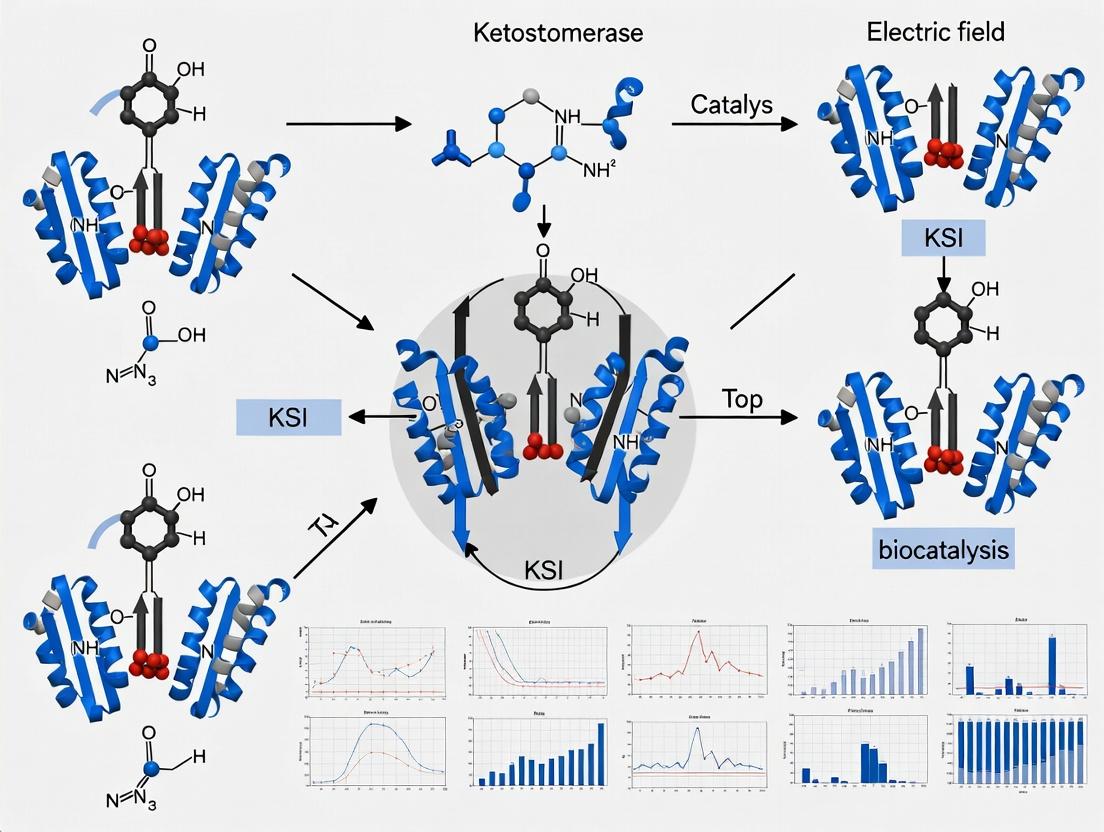
Ketosteroid Isomerase (KSI; EC 5.3.3.1) is a paradigm for understanding electrostatic catalysis in biological systems. This enzyme accelerates the allylic isomerization of Δ⁵-3-ketosteroids to their Δ⁴-conjugated isomers by over 10¹⁰-fold, primarily via stabilization of the enolate intermediate through a pre-organized, strong electric field generated by active-site residues. This whitepaper details the mechanistic principles, quantitative experimental evidence, and methodologies central to KSI research, framed within the ongoing thesis of elucidating electric field-driven enzymatic rate enhancement for applications in drug development and enzyme design.
KSI catalyzes a near diffusion-limited reaction via a diacid mechanism. Two key tyrosine residues (Tyr14 and Tyr55 in Pseudomonas putida KSI) act as a hydrogen-bonding diad. One tyrosine (Tyr14) donates a proton to the steroid carbonyl oxygen, while the other (Tyr55) abstracts the C4 proton. This concerted, yet asymmetric, process generates a short-lived, high-energy dienolate intermediate. The catalytic power derives from the enzyme's ability to pre-organize a strong electric field that stabilizes the negative charge developing on the carbonyl oxygen in the transition state, effectively lowering the activation barrier.
Diagram: KSI Catalytic Mechanism and Electric Field
Quantitative Evidence for Electrostatic Catalysis
Experimental data from kinetic isotope effects, site-directed mutagenesis, and advanced spectroscopy quantify KSI's catalytic prowess.
Table 1: Kinetic Parameters for Wild-Type and Mutant KSI
| Enzyme Variant (P. putida) | kcat (s⁻¹) | KM (μM) | kcat/KM (M⁻¹s⁻¹) | Relative Rate (kcat/KM) |
|---|---|---|---|---|
| Wild-Type KSI | ~ 1.4 x 10⁶ | ~ 50 | ~ 2.8 x 10¹⁰ | 1 |
| Tyr14Phe Mutant | ~ 1.4 x 10² | ~ 50 | ~ 2.8 x 10⁶ | 10⁻⁴ |
| Tyr55Phe Mutant | ~ 1.5 x 10³ | ~ 40 | ~ 3.8 x 10⁷ | ~1.4 x 10⁻³ |
| Asp38Leu Mutant | ~ 1.3 x 10¹ | ~ 70 | ~ 1.9 x 10⁵ | ~7 x 10⁻⁶ |
| Uncatalyzed Reaction | ~ 1.7 x 10⁻⁵ | N/A | N/A | ~6 x 10⁻¹⁶ |
Table 2: Physical Probes of Electric Field in KSI
| Experimental Technique | Key Measurement | Implication for Electric Field |
|---|---|---|
| Vibrational Stark Effect (VSE) | Frequency shift of nitrile probe at active site. | Measures field strength ~ 100-150 MV/cm directed toward catalytic diad. |
| ¹³C NMR Chemical Shift | Downfield shift of intermediate analog's carbonyl carbon. | Indicates strong polarization of the carbonyl bond due to field. |
| X-ray Crystallography | Precise atomic coordinates of active site with bound intermediate analogs. | Reveals pre-organized, rigid architecture optimizing electrostatic interactions. |
| Computational MD/QC | Calculated field vector and strength at reaction coordinate. | Predicts ~80% of catalytic rate enhancement from pre-organized electrostatics. |
Experimental Protocols
Protocol: Site-Directed Mutagenesis and Kinetic Assay for Catalytic Residues
Objective: To quantify the contribution of specific residues (Tyr14, Tyr55, Asp38) to catalysis.
- Mutagenesis: Using a plasmid encoding the KSI gene, perform PCR-based site-directed mutagenesis (e.g., QuikChange protocol) to generate Tyr→Phe and Asp→Leu mutants.
- Protein Expression & Purification: Transform mutants into E. coli BL21(DE3). Induce expression with IPTG. Purify via affinity chromatography (His-tag) and size-exclusion chromatography.
- Steady-State Kinetics: Assay activity spectrophotometrically by monitoring increase in absorbance at 248 nm (Δ⁴-product formation) in 10 mM potassium phosphate, pH 7.0, 25°C.
- Data Analysis: Use initial velocities with varying substrate (e.g., 5-androstene-3,17-dione) concentrations (1-100 μM). Fit data to Michaelis-Menten equation to extract kcat and KM.
Protocol: Measuring Electric Field via Vibrational Stark Effect
Objective: To experimentally determine the electric field magnitude and orientation in the KSI active site.
- Probe Incorporation: Introduce a non-perturbative vibrational reporter (e.g., a nitrile-modified steroid analog) into the active site via co-crystallization or soaking.
- FTIR Spectroscopy: Acquire high-resolution infrared spectra of the probe bound to KSI in D₂O buffer. Precisely measure the nitrile stretch frequency (ν~CN).
- Calibration: Determine the Stark tuning rate (Δμ) of the nitrile probe in solvents of varying dielectric constant (typically ~ 1 cm⁻¹/(MV/cm)).
- Field Calculation: The electric field projection along the nitrile bond axis is calculated: F = Δν / Δμ, where Δν is the frequency shift from the gas-phase reference.
Diagram: VSE Experimental Workflow
The Scientist's Toolkit: Key Research Reagent Solutions
Table 3: Essential Materials for KSI Electrostatic Catalysis Research
| Item | Function/Description | Example/Specification |
|---|---|---|
| Recombinant KSI Plasmid | Expression vector for wild-type and mutant KSI. | pET-28a(+) with ksi gene from P. putida; includes His-tag. |
| Ketosteroid Substrates | Native and analog substrates for kinetic and structural studies. | 5-Androstene-3,17-dione (5-AND); 19-Nor-5(10)-estene-3,17-dione. |
| Transition-State/Intermediate Analogs | High-affinity inhibitors for structural and field analysis. | Equilenin; Phenol (mimics dienolate). |
| Vibrational Stark Probes | Nitrile- or isotope-labeled steroids for FTIR/VSE. | 3-Cyano-5-androstene-17-dione. |
| Crystallization Screen Kits | For obtaining high-quality protein-ligand complex crystals. | Hampton Research Index or MCSG screens. |
| Deuterated Buffer | Solvent for FTIR and NMR to minimize water absorption interference. | 50 mM Potassium Phosphate, pD 7.0, in D₂O. |
| Stopped-Flow Apparatus | For measuring pre-steady-state kinetics of ultra-fast isomerization. | Applied Photophysics or KinTek models. |
| MD Simulation Software | To compute electric fields and model catalysis at atomic detail. | AMBER, CHARMM, or GROMACS with QM/MM modules. |
Implications for Drug Development
Understanding KSI's electrostatic catalysis provides a blueprint for:
- Drug Design: Mimicking transition-state stabilization by designing inhibitors that optimally engage pre-organized electric fields in target enzymes.
- Therapeutic Enzyme Engineering: Informing the design of catalytic antibodies (abzymes) or synthetic enzymes for novel chemistries by incorporating electrostatic active sites.
- Allosteric Modulation: Rational design of allosteric modulators that subtly alter the active-site electric field to fine-tune enzyme activity, offering new targeting strategies.
Within the enzyme Ketosteroid Isomerase (KSI), the precise spatial arrangement and chemical cooperation of Tyr14 and Asp99 form a catalytic dyad fundamental to its extraordinary proficiency. This dyad operates within a pre-organized, high-electric-field environment, facilitating ultrafast proton transfer critical for the isomerization of Δ⁵-3-ketosteroids to their Δ⁴-conjugated isomers. This whitepaper delves into the mechanistic role of this dyad, situating it within the broader context of KSI electric field catalysis research, which posits that the enzyme's active site is optimized to generate a strong electrostatic field that stabilizes key transition states and intermediates.
Mechanistic Role of Tyr14 and Asp99
The catalytic cycle hinges on a dienolate intermediate. Tyr14 acts as the general acid, donating its proton to the carbonyl oxygen (O1) of the steroid substrate. Concurrently, Asp99 acts as the general base, abstracting the proton from the steroid carbon (C4). This concerted, yet asynchronous, proton transfer is enabled by their precise orientation and the electrostatic environment.
- Tyr14 (Acid): Its phenolic OH group, with a perturbed pKₐ, forms a strong low-barrier hydrogen bond (LBHB) with the steroid's carbonyl oxygen. This interaction polarizes the carbonyl, stabilizing the developing negative charge on the dienolate oxygen.
- Asp99 (Base): Positioned to abstract the C4 proton, its carboxylate group is part of a hydrogen-bonding network that includes Tyr14's phenolic oxygen and often a conserved water molecule. This network serves as a proton shuttle and tuning element, modulating the pKₐ of both residues.
Their synergy lowers the activation energy for proton transfer by >10¹¹-fold compared to the uncatalyzed reaction in solution, making KSI a paradigm for proton transfer catalysis.
Quantitative Data on Dyad Function
Table 1: Key Biophysical and Kinetic Parameters for KSI Catalytic Dyad Mutants
| KSI Variant | kcat (s⁻¹) | ΔΔG‡cat (kcal/mol) | pKₐ Shift (Tyr14) | Key Observation | Primary Method |
|---|---|---|---|---|---|
| Wild-Type | ~1.4 x 10⁶ | 0.0 | ~6.5 (perturbed) | Optimal proton transfer network. | Stopped-flow, NMR |
| Y14F | ~2.0 x 10¹ | ~5.2 | N/A | Severe loss of acid catalysis; confirms Tyr as proton donor. | Kinetics, X-ray |
| D99A | ~3.0 x 10³ | ~3.4 | Shifts to ~9.5 | Impaired base catalysis; network disrupted, Tyr pKₐ elevates. | Kinetics, FTIR |
| D99N | ~1.0 x 10⁴ | ~2.8 | ~8.0 | Softer impairment; asparagine cannot fully replicate carboxylate function. | Kinetics, NMR |
| Y14F/D99A | < 1 | >10 | N/A | Catalysis virtually abolished; additive effect confirms synergy. | Kinetics |
Table 2: Electric Field Measurements at the KSI Active Site
| Measurement Target | Technique | Reported Electric Field (MV/cm) | Direction/Effect | Role of Dyad |
|---|---|---|---|---|
| C=O Bond of Substrate | Stark Spectroscopy / Vibrational Probe | ~ -140 to -170 | Aligns with C=O bond; stabilizes negative charge on O1. | Tyr14 H-bond is a primary source of this field. |
| Dienolate O1 | Computational (QM/MM) | ~ +100 (parallel to O-H bond) | Facilitates proton transfer from Tyr14. | Field from Asp99 and backbone dipoles tunes Tyr14 acidity. |
| C4-H Bond | Vibrational Frequency Shift | Implied strong field | Polarizes bond for proton abstraction. | Field from Asp99 carboxylate directly activates C-H bond. |
Detailed Experimental Protocols
Protocol: Site-Directed Mutagenesis and Purification of KSI Variants
Objective: Generate and purify Y14F, D99A, and other dyad mutants for functional analysis.
- Primer Design: Design complementary oligonucleotide primers containing the desired point mutation (e.g., TAC→TTC for Y14F).
- PCR Amplification: Perform PCR using a high-fidelity polymerase with the mutant primers and a plasmid containing the wild-type KSI gene (pcrA or KSI from P. putida).
- DpnI Digestion: Treat the PCR product with DpnI endonuclease to digest the methylated parental template DNA.
- Transformation: Transform the digested product into competent E. coli DH5α cells for plasmid propagation.
- Sequence Verification: Isolate plasmid DNA and confirm the mutation by Sanger sequencing of the entire KSI coding region.
- Protein Expression: Transform verified plasmid into E. coli BL21(DE3) expression strain. Induce expression with 0.5 mM IPTG at OD₆₀₀ ~0.6 for 4-6 hours at 30°C.
- Purification: Lyse cells and purify protein via anion-exchange chromatography (Q-Sepharose) followed by size-exclusion chromatography (Sephacryl S-200). Confirm purity by SDS-PAGE.
Protocol: Stopped-Flow Kinetic Assay of Proton Transfer
Objective: Measure the rate constant (kobs) for the chemical step catalyzed by dyad mutants.
- Sample Preparation: Prepare 10 µM KSI (wild-type or mutant) in 10 mM potassium phosphate buffer, pH 7.0. Prepare 100 µM 5-androstene-3,17-dione (5-AND) substrate in the same buffer with 2% (v/v) acetonitrile.
- Instrument Setup: Load enzyme and substrate solutions into separate syringes of a stopped-flow spectrophotometer thermostatted at 25°C.
- Data Acquisition: Rapidly mix equal volumes (typically 50 µL each). Monitor the increase in absorbance at 248 nm (characteristic of Δ⁴-product formation) over time (0-100 ms).
- Data Analysis: Fit the resulting single-exponential curve to the equation: At = A∞(1 - e-kobst), where kobs approximates kcat under these conditions.
Protocol: FTIR Spectroscopy for Probing Hydrogen-Bonding
Objective: Characterize the strength of the LBHB between Tyr14 and the substrate/intermediate.
- Sample Preparation: Generate the dienolate intermediate analog equilenin bound to KSI in D₂O buffer. Use a sealed demountable cell with CaF₂ windows and a path length of 50 µm.
- Spectrum Collection: Acquire FTIR spectra at 4 cm⁻¹ resolution on a spectrometer equipped with a liquid nitrogen-cooled MCT detector. Subtract buffer background.
- Analysis: Focus on the carbonyl stretching region (1600-1800 cm⁻¹). A significant red shift (~200 cm⁻¹) and broadening of the substrate C=O stretch upon binding to wild-type KSI indicates a strong LBHB. Compare the shift and lineshape for D99A and Y14F mutants to assess the dyad's role in tuning this bond.
Visualization of Mechanisms and Workflows
Diagram 1: KSI Catalytic Dyad Proton Transfer Mechanism (76 chars)
Diagram 2: Experimental Workflow for KSI Dyad Research (74 chars)
The Scientist's Toolkit: Research Reagent Solutions
Table 3: Essential Reagents and Materials for KSI Dyad Research
| Item Name / Reagent | Function / Purpose | Example Vendor / Specification |
|---|---|---|
| pET-KSI Plasmid | Expression vector containing the wild-type KSI gene for mutagenesis and overexpression. | In-house construct or Addgene repository derivative. |
| Phusion High-Fidelity DNA Polymerase | Accurate amplification during site-directed mutagenesis PCR to avoid unwanted mutations. | Thermo Fisher Scientific. |
| DpnI Restriction Enzyme | Selective digestion of the methylated template DNA post-PCR, enriching for mutant plasmids. | New England Biolabs. |
| 5-Androstene-3,17-dione (5-AND) | The primary native substrate for KSI, used in kinetic assays. | Sigma-Aldrich, >98% purity. |
| Equilenin | A stable dienolate intermediate analog for spectroscopic (FTIR, NMR) studies of the LBHB. | Steraloids Inc. |
| Stopped-Flow Spectrophotometer | Instrument for measuring rapid reaction kinetics (millisecond timescale) of KSI catalysis. | Applied Photophysics or Hi-Tech KinetAsyst. |
| FTIR Spectrometer with MCT Detector | For high-sensitivity infrared spectroscopy to characterize hydrogen bonds and electric field effects. | Bruker Vertex series, resolution ≤4 cm⁻¹. |
| Vibrational Stark Probe (e.g., 4-Cyanobenzyl) | A synthetic substrate modified with a nitrile group; its Stark shift reports local electric field. | Custom synthesis. |
| QM/MM Software Suite | For computational modeling of the active site electric field and proton transfer energetics. | Gaussian/AMBER or CHARMM/CHEMSHELL. |
Within the context of enzyme catalysis, the concept of preorganized internal electric fields posits that the enzyme's evolved, static architecture creates a precise, anisotropic electric field in its active site. This field is "preorganized"—established by the permanent arrangement of dipoles, charged residues, and hydrogen-bonding networks—prior to substrate binding. It directly stabilizes the transition state and polarizes substrate bonds, thereby accelerating the chemical transformation. This whitepaper explores this theoretical foundation, framed by seminal and ongoing research on Ketosteroid Isomerase (KSI), a paradigm for understanding electric field catalysis in biology and its implications for drug development.
Theoretical Framework and Quantitative Evidence from KSI
KSI catalyzes the isomerization of Δ⁵-3-ketosteroids to their Δ⁴-conjugated isomers. The reaction proceeds via a dienolate intermediate, where the rate-limiting step is the abstraction of a substrate proton by a catalytic aspartate (Asp-38 in Pseudomonas testosteroni KSI). The enzyme's electric field, preorganized by its structure, is critical for stabilizing this high-energy enolate intermediate.
Key Quantitative Findings from KSI Research:
| Experimental Parameter | Value / Observation | Theoretical Implication |
|---|---|---|
| Rate Enhancement (kcat/kuncat) | ~10⁹ to 10¹¹ | Demonstrates profound catalytic proficiency. |
| Contribution of Oriented Dipoles (ΔΔG) | ~5-6 kcal/mol stabilization of TS | Preorganized fields provide significant energy towards TS stabilization. |
| Field Strength in Active Site | ~ 100-150 MV/cm (calculated) | Comparable to fields in synthetic catalysts; sufficient to polarize bonds. |
| Mutation of Tyr-16 (H-bond donor) | ~10³-10⁴ reduction in kcat | Confirms critical role of preorganized H-bond network in field generation. |
| Electric Field Correlation (vibrational Stark) | Linear correlation between C=O frequency shift & Δkcat | Direct experimental proof of field-reaction rate relationship. |
Experimental Protocols for Measuring Internal Fields in KSI
Vibrational Stark Effect (VSE) Spectroscopy
This is the primary experimental method for quantifying electric fields in enzymes.
- Objective: Measure the electric field projected onto a specific bond (e.g., substrate's carbonyl) via its vibrational frequency shift.
- Protocol:
- Probe Incorporation: Introduce a spectroscopic probe (e.g., a nitrile- or carbonyl-containing substrate or inhibitor) into the KSI active site.
- FTIR/ Raman Measurement: Record high-resolution infrared or Raman spectra of the probe-enzyme complex. Precisely measure the vibrational frequency (e.g., C≡N or C=O stretch).
- Calibration: Determine the probe's Stark tuning rate (Δμ, in cm⁻¹/(MV/cm)) in a controlled environment (e.g., in solvents of known dielectric constant or under an external electric field).
- Field Calculation: The internal electric field (F) is calculated as: F = Δν / Δμ, where Δν is the observed frequency shift from a reference state (e.g., in nonpolar solvent).
- Correlation: Plot the measured field strength against catalytic rate (log kcat) for wild-type and mutant KSIs to establish a linear free energy relationship.
Structure-Function Analysis via Site-Directed Mutagenesis
- Objective: Dissect the contribution of specific residues to the preorganized field.
- Protocol:
- Target Identification: Select residues forming the active site dipolar network (e.g., Tyr-16, Asp-103, and oxyanion hole residues in KSI).
- Mutagenesis: Create plasmid constructs coding for KSI mutants (e.g., Y16F, D103L).
- Protein Expression & Purification: Express mutant proteins in E. coli and purify via affinity and size-exclusion chromatography.
- Kinetic Assay: Measure kcat and KM for the isomerization reaction using UV spectroscopy (shift in conjugated diene absorption at ~248 nm).
- Crystallography/Computational Modeling: Solve high-resolution crystal structures of mutants. Perform MD simulations and quantum mechanical calculations to compute the altered electric field.
The Scientist's Toolkit: Key Research Reagent Solutions
| Reagent / Material | Function in KSI Electric Field Research |
|---|---|
| Recombinant KSI (Wild-type & Mutants) | Catalytic protein scaffold for experimental measurement of fields and kinetics. |
| Site-Directed Mutagenesis Kit | For creating specific point mutations to disrupt the preorganized dipolar network. |
| Vibrational Probe (e.g., 5-Nitro-19-Nortestosterone) | A substrate analog with a nitrile (C≡N) or isotopically labeled carbonyl for VSE spectroscopy. |
| FTIR / Raman Spectrometer | High-sensitivity instrument for measuring vibrational frequency shifts of the probe. |
| Crystallization Screen Kits | For obtaining high-resolution protein crystals for structural analysis of mutants. |
| QM/MM Software (e.g., Gaussian, ORCA, Amber) | For performing quantum mechanical/molecular mechanics simulations to calculate electric fields. |
| Stark Tuning Rate Calibration Setup | Controlled environment (e.g., applied external field cell) to calibrate the probe's sensitivity. |
Visualization of the KSI Catalytic Mechanism and Field
Within the broader context of Ketosteroid Isomerase (KSI) electric field catalysis research, this whitepaper delves into the specific architectural features of KSI's active site that are optimized to generate a pre-organized electrostatic environment. This environment is crucial for catalyzing the rate-limiting enolization step in the isomerization of Δ⁵-3-ketosteroids to their Δ⁴-conjugated isomers. KSI serves as a paradigm for understanding how enzymes utilize electrostatic forces, rather than direct chemical participation, to achieve extraordinary rate enhancements (≥10¹¹-fold).
Active Site Architecture & Key Residues
The active site of bacterial KSI (from Pseudomonas putida) is a hydrophobic cavity containing two critical dyads of catalytic residues:
- Aspartate-38 (Asp38) and Aspartate-99 (Asp99): Positioned to interact with the carbonyl oxygen (O3) of the steroid substrate.
- Tyrosine-14 (Tyr14) and Tyrosine-55 (Tyr55): Positioned to act as hydrogen bond donors to the same carbonyl oxygen.
This architecture creates a unique, short, strong hydrogen-bonding network. The pKa of the active-site tyrosines is dramatically lowered (to ~4-6) due to the electrostatic influence of the aspartates, enabling them to act as strong acids. The precise geometry and electrostatic pre-organization of this network are the keys to catalysis.
The Electrostatic Catalytic Mechanism
The core thesis of modern KSI research posits that the enzyme's active site is evolutionarily tuned to generate a specific, optimal electrostatic field that stabilizes the high-energy dienolate intermediate formed during enolization.
| Catalytic Step | Role of Active Site Electrostatics | Quantitative Impact |
|---|---|---|
| Substrate Binding & Polarization | The Asp/Tyr dyad polarizes the substrate's carbonyl, increasing its electrophilicity. | Carbonyl bond order reduction observed via vibrational spectroscopy (Δν~30 cm⁻¹). |
| Proton Abstraction (Enolization) | Low-pKa Tyr14/Tyr55 donate a proton to the carbonyl oxygen, while the aspartate dyad stabilizes the developing negative charge. | Rate constant (k_cat) ~ 10⁴ s⁻¹; ΔG‡ reduction of ~15 kcal/mol compared to uncatalyzed reaction. |
| Intermediate Stabilization | The dienolate intermediate is stabilized via resonance and precise electrostatic interactions with the oxyanion hole (Asp38, Asp99). | Intermediate lifetime is microseconds; binding affinity for intermediate analogs (e.g., equilinin) K_d < 1 nM. |
| Product Formation & Release | The electrostatic environment facilitates reprotonation at C6 and product dissociation. | Overall catalytic proficiency (kcat/Km)/k_uncat ≥ 10¹¹ M⁻¹. |
Experimental Protocols for Probing Electrostatics
Vibrational Spectroscopy (FTIR/Raman)
Purpose: To directly measure electric field strength at the substrate's carbonyl bond. Protocol:
- Express and purify wild-type (WT) and site-directed mutant (e.g., D38N, Y14F) KSI.
- Prepare substrate analogs (e.g., 5-androstene-3,17-dione) or mechanism-based inhibitors (e.g., 19-nortestosterone acetate).
- Acquire FTIR spectra of the free substrate in a non-polar solvent (control).
- Acquire FTIR spectra of the substrate bound to KSI in a D₂O-based buffer (to avoid H₂O interference).
- Analyze the vibrational frequency shift (Δν) of the substrate's carbonyl (C=O) stretch. A downshift indicates bond weakening due to a strong electric field.
- Data Interpretation: The frequency shift (Stark tuning rate) is converted to an estimated electric field projection using the vibrational Stark effect (VSE) formula: Δν = -Δμ * E / hc, where Δμ is the difference dipole moment of the vibrational transition.
X-ray Crystallography of Intermediate Analog Complexes
Purpose: To visualize the precise geometry of the active site under conditions mimicking the transition state. Protocol:
- Co-crystallize KSI with a tight-binding intermediate analog (e.g., equilinin, which resembles the dienolate).
- Collect diffraction data at a synchrotron source (e.g., 1.0 Å resolution).
- Solve the crystal structure and refine the model.
- Analyze key metrics: O-O distances between Asp/Tyr oxygens and the ligand's O3, bond lengths of the ligand, and the overall polarity of the active site cavity.
- Compare with structures of apo-enzyme and product-bound complexes.
Double-Mutant Cycle Analysis
Purpose: To quantify the energetic coupling between key catalytic residues. Protocol:
- Create a series of KSI variants: WT, single mutants (Y14F, D38N), and the double mutant (Y14F/D38N).
- Measure the catalytic activity (k_cat) for each variant under identical conditions using a spectrophotometric assay (monitoring Δ⁴-product formation at 248 nm).
- Calculate the coupling energy (ΔΔG) between the two residues: ΔΔGint = ΔG(Y14F) + ΔG(D38N) - ΔG(Y14F/D38N) - ΔG(WT), where ΔG = -RT ln(kcat).
- A significant ΔΔG_int (|>1 kcal/mol|) indicates a strong functional interaction, often electrostatic in nature.
Diagram: KSI Catalytic Mechanism & Electrostatic Network
Title: KSI Catalytic Cycle with Electrostatic Residue Roles
Diagram: Experimental Workflow for Electric Field Analysis
Title: Integrated Workflow for KSI Electrostatics Research
The Scientist's Toolkit: Key Research Reagent Solutions
| Reagent / Material | Function in KSI Research |
|---|---|
| pET-KSI Plasmid (WT) | Expression vector for high-yield production of P. putida KSI in E. coli. |
| Site-Directed Mutagenesis Kit | For creating specific active-site variants (e.g., D38N, Y14F, Y55F). |
| Δ⁵-Androstene-3,17-dione | The canonical native substrate for standard enzymatic assays. |
| Equilinin (1,3,5(10),6,8-Estratetraene-3-ol-17-one) | A tight-binding intermediate analog for crystallography and binding studies. |
| 19-Nortestosterone Acetate | A mechanism-based inhibitor that forms a stable covalent intermediate. |
| Deuterated Buffer (D₂O pD 7.0) | For FTIR studies to avoid strong infrared absorption from H₂O. |
| Crystallization Screen Kits (e.g., PEG/Ion, Index) | For identifying initial conditions for co-crystallization of KSI-ligand complexes. |
| UV-Vis Spectrophotometer (248 nm filter) | For continuous kinetic assays monitoring product formation (Δε ~16,000 M⁻¹cm⁻¹). |
| High-Precision FTIR Spectrometer | For detecting subtle vibrational shifts in substrate carbonyl stretch upon binding. |
| Molecular Dynamics (MD) Software (e.g., AMBER, GROMACS) | For simulating the electric field vectors and dynamics within the KSI active site. |
Within the context of advanced electric field catalysis research, Ketosteroid Isomerase (KSI) stands as a paradigmatic enzyme. This whitepaper details the key historical experiments that have unequivocally demonstrated KSI's extraordinary catalytic prowess, providing a foundation for ongoing studies into the precise role of preorganized electric fields in enzyme function. These breakthroughs are critical for researchers and drug development professionals aiming to harness electrostatic principles in rational design.
Key Experimental Breakthroughs
The following table summarizes the quantitative data from seminal experiments that have defined our understanding of KSI catalysis.
Table 1: Key Quantitative Data from Historical KSI Experiments
| Experiment / Measurement | Key Value(s) | Catalytic Proficiency (kcat/kuncat) | Implication for Catalytic Mechanism |
|---|---|---|---|
| Primary Kinetic Isotope Effect (KIE) | D_KIE ~ 7 (for 5(10)-estrene-3,17-dione) | ~ 1 x 10¹¹ | Indicates C-H bond cleavage is (partially) rate-limiting, consistent with a dienolate intermediate. |
| Proton Affinity & pKa Shift | Substrate C-H acid pKa ~32; Active site Asp38 pKa ~4.7 (vs. ~4.0 in solution) | - | Shows enzyme active site dramatically increases substrate acidity by >10^27-fold via electric fields. |
| Double-Mutant Cycle Analysis (Asp38/Asn99) | Coupling energy (ΔΔG) ~ 4-5 kcal/mol | - | Demonstrates strong synergistic, cooperative electrostatic interaction between key residues. |
| Linear Free Energy Relationship (LFER) | Brønsted β value ~ 0.8 - 0.9 | - | Confirms transition state has substantial oxyanion character, indicating extensive proton transfer. |
| Electric Field Measurement (vibrational Stark effect) | Field along C=O bond: ~ -100 MV/cm (in D38N mutant) | - | Direct experimental measurement of the intense, preorganized electric field aligned for catalysis. |
Detailed Experimental Protocols
Measurement of Primary Kinetic Isotope Effect (KIE)
Objective: To determine if C-H bond breaking is a rate-limiting step in the KSI-catalyzed reaction. Methodology:
- Substrate Preparation: Synthesize the deuterated substrate, 5(10)-estrene-3,17-dione, with deuterium at the carbon-4 position ([4-²H]-substrate).
- Enzyme Purification: Express and purify wild-type KSI from Pseudomonas putida or a recombinant source.
- Kinetic Assays: Perform separate initial velocity measurements under identical conditions (e.g., 25°C, pH 7.0) using the protonated and deuterated substrates at saturation.
- Data Analysis: Calculate the KIE as the ratio of the maximal turnover numbers: DKIE = (kcat)H / (kcat)_D. A value significantly greater than 1 indicates bond cleavage is involved in the rate-determining step.
Double-Mutant Cycle Analysis for Electrostatic Cooperation
Objective: To quantify the energetic coupling between two active site residues (e.g., Asp38 and Asn99). Methodology:
- Construct Generation: Create four KSI variants: Wild-Type (WT), single mutants D38N and N99A, and the double mutant D38N/N99A.
- Steady-State Kinetics: Determine the catalytic efficiency (kcat/KM) for each variant with a standard substrate like 5-androstene-3,17-dione.
- Coupling Energy Calculation: Calculate the interaction energy (ΔΔG) using the formula: ΔΔG = -RT ln[(kcat/KM)WT * (kcat/KM)D38N/N99A] / [(kcat/KM)D38N * (kcat/KM)N99A]. A non-zero ΔΔG indicates cooperativity.
Vibrational Stark Effect Spectroscopy for Electric Field Measurement
Objective: To directly measure the magnitude of the electric field projected onto a substrate's carbonyl bond within the KSI active site. Methodology:
- Probe Incorporation: Use a substrate analogue containing a nitrile (-C≡N) reporter group, whose vibrational frequency (ν_C≡N) is sensitive to local electric fields.
- Spectroscopic Measurement: Obtain FTIR spectra of the nitrile probe bound to a mutant KSI active site (e.g., D38N, which binds but does not turn over the substrate).
- Calibration: Perform a separate Stark spectroscopy experiment on the nitrile probe in different external electric fields or solvents to establish the linear relationship between frequency shift (Δν) and field strength (Stark tuning rate).
- Field Calculation: Apply the Stark tuning rate to the observed frequency shift of the enzyme-bound probe relative to its frequency in a non-polar solvent to calculate the intrinsic electric field.
Mandatory Visualizations
Title: KSI Catalytic Cycle & Intermediate
Title: Electric Field Preorganization in KSI Active Site
The Scientist's Toolkit: Key Research Reagent Solutions
Table 2: Essential Reagents for KSI Catalysis Research
| Reagent / Material | Function & Rationale |
|---|---|
| 5-Androstene-3,17-dione | The canonical, high-affinity substrate for standard kinetic characterization of KSI activity and inhibition studies. |
| [4-²H]-5-Androstene-3,17-dione | Deuterated substrate essential for performing primary Kinetic Isotope Effect (KIE) experiments to probe the chemical mechanism. |
| Equilenin (5,7,9-estratrien-3-ol-17-one) | A stable dienolate intermediate analogue used for X-ray crystallography to capture the structure of the catalytic intermediate. |
| Nitrile-containing Substrate Analogues | Chemically synthesized probes (e.g., with -C≡N at the carbonyl position) for Vibrational Stark Effect spectroscopy to measure electric fields. |
| Site-Directed Mutagenesis Kits | Essential for generating specific KSI mutants (e.g., D38N, N99A) to dissect the role of individual residues via kinetics and structural biology. |
| High-Purity Expression System (e.g., pET vector in E. coli) | For recombinant production of large, homogeneous quantities of WT and mutant KSI enzymes for biophysical studies. |
| Isothermal Titration Calorimetry (ITC) Kit | Used to measure substrate binding affinities (K_D) and thermodynamics (ΔH, ΔS) for mutant enzymes, complementing kinetic data. |
The Marcus Theory and Quantum Tunneling Connection in KSI Catalysis
Ketosteroid Isomerase (KSI) catalyzes the allylic rearrangement of Δ5-3-ketosteroids to their Δ4-conjugated isomers, a fundamental step in steroid metabolism. The reaction involves the transfer of a proton from a carbon acid donor to a carbonyl oxygen acceptor via a dienolate intermediate. This proton transfer is exceptionally efficient, with rate accelerations exceeding 10¹¹-fold over the uncatalyzed reaction. Contemporary research frames this catalysis within the context of electric field effects and quantum mechanical phenomena. This whitepaper explores the synergistic connection between Marcus theory, which describes electron and proton transfer kinetics in a classical continuum, and quantum tunneling, a non-classical phenomenon where particles traverse energy barriers. In KSI, the pre-organized active site, featuring Asp38/99 as the catalytic base, generates a strong, oriented electrostatic field that optimizes both the classical reorganization energy (λ) and the tunneling probability, creating a paradigm for electric field-driven enzymatic catalysis.
Core Principles: Marcus Theory Applied to Proton Transfer
Marcus theory models proton transfer as a function of driving force (ΔG°), reorganization energy (λ), and the electronic coupling between reactant and product states. The rate constant k is given by: [ k = \frac{2\pi}{\hbar} |V|^2 \frac{1}{\sqrt{4\pi\lambda kBT}} \exp\left[-\frac{(\Delta G^\circ + \lambda)^2}{4\lambda kBT}\right] ] where |V| is the electronic coupling matrix element.
In KSI, the enzyme's primary role is to lower λ. The active site pre-organizes the substrate and catalytic residues, minimizing the solvent and intramolecular rearrangements required upon proton transfer. This reduction in λ brings the system closer to the "Marcus inverted region" for proton transfer, optimizing the rate. Recent electric field research demonstrates that the enzyme's interior field specifically stabilizes the charge-transfer transition state, effectively tuning both ΔG° and λ.
Table 1: Key Kinetic and Thermodynamic Parameters for KSI Catalysis
| Parameter | Uncatalyzed Reaction | KSI-Catalyzed Reaction | Experimental Method |
|---|---|---|---|
| Rate Constant (k) | ~10⁻⁶ s⁻¹ | ~10⁶ s⁻¹ | Stopped-flow spectrophotometry, NMR line-shape analysis |
| Activation Free Energy (ΔG‡) | ~24 kcal/mol | ~12 kcal/mol | Temperature-dependent kinetics (Arrhenius/Eyring plots) |
| Kinetic Isotope Effect (KIE) | ~3 (primary) | ~3-16 (primary, temp-dependent) | Comparison of rates with protium vs. deuterium substrate |
| Reorganization Energy (λ) | High (estimated >30 kcal/mol) | Significantly reduced (~10-15 kcal/mol) | Analysis of rate vs. driving force using substrate analogues |
Quantum Tunneling in KSI Proton Transfer
The observation of large, temperature-dependent primary KIEs and curved Arrhenius plots in KSI provides strong evidence for quantum tunneling. Tunneling allows the proton to transfer through the classical energy barrier rather than over it. The enzyme enhances tunneling probability by:
- Barrier Narrowing: Pre-organization and precise positioning of donor/acceptor atoms (C–H–O distance ~2.6-2.7 Å) reduce the width of the energy barrier.
- Barrier Compression: The electrostatic field and active site residues modulate the potential energy surface, creating a thinner barrier.
- Promoting Vibrations: Low-frequency protein vibrations (e.g., donor-acceptor distance fluctuations) transiently bring the reactant and product wavefunctions into greater overlap.
The connection to Marcus theory is explicit in models like "Marcus-like tunneling," where the classical free energy surface dictates the tunneling probability. The rate expression incorporates a tunneling correction factor (Γ). [ k{tun} = \Gamma(T) \cdot k{MT} ] where ( k_{MT} ) is the Marcus theory rate.
Experimental Protocols for Investigating the Connection
Protocol: Measuring Kinetic Isotope Effects (KIEs) and Activation Parameters
Objective: To detect and quantify quantum tunneling contributions. Methodology:
- Synthesize the native substrate (e.g., 5-androstene-3,17-dione) and its deuterated analogue at the transferring proton position.
- Perform kinetic assays using stopped-flow spectrophotometry (monitoring absorbance shift at ~248 nm) for both substrates across a temperature range (e.g., 5°C to 45°C).
- Determine ( kH ) and ( kD ) at each temperature. Calculate the primary KIE = ( kH/kD ).
- Plot ln(k) vs. 1/T (Arrhenius plot) for both H and D reactions. Curvature and a large difference in activation energies (( Ea^D - Ea^H > \text{RT} )) indicate tunneling.
- Calculate ΔH‡ and ΔS‡ from Eyring plots.
Protocol: Electric Field Measurement via Vibrational Stark Effect (VSE)
Objective: To quantify the magnitude and orientation of the active site electric field. Methodology:
- Incorporate a nitrile reporter group (as a vibrational probe) into a substrate analogue or inhibitor bound in the KSI active site.
- Use Fourier-transform infrared (FTIR) spectroscopy to measure the nitrile stretch frequency (( \nu_{CN} )).
- Relate the frequency shift (Δ( \nu{CN} )) to the electric field projection (F) along the nitrile bond using a previously calibrated Stark tuning rate: ( \Delta\nu{CN} = \Delta \vec{\mu} \cdot \vec{F} = |\Delta\mu| F_{||} ).
- Map the field vector and correlate its strength/orientation with catalytic rate parameters.
Protocol: Computational Analysis of Reorganization Energy
Objective: To calculate λ and model the reaction pathway. Methodology:
- Perform QM/MM (e.g., DFT/AMBER) simulations on the KSI-substrate complex.
- Constrain the reaction coordinate (e.g., donor-H distance) and perform potential energy surface scans.
- Use Marcus theory formulations to compute λ from the simulation data: ( \lambda = (\Delta E{RP}^f - \Delta E{RP}^i) ), where ( \Delta E_{RP} ) is the energy difference between reactant and product states.
- Calculate the tunneling probability using Wentzel–Kramers–Brillouin (WKB) approximation or instanton path integral methods.
Visualization of Concepts and Workflows
Title: Synergy of Marcus Theory and Tunneling in KSI
Title: Experimental-Comp. Workflow for KSI Proton Transfer
The Scientist's Toolkit: Research Reagent Solutions
Table 2: Essential Materials for KSI Electric Field & Tunneling Research
| Reagent / Material | Function & Role in Research |
|---|---|
| 5-Androstene-3,17-dione (and deuterated analogues) | Native KSI substrate; deuterated form is essential for primary KIE measurements to probe tunneling. |
| Site-Specific Nitrile-Modified Inhibitors (e.g., NO-Δ5-3-ketosteroid) | Contains a vibrational Stark probe (C≡N) for quantifying electric field strength/orientation via FTIR spectroscopy. |
| Recombinant KSI (Wild-Type & Mutants e.g., D38N, Y16F) | Catalytic protein. Mutants are used to dissect the role of specific residues in field generation and catalysis. |
| High-Resolution Stopped-Flow Spectrophotometer | For measuring fast catalytic rates (k~10⁶ s⁻¹) across a range of temperatures to obtain activation parameters. |
| FTIR Spectrometer with Cryostat | For sensitive detection of nitrile stretch frequency shifts of bound probes to calculate electric fields. |
| QM/MM Software (e.g., Gaussian, AMBER, CP2K) | For performing atomistic simulations to calculate reorganization energy (λ), barrier dimensions, and tunneling pathways. |
Measuring the Invisible: Computational and Experimental Tools for Quantifying KSI Electric Fields
Ketosteroid Isomerase (KSI) is a model enzyme in the study of electrostatic catalysis, where pre-organized electric fields are hypothesized to stabilize the reaction's enolate intermediate and transition state, accelerating the conversion of Δ⁵-3-ketosteroids to their Δ⁴-conjugated isomers. The central thesis in modern KSI research posits that the enzyme's catalytic power is derived not from chemical participation but from the precise alignment of electric fields within its active site. Vibrational Stark Effect (VSE) spectroscopy has emerged as a critical experimental technique to directly measure the magnitude and orientation of these electric fields, providing quantitative validation for theoretical models and offering a blueprint for rational drug and biocatalyst design.
Principles of the Vibrational Stark Effect
The VSE describes the linear shift in the frequency of a molecular vibrational mode (ν) in response to an external electric field (F). The relationship is given by: Δν = -Δμ · F / (hc) where Δμ is the Stark tuning rate, a vector representing the change in dipole moment of the bond upon excitation, h is Planck's constant, and c is the speed of light. By introducing a site-specific vibrational probe (e.g., a nitrile or carbonyl label) into a biological system, one can use the measured frequency shift to report on the local electrostatic environment experienced by the probe.
Experimental Protocol for VSE in KSI Studies
Step 1: Probe Incorporation.
- Method A (Genetic Incorporation): Introduce a non-canonical amino acid (e.g., p-cyanophenylalanine, CnF) via amber stop codon suppression at a specific site in the KSI active site or substrate.
- Method B (Chemical Labeling): Conjugate a synthetic nitrile-containing probe onto a cysteine residue introduced via site-directed mutagenesis.
- Protocol: The KSI mutant is expressed, purified, and either subjected to suppression technology for CnF incorporation or reacted with the nitrile probe under reducing conditions. Excess label is removed via size-exclusion chromatography.
Step 2: VSE Spectroscopy Measurement.
- FTIR Spectroscopy: Acquire high-resolution infrared spectra of the nitrile probe (~2230 cm⁻¹) for KSI in various states (apo, substrate-bound, transition state analog-bound).
- External Calibration: Record spectra of the probe in solvents of known dielectric constant or in a frozen glass under an applied external electric field to determine its intrinsic Stark tuning rate (|Δμ|).
- Protocol: Samples are loaded into a demountable liquid cell with CaF₂ windows and a fixed pathlength (e.g., 50 μm). Spectra are collected at a specific temperature (e.g., 25°C or 77K for frozen glasses) with high signal-to-noise (>1000 scans). Background subtraction and peak fitting are performed to determine the center frequency.
Step 4: Electric Field Calculation.
- Analysis: The observed frequency shift (Δνobs) relative to a reference state (e.g., in a nonpolar solvent) is used to calculate the projected electric field along the probe's transition dipole moment axis: *Fproj = -Δν_obs * hc / |Δμ|.
Key Quantitative Data from KSI VSE Studies
Table 1: Measured Electric Fields in KSI Active Site
| Probe Location / KSI State | Nitrile Frequency Shift Δν (cm⁻¹) | Calculated Projected Electric Field (MV/cm) | Key Reference (Conceptual) |
|---|---|---|---|
| CnF at Active Site (Apo KSI) | +0.5 to +1.5 | -5 to -15 | Fried et al., Science (2014) |
| CnF at Active Site (Bound to Phenolate TSA) | +4.0 to +5.0 | -40 to -50 | Fried et al., Science (2014) |
| Substrate C=O (Computational Prediction) | N/A | -140 (oriented to stabilize enolate) | Warshel et al., Biochemistry |
Table 2: Comparison of Catalytic Contributions
| Contribution Source | Estimated Energy Contribution (kcal/mol) | Method of Determination |
|---|---|---|
| Pre-organized Electric Field (from VSE) | 8 - 12 | VSE spectroscopy + linear response approximation |
| General Base Catalysis (Asp40) | 4 - 6 | Site-directed mutagenesis (D40N) |
| Desolvation of Substrate | ~3 | Comparison to solution reaction rate |
Visualizing the VSE Workflow and KSI Catalysis
VSE Experimental and Analysis Workflow
KSI Catalytic Mechanism and Field Stabilization
The Scientist's Toolkit: Key Research Reagents & Materials
Table 3: Essential Reagents for VSE Studies on KSI
| Reagent / Material | Function in Experiment | Key Consideration |
|---|---|---|
| p-Cyanophenylalanine (CnF) | Site-specific IR probe; genetically incorporated via amber codon suppression. | High Stark tuning rate; minimal perturbation to native structure. |
| KSI Expression Vector | Plasmid for recombinant KSI (wild-type and mutant) expression in E. coli. | Must contain selection marker (e.g., ampicillin resistance). |
| Phenolates (e.g., equilenin) | Transition state analog (TSA) mimics the enolate intermediate. | High-affinity binding (K_d in nM range) essential for field measurement in catalytically relevant state. |
| CaF₂ IR Windows | Windows for demountable liquid cell; transparent in mid-IR region. | Must be polished and cleaned meticulously to avoid scattering artifacts. |
| Polar Solvent Series (e.g., Dichloroethane to DMSO) | Used for in vitro calibration of the nitrile probe's sensitivity (Δμ). | Requires high purity, anhydrous conditions for accurate calibration. |
| FTIR Spectrometer | Equipped with liquid N₂-cooled MCT detector for high-sensitivity nitrile band detection. | Requires stable temperature control and purged with dry air/N₂ to reduce H₂O/CO₂ bands. |
Advanced Molecular Dynamics (MD) and QM/MM Simulations for Field Mapping
This technical guide details the application of advanced Molecular Dynamics (MD) and Quantum Mechanics/Molecular Mechanics (QM/MM) simulations for electric field mapping, framed within a broader thesis investigating the catalytic mechanism of Ketosteroid Isomerase (KSI). KSI catalyzes the isomerization of Δ⁵-3-ketosteroids to Δ⁴-3-ketosteroids at a diffusion-limited rate. The prevailing hypothesis is that KSI’s extraordinary catalytic proficiency (~10¹¹ rate enhancement) is driven by the pre-organized electrostatic environment of its active site, which stabilizes the enolate intermediate and its transition state through precise electric field alignment. This document provides a methodological framework for quantifying and mapping these catalytically critical electric fields using state-of-the-art simulation protocols.
Core Computational Methodologies
Enhanced Sampling Molecular Dynamics for KSI Conformational Analysis
Objective: To sample the conformational landscape of KSI (both apo and substrate-bound states) to identify dominant substates and assess active site pre-organization.
Protocol:
- System Preparation: Obtain the crystal structure of KSI (e.g., PDB ID 1OH0). Use the
pdb4ambertool to add missing hydrogen atoms. For substrate-bound simulations, dock the Δ⁵-androstene-3,17-dione (5-AND) substrate into the active site using induced-fit docking protocols. - Force Field Parameterization: Use the
antechamberandparmchk2modules from AmberTools to generate GAFF2 parameters for the substrate. Protein and ions are described with the ff19SB force field and OPC water model. - System Solvation and Equilibration: Solvate the system in a truncated octahedral water box with a 10 Å buffer. Neutralize with Na⁺/Cl⁻ ions and bring to physiological concentration (0.15 M). Follow a multi-step equilibration: (i) 5000 steps of steepest descent minimization restraining protein heavy atoms, (ii) 100 ps NVT heating to 300 K, (iii) 1 ns NPT equilibration at 1 bar with gradual restraint release.
- Production MD with Replica Exchange: Conduct Hamiltonian Replica Exchange MD (HREMD) or Temperature REMD (T-REMD). For 50 replicas, span a temperature range of 300-500 K. Run each replica for 200 ns, exchanging attempts every 2 ps. Use the
mpiruncommand withsander.MPIorpmemd.cuda.MPIfor execution. - Analysis: Cluster trajectories from the 300 K replica using the RMSD of active site residues (Asp-99, Tyr-14, Tyr-55, etc.) with a cutoff of 1.5 Å via the k-means algorithm. Perform Principal Component Analysis (PCA) on the Cα atoms to identify major conformational modes.
Key Quantitative Outputs:
- Root Mean Square Fluctuation (RMSF) of active site residues.
- Free energy landscape projected onto the first two principal components.
- Hydrogen bond occupancy (%) between substrate and catalytic residues.
Table 1: Representative H-bond Occupancy from KSI-Substrate MD
| Donor | Acceptor | Occupancy (%) | Average Distance (Å) |
|---|---|---|---|
| Substrate O3 | Tyr-14 OH | 98.7 | 2.65 ± 0.15 |
| Asp-99 OD1 | Substrate C4-H | 95.2 | 2.78 ± 0.20 |
| Tyr-55 OH | Substrate O1 | 89.5 | 2.71 ± 0.18 |
| Asp-38 OD2 | Tyr-16 OH | 99.1 | 2.62 ± 0.12 |
QM/MM Simulation for Reaction Pathway and Electric Field Calculation
Objective: To compute the reaction energy profile for the proton transfer steps in KSI and map the electric field projected onto key reaction coordinates.
Protocol:
- System Selection: Extract dominant conformational cluster representatives from the MD analysis as starting structures.
- QM/MM Partitioning: Define the QM region to include the substrate (5-AND) and the catalytic diad (Tyr-14 phenolatesubstrate O3H-Asp-99). Treat this region (≈40-50 atoms) with DFT (e.g., ωB97X-D/6-31G*) using the *ab initio QM/MM interface. The remaining protein and solvent constitute the MM region (ff19SB/GAFF2).
- Boundary Treatment: Use a hydrogen link atom scheme to saturate QM bonds cut at the boundary.
- Energy Minimization and Pathway Sampling: First, minimize the MM region with the QM region constrained. Then, perform a relaxed potential energy surface scan by driving the proton transfer coordinate (e.g., O3-H distance) in 0.1 Å increments, re-optimizing all other degrees of freedom at each step.
- Transition State Optimization: Use the QM/MM PES scan estimate as a starting point for transition state optimization using the partitioned rational function optimization (P-RFO) algorithm. Verify with frequency analysis (one imaginary frequency corresponding to the correct reaction coordinate).
- Electric Field Projection: At the optimized reactant, transition state, and intermediate geometries, compute the electric field vector (F) at the center of the key bond being broken/formed (e.g., the C-H bond of the substrate). The field is computed as the negative gradient of the electrostatic potential from all MM partial charges and QM electron density on the QM atoms: F = -∇V. The projection along the bond axis (μ̂) is the scalar quantity used for analysis: F_proj = F ⋅ μ̂.
Key Quantitative Outputs:
- QM/MM reaction energy profile (ΔG in kcal/mol).
- Electric field projection (in MV/cm) on key bonds at stationary points.
Table 2: QM/MM Reaction Energies and Field Projections for KSI Catalysis
| State | Relative Energy (kcal/mol) | Field on C-H Bond (MV/cm) | Field on O-H Bond (MV/cm) |
|---|---|---|---|
| Reactant (RS) | 0.0 | +25.4 ± 3.1 | -18.9 ± 2.5 |
| Transition State (TS1) | 12.3 ± 0.8 | +42.7 ± 4.5 | -35.6 ± 3.8 |
| Enolate Intermediate (INT) | -5.2 ± 1.2 | +15.1 ± 2.9 | N/A |
| Transition State (TS2) | 8.7 ± 1.0 | -20.3 ± 3.7 | +30.1 ± 4.2 |
Visualization of Workflows and Relationships
Diagram Title: KSI Electric Field Mapping Computational Workflow
Diagram Title: Electric Field Catalysis Logic in KSI
The Scientist's Toolkit: Key Research Reagent Solutions
Table 3: Essential Computational Tools and Materials for MD/QM/MM Field Mapping
| Item Name | Primary Function | Specific Application in KSI Research |
|---|---|---|
| AMBER/OpenMM | Molecular Dynamics Engine | Performing HREMD simulations for conformational sampling of KSI. |
| Gaussian/ORCA | Quantum Chemistry Software | Serving as the QM engine in QM/MM calculations for the active site. |
| CHARMM-GUI/tleap | System Builder | Preparing solvated, neutralized simulation boxes with correct force field parameters. |
| CP2K | Ab initio MD/DFT | Alternative for performing full DFT-level MD or QM/MM dynamics. |
| VMD/PyMOL | Molecular Visualization | Trajectory analysis, rendering electric field vectors onto protein structures. |
| MDTraj/MDAnalysis | Trajectory Analysis | Calculating RMSD, RMSF, H-bond occupancies, and distance/angle timeseries. |
| Python/Matplotlib | Custom Analysis & Plotting | Scripting electric field projection calculations and generating publication-quality figures. |
| Phenix/Refmac | Structure Refinement | Refining crystallographic data to obtain the initial high-quality KSI structure. |
The study of enzyme catalysis has been revolutionized by the recognition of the catalytic role of preorganized electric fields within active sites. This whitepaper details methodologies for incorporating explicit electric field analysis into rational mutagenesis studies, framed within the seminal research on Ketosteroid Isomerase (KSI). KSI serves as a paradigm for electric field catalysis, where its extraordinary rate enhancement (~10¹¹) is attributed not to chemical mechanisms but to a strong, preorganized electric field that stabilizes the charge-separated transition state. This guide provides a technical framework for extending this analysis to the rational design of enzymes and drug targets through mutagenesis informed by electric field computation and measurement.
Quantitative Electric Field Data in KSI & Mutants
Table 1: Key Quantitative Electric Field Metrics in Wild-Type KSI and Representative Mutants
| Metric | Wild-Type KSI | Mutant D103N | Mutant Y16F | Measurement Technique |
|---|---|---|---|---|
| Catalytic Rate (k~cat~, s⁻¹) | 1.4 x 10⁴ | ~1.1 x 10³ | ~6.0 x 10³ | Stopped-flow kinetics |
| Electric Field on C=O (MV/cm) | -144 | -85 (est.) | -120 (est.) | Vibrational Stark Effect (VSE) Spectroscopy |
| ΔG‡ (kcal/mol) | ~12.5 | Increased ~1.5 | Increased ~0.8 | Kinetic analysis |
| Field Projection on Reaction Axis | Strongly Aligned | Misaligned/Weakened | Moderately Weakened | MD/QC Calculation |
| pK~a~ Shift of Active Site Residue | Asp38 pK~a~ < 3 | Perturbed | Minimal Change | NMR Titration |
Core Experimental Protocols
Protocol: Vibrational Stark Effect (VSE) Spectroscopy for In Situ Field Measurement
Purpose: To measure the magnitude and direction of the electric field within a protein active site experimentally. Reagents: Purified wild-type/mutant enzyme, site-specific carbonyl probe (e.g., 4-Cyanobenzaldehyde, FTC-labeled ligand), appropriate buffer (e.g., 50 mM potassium phosphate, pH 7.0).
- Probe Incorporation: Introduce a nitrile or carbonyl vibrational probe via chemical modification of a ligand or via mutagenesis to a cyanophenylalanine residue at a strategic location.
- FTIR Spectroscopy: Collect absorption spectra of the probe-enzyme complex at cryogenic (77K) or room temperature.
- Stark Measurement: Apply a known external electric field (~10⁶ V/m) across the frozen sample and measure the shift in vibrational frequency (Δν).
- Calibration: Determine the Stark tuning rate (Δμ, change in dipole moment) of the probe in a simple solvent of known dielectric constant.
- Calculation: The internal electric field (F) is calculated as: F = -Δν / Δμ, where Δν is the difference between the probe's frequency in protein vs. reference state.
Protocol: Computational Electric Field Mapping (MD/Quantum Chemistry)
Purpose: To predict the electric field vector at a point in the active site. Software: AMBER/GROMACS (MD), Gaussian/ORCA (QM), VMD/MDAnalysis for analysis.
- System Preparation: Generate a solvated, neutralized simulation box for the protein-ligand complex.
- Molecular Dynamics: Run equilibration (NVT, NPT) followed by a >100 ns production MD simulation.
- Snapshot Sampling: Extract multiple structurally distinct snapshots from the trajectory.
- Electric Field Calculation: For each snapshot, compute the electric field vector at the point of interest (e.g., substrate carbonyl oxygen) using Coulomb's law: F = Σ (q~i~ * r~i~) / (4πε~0~ε~r~r~i~³), summing over all partial charges (q~i~) of protein and solvent atoms. Use a distance-dependent dielectric (ε~r~=4) or explicit water model.
- Analysis & Projection: Average field vectors. Project the field onto the relevant substrate reaction coordinate (e.g., C=O bond axis).
Protocol: Mutagenesis Cycle Informed by Field Analysis
- Initial Field Calculation/Measurement: Determine the field vector in the wild-type active site at the reaction critical point.
- Hypothesis-Driven Mutagenesis: Identify residues contributing significantly to the field. Design mutations (e.g., D103N, Y16F) predicted to alter charge, dipole, or orientation.
- Protein Expression & Purification: Perform site-directed mutagenesis, express in E. coli, and purify via affinity chromatography.
- Kinetic Assay: Measure k~cat~ and K~M~ to determine catalytic efficiency.
- Field Re-Assessment: Apply VSE spectroscopy and/or computation to the mutant to quantify field change.
- Correlation Analysis: Plot catalytic rate (log k~cat~) vs. measured/computed electric field strength. A linear Bronsted-type relationship validates the field mechanism.
Visualizations
Title: Rational Mutagenesis Cycle Driven by Electric Field Analysis
Title: VSE Spectroscopy Experimental Workflow
The Scientist's Toolkit: Research Reagent Solutions
Table 2: Essential Reagents & Materials for Electric Field-Guided Mutagenesis
| Item | Function & Application | Example/Supplier Note |
|---|---|---|
| Site-Directed Mutagenesis Kit | Introduces specific point mutations into the gene of interest. | NEB Q5 Site-Directed Mutagenesis Kit, Agilent QuikChange. |
| Vibrational Probe Reagents | Chemically synthesized labels for incorporating Stark-active probes. | 4-Cyanobenzaldehyde (for ligand labeling); p-Cyanophenylalanine (F~n~C) amino acid for nonsense suppression. |
| Polarizable Force Fields | MD force fields (e.g., AMOEBA) providing more accurate electric field calculations. | Implemented in Tinker, OpenMM. Superior to fixed-charge fields for field analysis. |
| Quantum Chemistry Software | Calculates partial charges, reaction coordinates, and field contributions from small active site clusters. | Gaussian 16, ORCA, Q-Chem. Used for calibration and cluster models. |
| Stark Spectroscopy Cell | Custom cuvette with parallel electrodes for applying high external fields to samples. | Home-built with sapphire windows, capable of ~1 MV/cm fields at 77K. |
| FTIR Spectrometer | Measures the infrared absorption spectrum of the vibrational probe. | Requires high sensitivity (e.g., liquid N~2~-cooled MCT detector). |
| Kinetic Assay Substrates | Enables rapid measurement of catalytic efficiency changes post-mutagenesis. | For KSI: 5-androstene-3,17-dione; coupled assays for other enzymes. |
| High-Performance Computing Cluster | Essential for running lengthy MD simulations and QM calculations. | Local or cloud-based (AWS, Google Cloud) GPU-accelerated clusters. |
Ketosteroid Isomerase (KSI) is a foundational model in enzymology for understanding the profound role of preorganized electrostatic environments in driving catalytic efficiency. Its mechanism, which involves the stabilization of a high-energy dienolate intermediate through a precise constellation of hydrogen-bond donors (notably, Tyr14, Asp103, and Asp40), demonstrates how enzymes use oriented internal electric fields to achieve rate enhancements exceeding 10¹¹-fold over the uncatalyzed reaction. This whitepaper details how the principles derived from KSI research—specifically, the strategic placement of charged and polar residues to generate catalytic electric fields—can be reverse-engineered into the de novo design of novel biocatalysts.
Core KSI Catalytic Principles for Design
The catalytic power of KSI is quantifiable and governed by distinct electrostatic principles.
Table 1: Quantitative Metrics of Wild-Type KSI Catalysis
| Parameter | Value | Significance |
|---|---|---|
| Rate Enhancement (kcat/kuncat) | ~10¹¹ | Magnitude of catalytic proficiency. |
| pKa of Substrate Carbonyl (in active site) | Lowered from ~13 to ~4.7 | Electric field stabilizes the dienolate anion. |
| Contribution of Tyr14 (ΔΔG‡) | ~5.8 kcal/mol | Major contributor to transition state stabilization. |
| Contribution of Asp103 (ΔΔG‡) | ~4.6 kcal/mol | Critical for orienting Tyr14 and direct stabilization. |
| Active Site Dielectric Constant (ε) | ~4-6 | Indicates a preorganized, water-excluded environment. |
The principle is not mere presence of dipoles, but their geometric preorganization and optimized field directionality relative to the reaction coordinate.
Computational Protocol for De Novo Enzyme Scaffold Design
This protocol translates KSI principles into a actionable computational workflow.
Workflow Title: De Novo Enzyme Design Using KSI Electrostatic Principles
Detailed Protocol:
Step 1: Reaction & Transition State (TS) Modeling: Using quantum chemistry software (Gaussian, ORCA), compute the geometry and electrostatic potential (ESP) of the target reaction's rate-limiting transition state. The negative ESP regions indicate where positive dipoles (NH groups) should be placed, and vice-versa, mimicking the KSI oxyanion stabilization.
Step 2: Catalytic Motif Placement: Using software like
RosettaEnzymes, search a de novo backbone scaffold library (e.g., helical bundles, TIM barrels fromSCUBA) for sites where the Cα positions can be aligned to host 2-3 residues that can replicate the KSI "catalytic triangle." The optimal geometry is defined by distances (2.6-3.2 Å H-bond lengths) and angles derived from KSI structures (PDB: 7CHO).Step 3: Scaffold Optimization & Field Calculation: After grafting putative Asp/Tyr/Ser/His networks, optimize the surrounding scaffold using Rosetta's
FastDesign. The internal electric field is then quantified using theValeLabPDB2PQR/APBSpipeline orAMBERABFEcalculations. The field vector along the reaction coordinate should exceed 50 MV/cm, aligned to stabilize the TS dipole.Step 4: In Silico Screening: Perform molecular dynamics (MD) simulations (100 ns) in explicit solvent (TIP3P) using
GROMACSorOpenMMto assess preorganization. Calculate the RMSF of catalytic sidechains (< 1.0 Å) and persistence of key H-bonds (>80% occupancy). Designs with high field strength but poor preorganization are discarded.
Experimental Validation Protocol for Designed Enzymes
Workflow Title: Experimental Pipeline for Validating De Novo Enzymes
Detailed Protocol: Kinetic & Biophysical Analysis
Expression & Purification: Clone synthetic genes into a pET vector. Express in E. coli BL21(DE3) with 0.5 mM IPTG induction at 18°C for 16h. Purify via Ni-NTA chromatography followed by size-exclusion chromatography (Superdex 75) in 20 mM Tris, 150 mM NaCl, pH 8.0.
Steady-State Kinetics: Assay activity using a substrate depletion or product formation assay (UV-Vis/LC-MS). Perform in 50 mM phosphate buffer, pH 7.5, 25°C. Fit initial rates to the Michaelis-Menten equation using
GraphPad Prismto extractk_catandK_M.Electric Field Measurement: Use substrate-titration FTIR on a designed enzyme bound to a substrate analog (e.g., a steroidal inhibitor). Monitor the vibrational frequency shift (Δν) of a carbonyl group localized in the active site. Apply the Stark effect formula:
Δμ = (h c Δν) / (ΔE), whereΔEis the electric field change, to quantify the field strength experienced by the substrate, directly testing the core KSI design principle.
Table 2: Expected Outcomes for a Successful KSI-Inspired Design
| Assay | Successful Result | Indication |
|---|---|---|
| Circular Dichroism | Minima at 208 nm & 222 nm | Proper α-helical/β-sheet folding. |
| kcat / kuncat | > 10⁶ | Meaningful catalytic proficiency achieved. |
| Tyr→Phe Mutant (k_cat) | Reduction by 10²-10⁴ fold | Confirms critical H-bond donation role. |
| FTIR C=O Stretch Shift | Red shift of 20-40 cm⁻¹ | Quantifies strong, stabilizing electric field. |
| X-ray Structure | Catalytic residue RMSD < 0.5 Å vs design | High-fidelity preorganization achieved. |
The Scientist's Toolkit: Key Reagents & Materials
Table 3: Essential Research Reagents for KSI-Inspired Design & Validation
| Item | Function & Specification |
|---|---|
| Rosetta Software Suite | Primary software for de novo protein scaffold generation and computational design. |
| PyMOL with APBS Plugin | Visualization and electrostatic surface/potential calculation of designed active sites. |
| pET-28a(+) Vector | Standard expression vector for E. coli with N-terminal His-tag for purification. |
| Ni-NTA Superflow Resin | Immobilized metal affinity chromatography resin for His-tagged protein purification. |
| Superdex 75 Increase 10/300 GL | Size-exclusion chromatography column for polishing and oligomeric state analysis. |
| 5-androsten-3,17-dione (and analogs) | Classic KSI substrate/inhibitors for experimental benchmarking and FTIR probes. |
| QuikChange Site-Directed Mutagenesis Kit | For creating point mutants (e.g., Tyr→Phe) to dissect catalytic contributions. |
| Deuterated Buffer Salts (e.g., d-Tris) | For FTIR/NMR experiments to reduce solvent interference in spectral windows. |
This technical guide explores the application of pre-organized electric fields as a quantum mechanical descriptor in computational drug discovery. Framed within the seminal research on Ketosteroid Isomerase (KSI) catalysis, we detail how the principles of electric field-driven catalysis can be translated into predictive models for ligand binding and activity. The intrinsic electric fields generated by protein active sites represent a fundamental, physically rigorous descriptor that can move beyond traditional, geometry-based scoring functions.
The catalytic power of Ketosteroid Isomerase (KSI) has been paradigm-shifting, demonstrating that pre-organized, static electric fields from the protein environment are a primary driver of enzymatic rate enhancement. This physical insight provides a direct link to molecular recognition in drug discovery: a drug target's active site generates a specific electric field landscape that a potential ligand must complement or modulate. By computing and analyzing these fields, we obtain a high-fidelity descriptor of binding interactions that captures electrostatic steering, polarization, and hydrogen-bonding dynamics more accurately than point-charge models.
Quantitative Foundations: Electric Field Theory and Measurement
The electric field E at a given point is a vector defined as the negative gradient of the electrostatic potential V: E = -∇V. In the context of proteins and ligands, fields are calculated via quantum mechanics/molecular mechanics (QM/MM) or full quantum chemical methods on optimized structures.
Table 1: Key Quantitative Metrics for Electric Field Analysis
| Metric | Formula/Description | Relevance in Drug Discovery |
|---|---|---|
| Field Projection | E · μ (where μ is the bond dipole moment) | Quantifies stabilization of transition state or ligand binding pose. |
| Field RMSD | √[Σ(Esite - Eref)² / N] | Compares field similarity between different protein conformations or mutant/wild-type. |
| Field Gradient | ∇E | Indicates direction and rate of field change, important for polarizable ligands. |
| Vibrational Stark Shift | Δν = -Δμ · E / hc | Experimental probe (e.g., using nitrile tags) to validate computed fields. |
Core Methodologies: Computational Protocols
Protocol: Computing the Protein Active Site Field
- System Preparation: Obtain a high-resolution crystal structure (e.g., PDB ID 7AHJ for KSI). Prepare the protein using standard molecular dynamics (MD) preparation tools (e.g.,
pdb4amber,LEaP). Perform protonation at physiological pH. - Geometry Optimization: Employ QM/MM geometry optimization. The active site residues and substrate/ligand are treated with a DFT method (e.g., ωB97X-D/6-31G*), while the protein environment is handled with a classical force field (e.g., AMBER ff14SB).
- Electric Field Calculation: Using the optimized structure, calculate the electric field vectors on a 3D grid encompassing the active site (grid spacing: 0.5 Å). Perform a single-point QM/MM calculation using an electronic structure code (e.g.,
Gaussian 16,ORCA) capable of electric field output via keyword (e.g.,Field=readin Gaussian). - Field Extraction & Analysis: Extract field vectors at critical points (e.g., along putative substrate reaction coordinates, or at key ligand atoms). Use analysis packages like
MultiwfnorVMDfor visualization and vector analysis.
Protocol: Using Field Descriptors in Virtual Screening
- Field Fingerprint Generation: For a library of target protein structures (or conformations from an MD ensemble), compute the active site electric field grid. Convert the grid to a fixed-length vector descriptor (Fieldprint).
- Ligand Field Complementarity Scoring: For a candidate ligand in its binding pose, compute its intrinsic molecular electrostatic potential (MEP). Derive the optimal complementary field. Score the ligand by calculating the correlation or vector dot product between the protein's active site field and the ligand's optimal complementary field.
- Machine Learning Integration: Use field descriptors (e.g., field magnitude at specific grid points) as features in a graph neural network or other ML model trained on binding affinity data.
Title: Computational Workflow for Electric Field Descriptors
The Scientist's Toolkit: Research Reagent Solutions
Table 2: Essential Tools for Electric Field-Based Drug Discovery
| Item | Function & Relevance |
|---|---|
| Quantum Chemistry Software (Gaussian, ORCA, Q-Chem) | Performs the core electronic structure calculations required to compute accurate electric fields from molecular wavefunctions. |
| QM/MM Interface (e.g., AMBER, CHARMM, QSite) | Enables the partitioning of the system for realistic modeling of the protein environment's effect on the active site field. |
| Vibrational Stark Probe Kit (e.g., 5-Cyanotryptophan) | Synthetic amino acid for experimental validation of computed fields via Infrared spectroscopy. |
| High-Throughput MD Engine (OpenMM, GROMACS) | Generates conformational ensembles of drug targets to account for field dynamics and flexibility. |
| Field Analysis Suite (Multiwfn, VMD with VolMap Tool) | Visualizes 3D field vectors and calculates key field projection metrics for analysis. |
| Specialized Force Fields (AMOEBA, DFTB) | Polarizable force fields that provide a more accurate description of field response in classical simulations. |
Application Case: Analyzing KSI Inhibitor Design
Applying this to KSI: The oxyanion hole of KSI generates a strong, pre-organized electric field (~140 MV/cm) that stabilizes the intermediate's dienolate. An effective inhibitor must not only occupy the binding pocket but also present a molecular electrostatic profile that either mimics this transition state complementarity or disrupts the field.
Table 3: Electric Field Analysis of KSI Wild-Type vs. Mutant
| System (PDB) | Field Strength at Oxyanion (MV/cm) | ΔGcalc (kcal/mol) | Experimental kcat/KM |
|---|---|---|---|
| KSI Wild-Type (with equilenin) | 142 | -12.7 | 4.6 x 10⁶ M⁻¹s⁻¹ |
| KSI Y16F Mutant | 98 | -9.1 | 1.8 x 10⁴ M⁻¹s⁻¹ |
| High-Affinity Inhibitor (designed) | N/A (Ligand Field Correlation: 0.92) | -11.2 | Ki = 8 nM |
Title: KSI Electric Field Catalysis & Inhibitor Design Logic
Electric fields, as quantitatively demonstrated in KSI research, offer a transformative descriptor for computational drug discovery. By directly encoding the fundamental physical interaction governing molecular recognition, field-based methods promise improved accuracy in virtual screening and lead optimization. Future work will focus on integrating dynamic field descriptors from simulation, leveraging machine learning on field-featurized datasets, and expanding the use of experimental field probes for cross-validation in drug target proteins.
1. Introduction This whitepaper details the integration of time-resolved spectroscopy and cryogenic electron microscopy (cryo-EM) as synergistic tools for analyzing dynamic electric fields within enzyme active sites. The catalytic proficiency of enzymes like Ketosteroid Isomerase (KSI) is critically governed by preorganized electric fields that stabilize transition states. While classical structural biology provides snapshots, understanding the temporal evolution of these fields during catalysis requires techniques that combine high spatial resolution with millisecond-to-picosecond temporal resolution. This guide elaborates on the methodologies to capture these dynamics, directly supporting advanced research in electric field catalysis, with KSI as the central model system.
2. Core Techniques: Principles and Synergy
2.1 Time-Resolved Vibrational Spectroscopy This technique probes electric field changes via the Stark effect, where the vibrational frequency of a bond (e.g., C=O of a substrate or probe) shifts linearly with the local electric field projection along its bond axis.
- Time-Resolved FTIR: Ideal for monitoring substrate conversion and proton transfer events on the microsecond to millisecond timescale. A photolabile "caged" substrate precursor is rapidly released via a laser pulse (flash photolysis), initiating synchronous catalysis.
- Ultrafast 2D-IR Spectroscopy: Accesses picosecond to nanosecond dynamics, revealing electric field fluctuations, solvation, and fast protein motions preceding chemistry. Cross-peak dynamics report on coupling between specific vibrational modes.
2.2 Time-Resolved Cryo-EM This method captures structural ensembles at defined time points after reaction initiation, providing spatial maps of conformational distributions. By trapping intermediates via rapid freezing (plunge-freezing) at precise delays post-trigger, one can statistically analyze electric field-producing features (e.g., oxyanion hole geometry, charged residue positions) across thousands of particles.
3. Integrated Experimental Protocol for KSI Field Analysis
Protocol 1: Time-Resolved FTIR with a Caged Substrate Objective: Measure electric field strength evolution at the KSI oxyanion hole during catalytic turnover. Materials: Purified KSI (wild-type and mutants), 3,5-dienosterone steroid substrate, caged 3,5-dienosterone (e.g., 1-(2-Nitrophenyl)ethyl ester), reaction buffer (50 mM potassium phosphate, pH 7.0). Procedure:
- Prepare a solution of KSI (0.5 mM) and caged substrate (2.0 mM) in a sealed, demountable IR cell with CaF2 windows.
- Equilibrate to 25°C. Acquire a background FTIR spectrum.
- Initiate reaction with a 5-ns, 355-nm laser pulse (flash photolysis) to uncage the substrate.
- Record rapid-scan or step-scan FTIR spectra at pre-defined delays (50 µs to 100 ms).
- Monitor the Stark shift of the substrate's carbonyl stretch (νC=O ~1660 cm⁻¹). The frequency shift (Δν) relates to field change: Δν = -ΔF • Δμ / hc, where Δμ is the Stark tuning rate (cm⁻¹/(MV/cm)) for the carbonyl probe.
- Compare trajectories for KSI mutants (e.g., D40N, Y16F) to wild type.
Protocol 2: Time-Resolved Cryo-EM of KSI Intermediates Objective: Determine structural heterogeneity of KSI's active site at key millisecond time points. Materials: KSI-substrate complex, Quench-Freeze instrument (e.g., Spotiton robot), cryo-EM grids (Au UltraUFoil, 300 mesh), liquid ethane. Procedure:
- Pre-form the Michaelis complex by incubating KSI (5 µM) with substrate analog 19-nor-5(10)-esten-3,17-dione (10 µM) for 5 min.
- Load complex onto the time-resolved spray device. The second syringe contains a reaction quench/buffer solution.
- Initiate catalytic turnover by rapid mixing of the enzyme-substrate complex with a proton-donor buffer (pH jump) within the mixing chip.
- Allow the reaction to age for a defined delay (e.g., 5 ms, 25 ms, 100 ms).
- Automatically spray the reacting solution onto the EM grid, which is immediately plunged into liquid ethane (~10 ms blot-to-vitrify time).
- Collect cryo-EM data (e.g., 300 keV, nominal magnification of 105,000x, dose of 50 e⁻/Ų).
- Perform standard single-particle analysis (motion correction, particle picking, 3D classification) to obtain conformational classes and their populations at each time point.
- Analyze active site geometries, focusing on distances between catalytic residues (D40, Y16) and the substrate oxyanion.
4. Quantitative Data Synthesis
Table 1: Comparative Analysis of KSI Electric Field Dynamics via Spectroscopy & Cryo-EM
| Technique | Observed Parameter | Time Resolution | Key Quantitative Result (Example) | Inferred Field Property |
|---|---|---|---|---|
| TR-FTIR | Δν of substrate νC=O | 50 µs - 100 ms | Δν = -12 cm⁻¹ at 5 ms post-trigger | Field strength of ~140 MV/cm at oxyanion hole |
| Ultrafast 2D-IR | Spectral diffusion decay | 1 ps - 10 ns | Decay constant τ = 3.5 ps | Fast field fluctuations due to solvent/protein dynamics |
| TR Cryo-EM | Distance D40(Oδ)-Oxyanion | 5 ms & 100 ms traps | Mean distance: 2.7 Å (5 ms) vs. 3.1 Å (100 ms) | Geometric preorganization & its relaxation post-chemistry |
| TR Cryo-EM | Conformational Population | 25 ms trap | 65% "Closed", 35% "Open" active site | Heterogeneity in field-producing architecture |
5. The Scientist's Toolkit: Research Reagent Solutions
Table 2: Essential Materials for Dynamic Field Experiments
| Item | Function/Application | Example Product/Specification |
|---|---|---|
| Photolabile "Caged" Substrates | Enables synchronous reaction initiation for time-resolved studies. | 1-(2-Nitrophenyl)ethyl-caged steroid; >95% purity, characterized by NMR. |
| Stark Reporter Probes | Vibrational probes with calibrated Stark tuning rates (Δμ). | Isotopically labeled carbonyl substrates (¹³C=O) or thiocyanate (SCN-) incorporated site-specifically. |
| Ultraflat Gold Cryo-EM Grids | Provide a uniform, non-reactive support film for high-resolution time-resolved cryo-EM. | Au UltraUFoil grids, 300 mesh, R 1.2/1.3. |
| Rapid Mixing/Spray Devices | Achieve millisecond reaction initiation and vitrification for cryo-EM. | Commercial "Spotiton" or "Chameleon" instruments with <15 ms mixing-to-vitrify delay. |
| Deuterated Buffers & Solvents | Minimize infrared absorption overlap in spectral regions of interest for TR-FTIR. | D₂O-based potassium phosphate buffer, pD 7.0 (pH 7.0 + 0.4). |
6. Visualizing Workflows and Relationships
Diagram 1: Integrated Dynamic Field Analysis Workflow (80 chars)
Diagram 2: KSI Catalysis & Electric Field Role (75 chars)
Resolving Ambiguities: Common Pitfalls and Optimization Strategies in Electric Field Studies of KSI
Troubleshooting Artifacts in Vibrational Probe Placement and Interpretation
This technical guide addresses critical artifacts encountered in vibrational Stark effect (VSE) spectroscopy within the context of ketosteroid isomerase (KSI) electric field catalysis research. Accurate mapping of electrostatic fields is paramount for elucidating KSI's remarkable catalytic proficiency, often attributed to preorganized active-site electric fields. Misinterpretation due to probe placement or environmental coupling can significantly skew conclusions relevant to drug design targeting enzymatic electrostatics.
Ketosteroid isomerase catalyzes the allylic isomerization of Δ⁵-3-ketosteroids to Δ⁴-3-ketosteroids via a dienolate intermediate. A prevailing hypothesis posits that KSI's active site, characterized by an oxyanion hole and catalytic dyad (Asp-40/Tyr-16 in Pseudomonas putida), provides a preorganized, intense electric field that stabilizes the transition state. Vibrational probes, such as nitriles or isotopically labeled carbonyls, are site-specifically introduced to report on this field via the VSE. Artifacts in this process directly compromise the validation of this catalytic model.
Common Artifacts & Troubleshooting Table
| Artifact Category | Specific Issue | Quantitative Impact Range | Diagnostic Test | Corrective Action |
|---|---|---|---|---|
| Probe Placement | Perturbation of local protein structure | Δν shift: 2-8 cm⁻¹ vs. expected | Compare mutant (probe-less) activity to WT. Perform MD simulations. | Use smaller probes (e.g., SeCN⁻ vs. SCN⁻). Test multiple insertion sites. |
| Altered H-bonding network | Δν up to 10-15 cm⁻¹ | H/D exchange experiments; 2D IR spectroscopy. | Employ non-H-bonding probes (e.g., ({}^{13})C({}^{18})O). | |
| Environmental Coupling | Local dielectric effects | Stark tuning rate (Δμ) error: ±20% | Measure in solvents of varying dielectric constant. | Use internal reference probes. Deconvolute via line shape analysis. |
| Transition dipole coupling | Apparent field shift > 5 MV/cm | Vary probe concentration/labeling ratio. | Use spatially isolated, single probes. | |
| Interpretation | Anharmonicity of vibration | Non-linear calibration error | Temperature-dependent studies. | Use anharmonicity-corrected calibration (ab initio). |
| Non-electrostatic contributions (e.g., solvation) | Indistinguishable from Stark shift | Decomposition via MD/continuum electrostatics. | Triangulate with multiple probe chemistries. |
Experimental Protocols for Key Validation Experiments
Protocol: Diagnostic H/D Exchange for H-bond Artifacts
- Sample Prep: Prepare identical samples of KSI site-specifically labeled with a nitrile probe (e.g., at position F54C using cyanylated cysteine).
- Control Spectrum: Acquire FTIR or 2D IR spectrum in H₂O-based buffer (pH 7.0, 25°C).
- Experimental Spectrum: Gently exchange buffer to D₂O-based equivalent, equilibrate for >2 hours, and re-acquire spectrum.
- Analysis: A significant (>2 cm⁻¹) isotope shift indicates the probe is participating in a strong H-bond network, complicating the pure electric field interpretation.
Protocol:In SituStark Calibration via Solvent Dielectric Variation
- Calibration Sample: Dissolve the free vibrational probe molecule (e.g., 4-cyanophenol) in a series of inert solvents with varying dielectric constants (ε), e.g., hexane (ε~2), chlorobenzene (ε~5.6), dichloromethane (ε~8.9).
- Measurement: Record the infrared absorption frequency (ν) of the nitrile stretch in each solvent.
- Plot & Fit: Plot ν vs. the solvent reaction field factor f(ε) = (ε-1)/(2ε+1). Perform linear regression.
- Determine Δμ: The slope equals -Δμ/4πε₀, providing the probe's Stark tuning rate (Δμ), critical for converting observed Δν to electric field (F = -Δν/Δμ).
Visualization of Workflows & Relationships
Diagram Title: VSE Experimental & Diagnostic Workflow
Diagram Title: Signal Decomposition in VSE Measurement
The Scientist's Toolkit: Essential Research Reagents & Materials
| Item | Function in KSI VSE Research | Key Consideration |
|---|---|---|
| Site-Directed Mutagenesis Kit | Creates cysteine or stop codon for probe incorporation. | Use high-fidelity polymerase to avoid secondary mutations. |
| Unnatural Amino Acid (e.g., pCN-Phe) | Enables "minimally invasive" nitrile placement via amber suppression. | Requires orthogonal tRNA/synthetase pair for KSI expression. |
| Cyanobenzothiazole (CBT) | Conjugates with 1,2-aminothiol (e.g., on Cys) for specific nitrile labeling. | Ensure reducing conditions to prevent disulfide formation. |
| Isotopically Labeled Substrate (e.g., 3-^{13}C-Δ⁵-androstenedione) | Provides a native carbonyl vibrational probe. | Synthesize with high isotopic purity (>99%). |
| Deuterium Oxide (D₂O) | For diagnosing H-bonding artifacts via H/D exchange. | Account for pD correction (pD = pH + 0.4). |
| Non-Perturbing Buffer Salts (e.g., NaCl) | Maintains ionic strength without interfering IR signals. | Avoid anions with strong IR bands (e.g., phosphate, sulfate). |
| Stark Cell with Adjustable Electrode Gap | Applies external field for in situ Stark calibration. | Precisely measure gap width for accurate field calculation. |
| MD Simulation Software (e.g., GROMACS/AMBER) | Models probe environment and calculates predicted fields. | Force field parameterization for the probe is critical. |
The catalytic proficiency of Ketosteroid Isomerase (KSI) has been a paradigm for understanding enzyme catalysis, particularly the role of pre-organized electric fields in stabilizing reaction intermediates. Computational protocols are indispensable for quantifying these fields, but they demand a careful balance between quantum mechanical (QM) accuracy and the computational cost required for simulating biologically relevant systems. This guide provides a framework for optimizing these protocols within the context of KSI research, enabling reliable predictions for drug development targeting related steroid-processing enzymes.
Computational Methodologies: A Tiered Approach
Protocol Hierarchy and Cost-Accuracy Trade-offs
A tiered strategy allows researchers to screen systems with lower-cost methods before applying high-accuracy protocols to critical questions.
Table 1: Comparison of Computational Methods for Electric Field Calculation
| Method | Approx. Cost (CPU-hrs) | Typical System Size | Key Accuracy Metric (Field Error) | Best Use Case in KSI Research |
|---|---|---|---|---|
| Molecular Mechanics (MM) | 10-100 | 50k+ atoms | ± 50-100 MV/cm | Long-timescale dynamics of full solvated enzyme |
| QM/MM (Semi-empirical) | 100-1,000 | 1,000-5,000 atoms | ± 20-50 MV/cm | Reactive pathway sampling with explicit environment |
| Density Functional Theory (DFT) | 1,000-10,000 | 50-200 atoms | ± 5-15 MV/cm | Benchmark field at active site for specific snapshots |
| Ab Initio (e.g., CCSD(T)) | 10,000+ | <50 atoms | ± 1-5 MV/cm (Benchmark) | Final validation on minimal model cluster |
Detailed Experimental Protocol: QM/MM Electric Field Mapping for KSI
This protocol calculates the electric field vector projected onto a key bond (e.g, the O-H bond of the dienolate intermediate) in the KSI active site.
Materials & Software:
- Initial Structure: PDB ID 7AHG (KSI with intermediate analog).
- Classical MD Engine: GROMACS or OpenMM for equilibration.
- QM/MM Software: CP2K or ORCA for single-point calculations.
- Analysis Code: In-house Python scripts using MDAnalysis and NumPy.
Procedure:
- System Preparation: Protonate the protein structure at pH 7. Solvate in a TIP3P water box with 10 Å buffer. Add 0.15 M NaCl ions.
- Equilibration: Perform energy minimization, followed by 100 ps NVT and 1 ns NPT equilibration using an MM force field (e.g., CHARMM36).
- Sampling: Run a 100 ns production MD simulation. Extract 100-500 snapshots at regular intervals from the stable trajectory.
- QM/MM Partitioning: For each snapshot, define the QM region (dienolate intermediate + key Asp/tyrosine residues, ~50 atoms). Treat with DFT (e.g., B3LYP-D3/def2-SVP). The remainder is the MM region.
- Field Calculation: Perform a single-point QM/MM energy calculation. Compute the electric field vector E at the midpoint of the target bond using the charges of the MM atoms and the QM electron density: E = Σ (qi * ri) / (4πϵ0 ri³). Project E onto the bond axis.
- Statistical Analysis: Report the mean field projection and its standard deviation across all snapshots.
The Scientist's Toolkit: Essential Research Reagent Solutions
Table 2: Key Computational Tools for KSI Electric Field Studies
| Item/Category | Function & Relevance |
|---|---|
| AMBER/CHARMM Force Fields | Provide the classical potential for MD simulations of the full protein-solvent system. |
| DFT Functionals (e.g., ωB97X-D, B3LYP-D3) | Model QM region electron correlation and dispersion critical for accurate field generation. |
| Plotted Electric Field Analysis (PEFA) Code | Custom software to process QM/MM outputs and compute bond-field projections. |
| Continuum Solvation Models (PCM, SMD) | Implicitly model bulk solvent effects, reducing system size for pure QM benchmarks. |
| Machine Learning Potentials (ANI, NequIP) | Emerging tool to achieve near-QM accuracy at MM cost for enhanced sampling. |
Data Integration and Validation
Correlating Calculated Fields with Experimental Data
Ultrafast spectroscopy and NMR chemical shifts provide experimental benchmarks.
Table 3: Validation Metrics for KSI Computational Protocols
| Experimental Observable | Computational Proxy | Target Agreement | Informational Value |
|---|---|---|---|
| NMR Δδ (¹³C of intermediate) | EFG (Electric Field Gradient) at nucleus | ± 5 ppm | Validates field orientation and magnitude near probe. |
| Vibrational Stark Shift (IR) | Field projection on C=O bond | ± 5 cm⁻¹ / (100 MV/cm) | Direct measure of field strength along bond. |
| Catalytic Rate (k_cat) | Activation barrier vs. field correlation | Linear R² > 0.9 | Tests predictive power of the computational model. |
Optimized Workflow and Decision Pathways
The following diagram illustrates the decision-making process for selecting a computational protocol based on research goals and resources.
Title: Protocol Selection Workflow for KSI Field Calculations
The following diagram outlines the core QM/MM electric field calculation workflow applied to KSI.
Title: QM/MM Electric Field Calculation Workflow
Optimizing computational protocols for electric field calculations in KSI necessitates a judicious, question-driven selection of methods. By employing a tiered strategy—using MM and semi-empirical QM/MM for sampling and dynamics, and reserving high-level QM for definitive benchmarks—researchers can maximize physical insights while managing costs. The integration of machine-learning potentials and enhanced electrostatic embedding schemes promises to further shift the accuracy-cost frontier, offering deeper mechanistic understanding to guide the design of electric-field-based therapeutics.
Addressing Solvent and Dielectric Effects in Active Site Field Modeling
The central thesis of our ongoing research posits that the catalytic proficiency of Ketosteroid Isomerase (KSI) is fundamentally governed by the precise preorganization of electric fields within its hydrophobic active site. This field stabilizes the enolate intermediate, drastically lowering the activation barrier for the 1,3-proton transfer from C4 to C6 of the steroidal substrate. However, a critical challenge in quantitatively validating this thesis via computational Active Site Field Modeling is the accurate representation of the protein-solvent dielectric boundary. The low-dielectric active site, housing the dihydroxybenzene fragment of Tyr16 and Asp103, is embedded within a high-dielectric aqueous solvent. This work provides a technical guide for modeling these effects, essential for translating KSI field insights into predictive drug design for related enzymatic targets.
Core Computational Methodologies
2.1 Explicit Solvent Molecular Dynamics (MD) with Electric Field Analysis
- Protocol: A solvated, neutralized simulation system of KSI (e.g., PDB: 7AHG) is prepared. After minimization and equilibration, an ~100 ns production run is performed using AMBER or CHARMM. The electric field vector (in MV/cm) along the reaction coordinate (e.g., the C–H---O bond axis) is calculated for every frame using the
atomic multipole momentsderived from QM/MM or force fields like AMOEBA. The field distribution is statistically analyzed. - Purpose: Generates a dynamic, atomistically detailed reference for the electric field, incorporating explicit solvent polarization and protein fluctuations.
2.2 Continuum Solvent Modeling with Variable Dielectrics
- Protocol: Using a Poisson-Boltzmann Solver (e.g., in APBS), the electrostatic potential is calculated for the KSI active site. Key steps:
- Assign charges and radii to the protein and substrate.
- Define the dielectric map:
ε_in = 2-4for the protein interior/active site cavity,ε_out = 78-80for bulk solvent. - Model the ion-exclusion (Stern) layer.
- Solve the Poisson-Boltzmann equation numerically.
- Purpose: Computes the mean electrostatic field, highlighting the contribution of the solvent as a continuous dielectric medium.
2.3 QM/MM with Polarizable Embedding
- Protocol: The reacting substrate and key catalytic residues (Tyr16, Asp103) are treated quantum mechanically (e.g., DFT/B3LYP). The surrounding protein and solvent are treated with a polarizable MM force field (e.g., Drude oscillator model). Geometry optimization and frequency calculations are performed on the enolate intermediate.
- Purpose: Directly computes the effect of the polarizable environment on the electronic structure and stability of the reaction intermediate.
Table 1: Comparison of Electric Field Strengths (in MV/cm) at the KSI O1 Reaction Center via Different Solvent Models
| Model Type | Specific Method | Mean Field Magnitude | Field Direction (Relative to C=O bond) | Key Assumption/Parameter |
|---|---|---|---|---|
| Explicit Solvent | MD/AMOEBA (100 ns) | -142 ± 25 | Anti-parallel (Stabilizing) | Explicit water polarization |
| Continuum (Fixed ε) | PB/APBS (ε_in=4) | -115 | Anti-parallel | Homogeneous protein dielectric |
| Continuum (Variable ε) | PB/APBS (Cavity ε=2) | -165 | Anti-parallel | Low-dielectric active site cavity |
| Vacuum Reference | Gas-Phase QM | +15 | Parallel (Destabilizing) | No environment |
Table 2: Impact of Dielectric Constant (ε_in) on Calculated Activation Energy (ΔG‡)
| Active Site Dielectric (ε_in) | Calculated ΔG‡ (kcal/mol) | Experimentally Measured ΔG‡ (kcal/mol) | Deviation |
|---|---|---|---|
| 2 (Hydrophobic Cavity) | 12.1 | 12.4 | -0.3 |
| 4 (Typical Protein) | 9.5 | 12.4 | -2.9 |
| 8 (Polar Environment) | 6.3 | 12.4 | -6.1 |
Experimental Validation Protocols
4.1 Vibrational Stark Effect (VSE) Spectroscopy
- Objective: Measure the in situ electric field within the KSI active site experimentally.
- Protocol:
- Probe Incorporation: A carbon-deuterium (C–D) or a nitrile (C≡N) bond is site-specifically introduced into a substrate analog or inhibitor via synthetic chemistry.
- Sample Preparation: The probe molecule is bound to wild-type or mutant (e.g., Y16F) KSI in a suitable buffer.
- FTIR Measurement: Infrared spectra are collected. The vibrational frequency shift (Δν) of the probe is measured relative to its frequency in a non-polar solvent reference.
- Field Calculation: The field is calculated using the Stark tuning rate (Δμ) of the probe: F = -Δν / Δμ. The measured field can be directly compared to model predictions.
Visualizations
The Scientist's Toolkit: Key Research Reagents & Materials
| Item | Function in KSI Electric Field Research |
|---|---|
| Polarizable Force Field (e.g., AMOEBA) | Enables MD simulations with atoms that possess induced dipoles, critical for modeling electronic polarization in the active site. |
| Poisson-Boltzmann Solver Software (e.g., APBS) | Computes electrostatic potentials and fields in biomolecules using continuum dielectric models. |
| Vibrational Stark Probe (e.g., 4-Cyanotryptophan) | Genetically encodable IR probe; replaces Trp in KSI to report on local electric fields via its nitrile stretch frequency. |
| Isotopically Labeled Substrate (e.g., [4-2H]-Δ5-3-Ketosteroid) | Contains a C–D bond as a site-specific vibrational reporter for the electric field at the critical reaction center. |
| Polarizable QM/MM Software (e.g., TeraChem, Q-Chem/DivCon) | Performs quantum mechanical calculations on the active site while incorporating polarizable environmental effects. |
| High-Throughput MD Analysis Suite (e.g., MDAnalysis, VMD) | Scriptable tools for extracting and statistically analyzing electric field vectors from large-scale MD trajectory data. |
Decoupling Electric Field Effects from Other Catalytic Contributions (e.g., Strain, Desolvation)
This technical guide is framed within a broader thesis investigating the role of pre-organized electric fields in the catalytic mechanism of Ketosteroid Isomerase (KSI). KSI catalyzes the isomerization of Δ⁵-3-ketosteroids to Δ⁴-3-ketosteroids via a dienolate intermediate, achieving a rate enhancement of ~10¹¹. A central hypothesis in modern enzymology posits that a significant portion of this catalytic power originates from the enzyme's ability to generate a strong, pre-organized electrostatic environment that stabilizes the transition state. However, experimentally deconvoluting the direct effect of the electric field from other intertwined catalytic strategies—such as substrate strain, geometric destabilization of the ground state, and desolvation of the active site—presents a formidable challenge. This whitepaper provides an in-depth guide to the experimental and computational methodologies required to isolate and quantify electric field effects in KSI, serving as a model system for biocatalysis and drug design, where understanding precise catalytic contributions can inform the design of enzyme inhibitors and artificial catalysts.
Theoretical Framework: Catalytic Contributions in KSI
The overall observed catalytic rate acceleration (k_cat/k_uncat) in KSI is a multiplicative product of several contributing factors:
Catalytic Rate Acceleration = (Electric Field Effect) × (Strain/Desolvation Effect) × (Other Effects)
The electric field effect is defined as the stabilization energy provided by the permanent dipoles and charges within the enzyme's active site, oriented to preferentially stabilize the charge distribution of the transition state over the ground state. Strain refers to the distortion of the substrate towards the transition state geometry upon binding. Desolvation involves the removal of the substrate from bulk water, which can differentially destabilize the ground state relative to the less polar transition state. These factors are non-additive and often synergistic.
Table 1: Key Catalytic Parameters for Wild-Type KSI and Relevant Mutants
| Parameter | Wild-Type KSI | D38N Mutant (Reduced Field) | Oxyanion Hole Mutant (e.g., Y16F) | Reference (Uncatalyzed Reaction) |
|---|---|---|---|---|
| k_cat (s⁻¹) | ~1.4 × 10⁶ | ~2.8 × 10⁴ | ~1.5 × 10⁵ | - |
| K_M (μM) | ~15 | ~50 | ~30 | - |
| ΔG‡ (kcal/mol) | ~12.7 | ~15.2 | ~14.0 | ~24.5 |
| Rate Acceleration (kcat/kuncat) | ~1 × 10¹¹ | ~2 × 10⁹ | ~1 × 10¹⁰ | 1 |
| Theoretical Electric Field (MV/cm)* | +140 (towards O1/O2) | ~60% Reduction | Minor Reduction | 0 |
| Primary Contribution Affected | Baseline | Electric Field | Hydrogen Bonding/Partial Field | N/A |
*Estimated from vibrational Stark effect spectroscopy or quantum calculations. Data is representative and compiled from recent literature.
Table 2: Techniques for Decoupling Catalytic Contributions
| Technique | What it Measures | How it Decouples Electric Field | Key Observables |
|---|---|---|---|
| Vibrational Stark Effect (VSE) Spectroscopy | Local Electric Field Projection | Directly measures field strength at a specific bond via probe frequency shift. | Stark tuning rate (Δμ), vibrational frequency (cm⁻¹). |
| Double-Mutant Cycle Analysis | Coupling Energy between Residues | Tests additivity of mutations; non-additivity implies synergy (e.g., field + desolvation). | Coupling energy ΔΔG (kcal/mol). |
| Isotope-Edited IR/FTIR | Electrostatic Environment of Specific Atoms | Uses ¹³C=¹⁸O labels to isolate substrate modes from protein background. | Frequency shift (Δν) upon mutation or binding. |
| Quantum Mechanics/Molecular Mechanics (QM/MM) | Energy Decomposition | Computationally partitions total stabilization into field, strain, and vacuum terms. | Electric Field Energy Contribution (kcal/mol). |
| Non-Natural Amino Acid Incorporation | Specific Chemical Functionality | Replaces e.g., Tyr with fluorinated variants to perturb dipole without major structural change. | kcat, KM, vibrational frequencies. |
Experimental Protocols
Protocol: In-Situ Vibrational Stark Effect Spectroscopy of KSI-Substrate Analog Complex
Objective: To measure the magnitude and direction of the electric field exerted on the carbonyl bond of a steroidal substrate analog bound in the KSI active site. Materials: Purified KSI, 5(10)-estrene-3,17-dione (a non-isomerizable substrate analog), D₂O-based buffer (pD 7.0). Procedure:
- Probe Incorporation: The substrate analog is site-specifically labeled with a nitrile (-C≡N) reporter group at the C3 position. The nitrile's vibrational frequency is exquisitely sensitive to local electric fields via the Stark effect.
- Sample Preparation: Prepare a 1 mM solution of the nitrile-labeled analog in D₂O buffer. Titrate with concentrated KSI stock to achieve full complex formation (monitored by UV-Vis or FTIR). Final protein concentration should be ~2 mM in complex.
- FTIR Data Collection: Acquire high-resolution FTIR spectra of the free analog and the KSI-analog complex at 4 cm⁻¹ resolution, 256 scans, 25°C.
- External Electric Field Application: For calibration, place the free analog in a frozen organic glass (e.g., 2-methyltetrahydrofuran) at 77K. Collect spectra while applying a known external electric field (0-10 MV/cm) across the sample using a Stark cell.
- Data Analysis:
- Determine the peak frequency of the nitrile stretch for the free analog (νfree) and the bound analog (νbound). Calculate Δν = νbound - νfree.
- From the Stark calibration experiment, determine the Stark tuning rate (Δμ), the change in dipole moment upon excitation, from the slope of the frequency shift vs. applied field plot.
- Calculate the experienced electric field: F = -Δν / Δμ.
Protocol: Double-Mutant Cycle Analysis for Synergy Between Asp38 and Tyr16
Objective: To test whether the catalytic effects of the catalytic base (Asp38) and an oxyanion hole residue (Tyr16) are independent or synergistic, indicating coupled contributions (e.g., field + desolvation). Materials: Purified KSI variants: Wild-Type, D38N, Y16F, D38N/Y16F (double mutant). Procedure:
- Steady-State Kinetics: For each enzyme variant, perform Michaelis-Menten kinetics using a standard substrate (e.g., 5-androstene-3,17-dione). Measure initial velocities across a range of substrate concentrations ([S]) in appropriate buffer (e.g., 10 mM Tris, pH 7.0, 25°C).
- Data Fitting: Fit the data (v vs. [S]) to the Michaelis-Menten equation to extract kcat and KM for each variant.
- Coupling Energy Calculation:
- Calculate the transition-state stabilization energy for each mutant relative to WT: ΔΔG‡X = -RT ln[(kcat/KM)X / (kcat/KM)WT]
- Calculate the coupling energy (Ω) between mutations D38N and Y16F: Ω = ΔΔG‡D38N/Y16F - (ΔΔG‡D38N + ΔΔG‡Y16F)
- A statistically significant Ω ≠ 0 indicates synergistic (Ω < 0) or antagonistic (Ω > 0) interactions, meaning their contributions (e.g., electric field from Asp38 and H-bond from Tyr16) are not separable and independent.
Protocol: QM/MM Energy Decomposition Analysis
Objective: To computationally decompose the total catalytic stabilization into electric field, strain, and desolvation components. Procedure:
- System Setup: Generate atomic coordinates from a high-resolution KSI crystal structure (e.g., PDB: 1OH0). Solvate the protein in a explicit water box, add counterions, and minimize/equilibrate using classical MD.
- QM Region Selection: Define the QM region to include the full substrate (e.g., equilenin) and key active site residues (Asp38, Tyr16, Asp103). Treat with DFT (e.g., B3LYP/6-31G*). The remainder is the MM region.
- Energy Decomposition (Morokuma-like Analysis): a. "Enzyme" Calculation: Perform QM/MM optimization of the reactant and transition state complexes. Compute the QM energy (EQM(ENZ)). b. "Reference in Solution" Calculation: In a separate simulation, place the QM region (substrate + truncated side chains) in a box of explicit water, without the protein. Re-optimize and compute the QM energy (EQM(SOL)). c. "Gas-Phase" Calculation: Optimize the QM region in vacuum and compute its energy (E_QM(GAS)).
- Component Assignment:
- Total Catalytic Effect: ΔEtotal = EQM(ENZ)TS - EQM(ENZ)RS
- Desolvation Penalty: ΔEdesolv = (EQM(SOL)RS - EQM(GAS)RS) - A simulated penalty from bulk water removal.
- Strain Energy: ΔEstrain = EQM(GAS)RS(distorted) - EQM(GAS)_RS(optimal).
- Electric Field Effect: ΔEfield = ΔEtotal - (ΔEdesolv + ΔEstrain). This represents the pure electrostatic stabilization from the pre-organized protein environment.
Mandatory Visualizations
Diagram 1: Multiplicative Catalytic Contributions in KSI
Diagram 2: VSE Spectroscopy Protocol for Field Measurement
Diagram 3: Double-Mutant Cycle for Decoupling Synergy
The Scientist's Toolkit: Research Reagent Solutions
Table 3: Essential Materials for Decoupling Experiments in KSI Research
| Item / Reagent | Function & Relevance |
|---|---|
| Nitrilated Steroid Analogs (e.g., 3-Cyano-5(10)-estrene-17-dione) | Non-reactive substrate analogs with a Vibrational Stark Effect (VSE) probe for direct electric field measurement via FTIR. |
| Site-Directed Mutagenesis Kit (e.g., Q5) | For creating precise single and double mutants (D38N, Y16F, etc.) to dissect individual residue contributions. |
| Deuterated Buffer Salts & D₂O | Required for FTIR spectroscopy to avoid spectral interference from H₂O's O-H stretching band. |
| Stark Cell with Calibrated Electrodes | Apparatus for applying a known, uniform external electric field to calibrate the Stark tuning rate (Δμ) of vibrational probes. |
| Non-Natural Amino Acids (e.g., 3-Fluorotyrosine, p-Cyanophenylalanine) | For selective incorporation via nonsense suppression to subtly perturb dipoles/fields without major structural disruption. |
| High-Pressure Liquid Chromatography (HPLC) System with Chiral Column | For purification of steroidal substrates and products, and for precise kinetic assay measurements. |
| QM/MM Software Suite (e.g., Gaussian/Amber or ORCA/GROMACS) | For performing advanced energy decomposition calculations to partition catalytic effects computationally. |
| Isotopically Labeled Substrates (¹³C=¹⁸O at C3) | For isotope-edited IR studies to isolate substrate carbonyl frequency from overlapping protein amide I bands. |
Best Practices for Mutant Design to Isolate Specific Electrostatic Contributions
Within the framework of ketosteroid isomerase (KSI) electric field catalysis research, isolating the specific electrostatic contributions of individual residues is paramount. KSI catalyzes the allylic rearrangement of Δ⁵-3-ketosteroids to Δ⁴-3-ketosteroids at a rate approaching the diffusion limit, with a significant portion of its catalytic prowess attributed to pre-organized electrostatic environments. This guide details best practices for designing mutants to deconvolute these complex electrostatic networks, moving beyond simple Ala-scanning to strategically probe field effects.
Core Principles of Electrostatic Mutant Design
The goal is to create mutations that alter the electrostatic potential at a precise point in the active site while minimizing structural and dynamic perturbations. Key design principles include:
- Conservative vs. Non-conservative Changes: Use conservative mutations (e.g., Asp to Glu) to subtly alter charge geometry, and non-conservative ones (e.g., Asp to Asn) to remove a charge.
- Steric Mimicry: Select mutant side chains that closely mimic the wild-type volume to avoid cavity formation or steric clashes.
- Preservation of Hydrogen-Bonding Networks: Where possible, use mutations that maintain H-bond capacity if the target residue's primary role is not electrostatic.
- Double-Mutant Cycles: Essential for isolating pairwise electrostatic interactions from background effects.
Strategic Mutant Libraries for KSI
The following table outlines a targeted mutant strategy for probing KSI's active site (exemplified by Pseudomonas putida KSI with key residues Asp40, Asp103, Tyr16, Tyr57, and Tyr32).
Table 1: Strategic Mutant Design for KSI Electrostatic Analysis
| Target Residue | Proposed Mutation | Rationale for Electrostatic Isolation | Expected Perturbation |
|---|---|---|---|
| Asp40 (Catalytic Diad) | D40N | Removes negative charge while preserving H-bonding side chain length and volume. Isolates charge contribution from H-bond. | Major decrease in k_cat; minimal structural change. |
| D40E | Shifts negative charge by ~1.5 Å (Cγ to Cδ). Probes sensitivity of field to exact charge position. | Moderate rate change; reveals geometric constraint of field. | |
| Asp103 (Catalytic Diad) | D103N | Removes negative charge. Used in conjunction with D40N for double-mutant cycle analysis. | Major decrease in k_cat; used in energetic coupling analysis. |
| Tyr16 (Oxyanion Hole) | Y16F | Removes phenol -OH, eliminating its dipole and H-bond but preserving aromatic ring π-stacking/field. Isolates dipole contribution. | Modest rate change; reveals role of dipole vs. π-system. |
| Tyr57 (Active Site) | Y57F | As above, isolates dipole effect of a specific phenol group in the cluster. | Modest rate change; helps map field vector contributions. |
| Tyr32 (Hydrogen Bond) | Y32F | Removes H-bond to substrate carbonyl, testing electrostatic vs. chemical catalytic role. | Significant rate change if H-bonding is critical. |
Quantitative Analysis via Double-Mutant Cycles
The double-mutant cycle (DMC) is the definitive tool for measuring electrostatic coupling between two residues. For KSI residues i and j:
ΔΔG(int) = ΔG(i mut) + ΔG(j mut) - ΔG(i/j double mut) - ΔG(WT)
Where ΔG = -RTln(kcat/KM). A non-zero ΔΔG(int) indicates a direct electrostatic or cooperative interaction.
Table 2: Illustrative Double-Mutant Cycle Data for KSI Asp40 and Asp103
| Enzyme Variant | k_cat (s⁻¹) | K_M (μM) | kcat/KM (M⁻¹s⁻¹) | ΔΔG (kcal/mol)* |
|---|---|---|---|---|
| Wild-Type | 1.1 x 10⁶ | 80 | 1.38 x 10¹⁰ | 0.00 (Reference) |
| D40N | 2.5 x 10² | 150 | 1.67 x 10⁶ | 5.43 |
| D103N | 3.8 x 10² | 120 | 3.17 x 10⁶ | 5.14 |
| D40N/D103N | 1.1 x 10² | 140 | 7.86 x 10⁵ | 5.74 |
*ΔΔG = -RTln[(k_cat/K_M)_mut / (k_cat/K_M)_WT]; T = 298K. ΔΔG(int) for cycle = 5.43 + 5.14 - 5.74 - 0.00 = 4.83 kcal/mol. This large coupling energy confirms a strong, direct electrostatic interaction between the two aspartates.
Experimental Protocols
Site-Directed Mutagenesis (QuickChange Protocol)
Materials: Template plasmid (e.g., pET- KSI), PfuUltra High-Fidelity DNA Polymerase, forward and reverse mutagenic primers (designed per strategy in Table 1), DpnI restriction enzyme. Method:
- Set up PCR reaction: 50 ng template, 125 ng each primer, 1X PfuUltra buffer, 200 μM dNTPs, 2.5 U PfuUltra polymerase.
- Thermocycle: 95°C/30s; 18 cycles of 95°C/30s, 55°C/1min, 68°C/6min (2 min/kb); final 68°C/10min.
- Digest parental DNA: Add 10 U DpnI directly to PCR product, incubate at 37°C for 1 hour.
- Transform 2 μL of DpnI-treated DNA into competent E. coli, plate on selective agar.
- Sequence entire KSI gene to confirm mutation and exclude second-site mutations.
Kinetic Assay for KSI Activity
Materials: Purified KSI variants, 5-androstene-3,17-dione (substrate) in DMSO, potassium phosphate buffer (pH 7.0), UV-transparent plate or cuvette. Method:
- Prepare substrate in assay buffer (final [DMSO] ≤ 1%).
- Monitor reaction at 248 nm (Δε ≈ 12,000 M⁻¹cm⁻¹ for Δ⁵ to Δ⁴ isomerization) for 60s.
- Use initial linear rates (v0) from substrate concentrations 0.2-5 x K_M.
- Fit v0 vs. [S] to the Michaelis-Menten equation using nonlinear regression to extract kcat and KM.
The Scientist's Toolkit: Key Research Reagent Solutions
Table 3: Essential Reagents for KSI Electrostatic Mutagenesis Studies
| Reagent / Material | Function in Research | Notes for Electrostatic Studies |
|---|---|---|
| PfuUltra HF DNA Polymerase | High-fidelity PCR for mutagenesis. | Critical for error-free introduction of subtle codon changes (e.g., GAC→AAC for D→N). |
| DpnI Restriction Enzyme | Digests methylated parental plasmid post-PCR. | Essential step in site-directed mutagenesis to lower background. |
| Ni-NTA Agarose Resin | Affinity purification of His-tagged KSI variants. | Ensures >95% purity for accurate kinetic and structural comparison. |
| 5-Androstene-3,17-dione | Prototypical Δ⁵-3-ketosteroid substrate for KSI. | Must be high-purity; prepare fresh stock solutions in DMSO to avoid hydrolysis. |
| Potassium Phosphate Buffer | Standard assay buffer (pH 7.0-7.5). | Low ionic strength (e.g., 10-50 mM) is crucial to avoid shielding electrostatic effects. |
| Stopped-Flow Spectrometer | Measures pre-steady-state kinetics of fast reactions. | Required for KSI due to near-diffusion-limited rates; captures true k_cat. |
| Vibrational Stark Effect (VSE) Probes (e.g., CN-modified steroids) | Reports on the local electric field in the active site. | Direct experimental validation of computed field changes from designed mutants. |
Visualizing the Mutant Design and Analysis Workflow
Title: Mutant Design Workflow for KSI Electrostatics
Title: Double-Mutant Cycle for Energy Coupling
Data Reproducibility and Benchmarking Across Different Research Groups
Research into Ketosteroid Isomerase (KSI) and its reliance on pre-organized electric fields for catalytic proficiency presents a quintessential case study for data reproducibility challenges. The precise measurement of electric field strengths, binding constants (Kd), and catalytic rate enhancements (kcat/kuncat) across different laboratories using varying spectroscopic techniques, force fields, and expression systems has led to significant discrepancies in reported values. This whitepaper outlines a rigorous framework for benchmarking and ensuring reproducibility in KSI electric field catalysis research, with applications to broader enzymology and drug discovery.
Core Challenges in Reproducing KSI Electric Field Data
The primary obstacles to reproducibility in this field stem from methodological and reporting variances.
Table 1: Sources of Discrepancy in Reported KSI Catalytic Parameters
| Parameter | Source of Variability | Reported Range in Literature | Impact on Reproducibility |
|---|---|---|---|
| Electric Field Strength | Vibrational Stark Effect (VSE) probe placement, QM/MM method, solvent model. | 50 - 150 MV/cm for active site. | High; direct comparison impossible without identical computational/experimental setup. |
| ΔG‡ (Activation Free Energy) | Choice of reaction coordinate, level of QM theory (DFT functional), sampling time. | 10 - 13 kcal/mol. | Medium-High; trends more reproducible than absolute values. |
| Kd (Substrate Binding) | Substrate purity, assay buffer (ionic strength, pH), method (ITC vs. fluorescence). | 0.1 - 10 µM for Δ5-3-ketosteroids. | Medium; sensitive to exact experimental conditions. |
| kcat | Enzyme preparation (tag, purification protocol), assay temperature control, substrate solubility. | 1 x 10⁴ - 6 x 10⁴ s⁻¹. | Medium; requires strict protocol adherence. |
Standardized Experimental Protocols for Benchmarking
To enable direct comparison, research groups must adopt core standardized protocols.
Protocol: Expression and Purification of Wild-TypePseudomonas putidaKSI
- Expression: Transform BL21(DE3) E. coli with pET vector containing Pseudomonas putida KSI gene (UniProt P07445). Grow in LB+AMP at 37°C to OD600=0.6. Induce with 0.5 mM IPTG for 16-18 hours at 18°C.
- Lysis: Pellet cells, resuspend in Lysis Buffer (50 mM Tris, 150 mM NaCl, pH 7.5, 1 mM PMSF). Lyse by sonication (5 cycles of 30s on/off on ice). Clarify by centrifugation (40,000 x g, 45 min, 4°C).
- Purification: Pass supernatant over Ni-NTA column (for His-tagged construct). Wash with 10 column volumes (CV) Wash Buffer (Lysis Buffer + 25 mM imidazole). Elute with Elution Buffer (Lysis Buffer + 250 mM imidazole).
- Buffer Exchange & Storage: Dialyze into Standard Assay Buffer (10 mM Potassium Phosphate, pH 7.0, 1 mM DTT). Concentrate to >10 mg/mL, aliquot, flash-freeze in LN₂, store at -80°C. Report exact concentration (A280, ε = 16,460 M⁻¹cm⁻¹) and yield.
Protocol: Vibrational Stark Effect (VSE) Measurement of Electric Fields
- Probe Incorporation: Introduce a single cyanophenylalanine (CNF) probe via site-directed mutagenesis at a defined active site position (e.g., D40CNF).
- FTIR Spectroscopy: Acquire FTIR spectra of enzyme (in Standard Assay Buffer) and enzyme-substrate complex. Use a temperature-controlled cell at 25.0 ± 0.1°C.
- Data Analysis: Fit the CN stretch band (~2230 cm⁻¹) to a Gaussian. The Stark tuning rate (Δµ, change in dipole moment) is probe-dependent. The electric field projection is: Δν = -Δµ · ΔE / hc, where Δν is the frequency shift upon binding.
- Reporting: Must report full FTIR spectrum, fitting parameters, Δν, assumed Δµ value (from model compound calibration), and calculated field strength (MV/cm).
Protocol: Standard Kinetic Assay for kcat and KM
- Substrate Preparation: Prepare 5-androstene-3,17-dione in anhydrous ethanol as a 10 mM stock. Verify concentration by UV-Vis (ε248 = 16,800 M⁻¹cm⁻¹ in EtOH).
- Assay Conditions: Use a stopped-flow or rapid-mixing spectrophotometer thermostatted at 25.0°C. Standard Assay Buffer: 10 mM Potassium Phosphate, pH 7.0.
- Reaction Monitoring: Initiate reaction by mixing equal volumes of enzyme (final [E] = 100 nM) and substrate (final [S] = 1-100 µM). Monitor decrease in absorbance at 248 nm (Δε = 12,000 M⁻¹cm⁻¹).
- Analysis: Fit initial velocity data (v0) to the Michaelis-Menten equation: v0 = (kcat [E]_0 [S]) / (KM + [S]). Report kcat, KM, R² of fit, and raw absorbance traces.
The Scientist's Toolkit: Key Research Reagent Solutions
Table 2: Essential Materials for Reproducible KSI Electric Field Research
| Item | Function / Role | Critical Specification for Reproducibility |
|---|---|---|
| pET-28a(+)-KSI Plasmid | Standardized expression vector for P. putida KSI. | Exact sequence (wild-type or mutant), location of affinity tag (e.g., N-terminal 6xHis). |
| 5-Androstene-3,17-dione | Canonical substrate for kinetic assays. | Purity (≥99%, HPLC), supplier lot number, storage conditions (desiccated, -20°C). |
| CNF (Cyanophenylalanine) | Vibrational Stark effect probe. | Chemical purity, method of incorporation (solid-phase synthesis vs. in vivo). |
| Standard Assay Buffer | Universal buffer for all assays. | Exact composition (10 mM KPi, pH 7.00 ± 0.02 @ 25°C), ionic strength, source of water (e.g., 18.2 MΩ·cm). |
| Reference KSI (Wild-Type) | Benchmark protein for inter-lab comparison. | Shared source (e.g., from a central repository) with agreed-upon activity range (kcat = 3.0 ± 0.3 x 10⁴ s⁻¹ under Standard Protocol). |
Visualization of Workflows and Relationships
Diagram 1: KSI Reproducibility Workflow Cycle
Diagram 2: KSI Electric Field Catalysis Mechanism
A Framework for Collaborative Benchmarking
We propose a consortium model for KSI research:
- Shared Repository: Establish a public database for raw data (FTIR spectra, kinetic traces, MD topology files).
- Blind Benchmark Studies: Circulate a standardized KSI mutant protein for independent analysis by participating labs.
- Result Reporting Template: Mandatory table summarizing all experimental conditions (Table 3) alongside results.
Table 3: Mandatory Metadata for Reporting KSI Results
| Category | Required Information |
|---|---|
| Protein | Sequence, expression system, purification protocol, final buffer, concentration method. |
| Assay Conditions | Buffer (pH, temp, ionic strength), substrate source & preparation, instrument model. |
| Computational | Software/version, force field, QM method, solvation model, simulation time/convergence. |
| Data & Code | DOI for raw data, GitHub link for analysis scripts, fitting function used. |
Adherence to these detailed protocols, use of standardized reagents, and transparent reporting through structured tables and diagrams are essential for achieving true reproducibility. This allows the KSI field to move beyond conflicting numbers towards a consensus understanding of electric field catalysis, with profound implications for computational enzyme design and pharmaceutical development.
Beyond KSI: Validating and Comparing Electrostatic Catalysis Across Enzyme Superfamilies
Validation Through Double-Mutant Cycles and Linear Free Energy Relationships
Within the study of Ketosteroid Isomerase (KSI) and its remarkable proficiency in catalyzing allylic isomerizations via electric field effects, rigorous thermodynamic and mechanistic validation is paramount. This guide details the application of double-mutant cycle analysis and Linear Free Energy Relationships (LFERs) to dissect cooperative interactions and transition state stabilization in KSI, providing a framework for quantitative enzymology and electric field catalysis research relevant to drug discovery.
Ketosteroid Isomerase (KSSI) serves as a paradigm for understanding catalysis by preorganized electric fields. The enzyme accelerates the reaction by ~10¹¹-fold, primarily by stabilizing the enolate intermediate through oriented electrostatic interactions from active-site residues (e.g., Asp40, Tyr16, Tyr57 in P. putida KSI). Validating the precise energetic contributions and cooperativity of these residues requires sophisticated thermodynamic dissection.
Double-Mutant Cycles allow the quantification of coupling energies between two residues, isolating their interaction energy from their individual contributions to catalysis. Linear Free Energy Relationships (LFERs), such as Brønsted or Hammett plots, correlate changes in substrate reactivity with changes in enzyme rate constants, providing evidence for the nature of the transition state.
Double-Mutant Cycle Analysis: Theory and Application to KSI
Theoretical Foundation
A double-mutant cycle involves measuring the catalytic activity (e.g., k_cat/K_M or ΔΔG‡) for four species: the wild-type enzyme (WT), two single mutants (A and B), and the double mutant (AB). The coupling energy, ΔΔGint, is calculated as: ΔΔGint = ΔΔG‡A→AB - ΔΔG‡WT→B = ΔΔG‡B→AB - ΔΔG‡WT→A
A non-zero ΔΔG_int indicates a functional interaction between the two residues, which can be direct (steric, electrostatic) or indirect (through water, shared conformational change).
Protocol for Double-Mutant Cycle in KSI Electric Field Studies
- Protein Preparation: Clone, express, and purify WT KSI, single mutants (e.g., D40A, Y16F), and the double mutant (D40A/Y16F). Ensure high purity via affinity chromatography and SEC.
- Steady-State Kinetics: Perform assays using a spectrophotometer to track product formation (e.g., dienolate absorption at ~248 nm). Use a range of substrate (e.g., 5-androstene-3,17-dione) concentrations in appropriate buffer (e.g., 10 mM Tris, pH 7.0).
- Data Analysis: Fit data to the Michaelis-Menten equation to obtain k_cat and K_M for each enzyme variant.
- Free Energy Calculation: Calculate the activation free energy difference (ΔΔG‡) for each mutation relative to WT: ΔΔG‡ = -RT ln[( (k_cat/K_M)mut / (k_cat/K_M)WT )].
- Cycle Construction & Coupling Energy: Construct the 2x2 cycle and calculate ΔΔG_int using the formula above.
Example Data from KSI Studies
Table 1: Hypothetical Kinetic Parameters for a KSI Double-Mutant Cycle
| Enzyme Variant | k_cat (s⁻¹) | K_M (μM) | k_cat/K_M (μM⁻¹s⁻¹) | ΔΔG‡ (kcal/mol) |
|---|---|---|---|---|
| WT | 1.0 x 10⁴ | 20 | 500 | 0.00 |
| D40A | 2.0 x 10² | 50 | 4.0 | 2.87 |
| Y16F | 1.5 x 10³ | 25 | 60 | 1.31 |
| D40A/Y16F | 5.0 x 10¹ | 80 | 0.625 | 4.03 |
Calculation: ΔΔGint = (ΔΔG‡{D40A→D40A/Y16F}) - (ΔΔG‡_{WT→Y16F}) = (4.03 - 2.87) - (1.31) = -0.15 kcal/mol. This small, near-zero coupling energy suggests the electrostatic contributions of Asp40 and Tyr16 to transition state stabilization are largely additive, indicating independent roles in orienting the electric field or stabilizing different aspects of the intermediate.
Linear Free Energy Relationships for Probing KSI Mechanism
Theory and Relevance
LFERs test how changes in substrate structure (modulated by substituent constants like σ) correlate with enzymatic log(k_cat) or log(k_cat/K_M). A strong linear correlation indicates a similar mechanism across substrates and reveals the sensitivity of the transition state to electronic effects—critical for validating electric field catalysis.
Protocol for Brønsted Analysis in KSI
- Substrate Series: Synthesize or acquire a series of substituted steroid substrates with varying pK_a at the reacting oxygen.
- Kinetic Measurements: Determine k_cat and K_M for WT KSI with each substrate under identical conditions.
- Plotting LFER: Plot log(k_cat) or log(k_cat/K_M) versus the pK_a of the substrate's conjugate acid. The slope of the line is the Brønsted coefficient (β).
- Interpretation: A large negative β value (e.g., ~ -0.8 to -1.0) suggests a highly charged, enolate-like transition state, consistent with KSI's electric field stabilization mechanism.
Example LFER Data
Table 2: Hypothetical Brønsted Data for KSI with Substituted Steroids
| Substrate Analog | Substituent | pK_a | log(k_cat/K_M) |
|---|---|---|---|
| 5-androstene-3,17-dione | H | 13.0 | 2.70 |
| 19-nor derivative | CH3 | 12.8 | 2.85 |
| 6-Fluoro derivative | F | 13.5 | 2.30 |
| 6-Nitro derivative | NO2 | 14.2 | 1.65 |
Analysis: Plotting log(k_cat/K_M) vs. pK_a yields a slope (β) of ~ -1.2, indicating a highly developed negative charge on the enolate oxygen in the transition state, which is stabilized by the enzyme's preorganized electric field.
The Scientist's Toolkit: Key Reagents and Materials
Table 3: Essential Research Reagents for KSI Mechanistic Studies
| Reagent / Material | Function in Experiment | Notes / Key Property |
|---|---|---|
| Recombinant KSI (WT & Mutants) | Core enzyme for kinetic and structural studies. | Clone from P. putida or C. testosteroni; His-tag facilitates purification. |
| 5-Androstene-3,17-dione | Native substrate for standard KSI assays. | UV-active product allows continuous spectrophotometric assay. |
| Series of Substituted Steroid Analogs | For LFER studies to probe transition state. | Must vary systematically in electron-withdrawing/donating ability. |
| Site-Directed Mutagenesis Kit | Generation of single and double mutants. | Essential for constructing double-mutant cycles. |
| High-Performance Liquid Chromatography (HPLC) | Purification of steroid substrates and analysis of product purity. | Reverse-phase C18 columns commonly used. |
| Stopped-Flow Spectrophotometer | Measurement of pre-steady-state kinetics for rapid catalytic steps. | Crucial for detecting transient intermediates in fast catalysis. |
| Isothermal Titration Calorimetry (ITC) | Direct measurement of substrate binding thermodynamics. | Provides ΔH and ΔS, complementing kinetic ΔΔG. |
| Vibrational Spectroscopy (FTIR) | Probing electric field strength via substrate frequency shifts. | Direct experimental measure of internal electric fields. |
Integrated Validation: Combining Cycles and LFERs
The most powerful insights emerge from combining these approaches. For example, performing double-mutant cycles on KSI with a series of substrates characterized by LFER can reveal whether the coupling energy between two catalytic residues changes with transition state character. This integrated approach solidifies mechanistic models, directly linking atomic-level interactions to the thermodynamic forces driving catalysis—a cornerstone for informed drug design targeting enzymatic mechanisms.
This whitepaper provides a comparative analysis of the role of pre-organized electric fields in the catalytic mechanisms of serine proteases and Ketosteroid Isomerase (KSI). Framed within ongoing thesis research on electric field catalysis in KSI, this guide details the physical principles, quantitative measurements, experimental methodologies, and implications for enzyme engineering and drug design. The convergent use of oriented electric fields to stabilize key transition states is a fundamental paradigm in biocatalysis.
Enzymes achieve extraordinary rate accelerations by pre-organizing their active sites to create strong, directional electric fields that stabilize charge redistribution in transition states. This analysis contrasts two canonical examples: the serine protease triad (a paradigm of covalent catalysis) and KSI (a paradigm of non-covalent, ultra-efficient catalysis involving enolate intermediates).
Fundamental Mechanisms & Electric Field Alignment
Serine Protease Catalytic Cycle
The classic catalytic triad (Asp-His-Ser) generates a nucleophilic serine. The key electric field effect involves the "oxyanion hole," which uses backbone NH groups (e.g., Gly193 and Ser195 in chymotrypsin) to provide a strong, positive electrostatic potential to stabilize the negatively charged tetrahedral intermediate/transition state.
KSI Catalytic Cycle
KSI catalyzes the isomerization of Δ⁵-3-ketosteroids to Δ⁴-3-ketosteroids via a dienolate intermediate. The rate-limiting step is proton transfer. The active site orients key aspartate/tyrosine residues to create an intense electric field (>100 MV/cm) that stabilizes charge separation in the transition state, effectively lowering the pKₐ of the substrate by >10 units.
Quantitative Data Comparison
Table 1: Comparative Electric Field & Catalytic Parameters
| Parameter | Serine Protease (e.g., Chymotrypsin) | Ketosteroid Isomerase (KSI from P. putida) |
|---|---|---|
| Primary Catalytic Strategy | Covalent (acyl-enzyme intermediate) | Non-covalent, concerted acid-base |
| Key Electric Field Source | Oxyanion hole (dipole from backbone amides) | Pre-oriented Asp38 (or Asp99) and Tyr16 (or Tyr57) side chains |
| Estimated Field Strength | ~50 - 100 MV/cm (at oxyanion hole) | ~140 MV/cm (calculated/measured at C=O of substrate) |
| Rate Acceleration (kcat/kuncat) | ~10¹⁰ | ~10¹¹ |
| Key Physical Technique for Measurement | Vibrational Stark effect (VSE) spectroscopy, X-ray crystallography of intermediates | VSE, NMR, Kinetic Isotope Effects, Computational (MD/QC) |
| Role of Field | Stabilize anionic tetrahedral intermediate | Stabilize dienolate transition state, lower substrate pKₐ |
| Impact of Mutation | Loss of oxyanion hole H-bond donors reduces k_cat by 10³-10⁴ fold | Mutation of Asp38 to Asn reduces k_cat by 10⁵ fold |
Table 2: Key Experimental Observations from Recent Studies (2020-2024)
| Observation | Serine Protease Field Research | KSI Field Research (Thesis Context) |
|---|---|---|
| Direct Field Measurement | VSE using carbonyl probes in engineered substrates confirms field directionality towards oxyanion hole. | VSE with nitrile probes incorporated into steroid analogs confirms intense, pre-organized field from Asp to carbonyl. |
| Computational Insight | QM/MM shows field contribution of ~15 kcal/mol to transition state stabilization. | QM calculations show >90% of catalytic effect from pre-organized electrostatics, not chemical coupling. |
| Enzyme Engineering | Attempts to redesign novel proteases focus on installing optimal oxyanion hole geometry. | KSI is a model for "designed electric field" catalysts; efforts aim to transplant its field principles into synthetic scaffolds. |
| Drug Design Implication | Inhibitors designed to optimally engage the oxyanion hole (e.g., protease inhibitors for HIV, HCV). | Understanding field-assisted proton transfer informs design of inhibitors for steroid-processing enzymes in cancer. |
Detailed Experimental Protocols
Protocol: Measuring Electric Fields via Vibrational Stark Effect (VSE) Spectroscopy
This protocol is applicable to both enzyme classes.
Objective: To quantify the magnitude and direction of the electric field projected onto a specific bond of a substrate or probe within the enzyme active site.
Reagents & Materials: See Scientist's Toolkit (Section 6).
Procedure:
- Probe Incorporation: Synthesize a substrate analog containing a vibrational Stark probe (e.g., a nitrile or carbonyl group with a known Stark tuning rate). For KSI, synthesize 5-androsten-3,17-dione with a nitrile at the C3 position. For proteases, use a peptide substrate with a carbonyl replaced by a nitrile.
- Sample Preparation: Purify enzyme to homogeneity. Prepare complex by incubating enzyme with probe-containing ligand (under non-reactive conditions if necessary, e.g., using a substrate analog or at low temperature). Use appropriate buffer (e.g., 50 mM phosphate, pH 7.0).
- FTIR Spectroscopy: Acquire infrared spectra of the free probe in solution and the enzyme-bound probe using a high-sensitivity FTIR spectrometer with a cryostat (to reduce thermal noise). Use a CaF₂ cell.
- Spectral Analysis: Measure the frequency shift (Δν) of the probe's stretching vibration upon binding. Deconvolute any overlapping peaks.
- Field Calculation: Apply the Stark tuning rate (Δμ, determined from solvatochromic model studies) using the linear relationship: Δν = -Δμ ⋅ F / hc, where F is the electric field, h is Planck's constant, and c is the speed of light.
- Control: Perform identical measurements with active-site mutants (e.g., KSI D38N; Protease oxyanion hole Gly→Ala mutant) to confirm the field's origin.
Protocol: Kinetic Isotope Effect (KIE) Analysis for KSI Proton Transfer
Objective: To determine the contribution of proton tunneling and the nature of the transition state in KSI-catalyzed reaction, indicative of electric field assistance.
Procedure:
- Substrate Preparation: Prepare natural abundance and deuterium-labeled Δ⁵-3-ketosteroid (e.g., deuterated at the transferring carbon).
- Stopped-Flow Kinetics: Use a stopped-flow spectrophotometer thermostatted at 25°C. Load one syringe with enzyme, the other with substrate (in identical buffers, e.g., 10 mM Tris, pH 7.5).
- Data Acquisition: Mix rapidly and monitor the increase in absorbance at 248 nm (formation of Δ⁴-isomer) over milliseconds.
- Analysis: Fit the time course to a first-order rate equation to obtain k_cat. Perform identical experiments with deuterated substrate.
- KIE Calculation: Compute the KIE as kcat(H) / kcat(D). A large, temperature-independent KIE (>10) suggests a dominant contribution from quantum mechanical tunneling, enhanced by the pre-organized electric field that narrows the proton transfer barrier.
Visualizations
Diagram 1: Serine Protease Cycle & Oxyanion Field
Diagram 2: KSI Catalytic Cycle & Field Assist
Diagram 3: VSE Field Measurement Workflow
The Scientist's Toolkit: Research Reagent Solutions
| Item | Function in Electric Field Studies | Example/Supplier Note |
|---|---|---|
| Vibrational Stark Probes | Nitrile or isotopically labeled carbonyl groups act as molecular voltmeters reporting local electric field. | 5-Androsten-3,17-dione-3-cyano derivative (custom synthesis required). |
| Ultra-Pure Enzyme Prep Kits | Ensures enzyme homogeneity for accurate spectroscopy and kinetics. | His-tag purification kits (Ni-NTA) for recombinant KSI/proteases. |
| Stopped-Flow Spectrometer | Measures rapid kinetic phases (ms) for KIE and pre-steady-state analysis. | Applied Photophysics or Hi-Tech KinetAsypt models. |
| FTIR Spectrometer with Cryostat | High-sensitivity detection of small vibrational frequency shifts. | Bruker Vertex series with liquid N₂ cooled MCT detector. |
| Isotopically Labeled Substrates | For KIE experiments to dissect proton transfer mechanisms. | Deuterated ketosteroids (CIL); ¹⁵N/¹³C-labeled peptides (Sigma). |
| QM/MM Software Suite | Computes electric fields and catalytic contributions in silico. | Gaussian/ORCA (QM) + AMBER/CHARMM (MM). |
| X-ray Crystallography Reagents | For solving high-resolution structures of enzyme-transition state analogs. | Jena Bioscience TS analogs (e.g., boronic acids for proteases). |
| pH & Ionic Strength Controls | Critical for electrostatic measurements; buffers must be precisely prepared. | Use low-ionic strength buffers (e.g., <50 mM) for field studies. |
This whitepaper provides a comparative analysis of catalytic antibody (abzyme) design principles and the mechanistic insights derived from the study of Ketosteroid Isomerase (KSI), a paradigm of electrostatic catalysis. Framed within ongoing research on KSI's preorganized electric field catalysis, this document details how lessons from this natural enzyme inform the rational design of artificial biocatalysts for therapeutic and industrial applications.
Ketosteroid Isomerase (KSI) catalyzes the allylic isomerization of Δ⁵-3-ketosteroids to Δ⁴-3-ketosteroids at a diffusion-limited rate. The core catalytic strategy involves a diad of catalytic residues (typically Tyr16/Asp103 in Pseudomonas testosteroni KSI) that generate a preorganized, intense electric field. This field stabilizes the enolate intermediate's oxyanion and lowers the reaction's activation barrier primarily through electrostatic transition-state stabilization, rather than chemical participation.
The quantitative study of KSI's electric field, via vibrational spectroscopy and computational analysis, provides a blueprint for designing catalysts that exploit physical principles over reactive chemistry. This is directly relevant to the challenge of generating catalytic antibodies, which are often raised against transition-state analogs (TSAs) but frequently lack the precise preorganization and optimal electrostatic environments of natural enzymes.
Core Principles: Catalytic Antibodies vs. KSI Mimicry
Design Philosophy
- Catalytic Antibodies: Utilize the immune system's ability to generate complementary shapes. A hapten mimicking the transition state (TSA) is used to elicit antibodies with binding pockets complementary to the reaction's transition state, theoretically providing stabilization.
- KSI-Informed Mimicry: Focuses on installing a preorganized, functionally anisotropic electric field within a binding pocket. The design goal is not just shape complementarity, but the precise positioning of dipoles and charges to stabilize charge redistribution along the reaction coordinate.
Quantitative Comparison of Catalytic Parameters
The following table summarizes key performance metrics, highlighting the efficiency gap and the role of electric field optimization.
Table 1: Comparative Catalytic Performance Metrics
| Parameter | Natural KSI (P. testosteroni) | Typical Catalytic Antibody (e.g., for ester hydrolysis) | KSI-Informed Synthetic Catalyst |
|---|---|---|---|
| kcat / kuncat | ~10¹¹ | 10³ - 10⁶ | 10² - 10⁵ (Theoretical) |
| Rate Acceleration | Diffusion-limited (~10⁹ M⁻¹s⁻¹) | Moderate (10² - 10⁵ M⁻¹s⁻¹) | Variable |
| Primary Mechanism | Preorganized electric field (Oxyanion stabilization) | Transition-State Shape Complementarity | Designed electrostatic stabilization |
| ΔΔG‡ (TS Stabilization) | ~12-15 kcal/mol | 4-8 kcal/mol | Target: >10 kcal/mol |
| Preorganization Energy Cost | High (paid in folding) | Often low (induced fit) | Deliberately engineered |
Lessons from KSI for Abzyme Design
- Electric Field over Lock-and-Key: Perfect geometric complementarity to a TSA is insufficient. The binding pocket must generate a strong, directional electric field.
- Preorganization is Critical: Catalytic groups must be rigidly positioned, as in KSI's Asp-Tyr diad. Antibody flexibility (induced fit) dissipates catalytic energy.
- Substrate Alignment: Beyond binding, the catalyst must enforce the exact substrate orientation for optimal field alignment, akin to KSI's steroid binding orientation.
Experimental Protocols & Methodologies
Protocol: Measuring Electric Field Strength in a Protein Pocket (FTIR/Vibrational Spectroscopy)
This method, pivotal in KSI research, can be adapted to characterize catalytic antibody candidates.
- Probe Incorporation: Introduce a vibrational reporter (e.g., a nitrile or carbonyl) at a strategic location within the antibody binding site or bound substrate.
- FTIR Measurement: Record high-resolution infrared spectra of the probe in the protein-substrate complex versus in solution.
- Stark Effect Analysis: The vibrational frequency shift (Δν) of the probe is directly proportional to the projected electric field (F) along the bond axis: Δν = Δμ * F / hc, where Δμ is the difference dipole moment.
- Field Calculation: Use calibrated Δμ values to calculate the electric field strength in MV/cm.
Protocol: Evaluating Catalytic Antibody Efficiency
- TSA Synthesis & Conjugation: Design and synthesize a stable analog of the reaction's transition state. Conjugate it to a carrier protein (e.g., KLH).
- Immunization & Hybridoma Generation: Immunize mice with the TSA-conjugate. Perform cell fusion to generate hybridoma libraries.
- High-Throughput Screening: Screen monoclonal antibodies for:
- Binding: Affinity to TSA vs. substrate/product (ELISA, SPR).
- Catalysis: Direct activity assay (e.g., spectrophotometric, fluorogenic turnover).
- Kinetic Characterization: For hits, determine Michaelis-Menten parameters (kcat, KM) and calculate rate acceleration (kcat / kuncat).
Protocol: Computational Design of a KSI-Inspired Catalytic Pocket
- Target Reaction Analysis: Identify the key charge development/redistribution in the transition state using QM calculations.
- Scaffold Selection: Choose a stable, designable protein scaffold (e.g., thioredoxin, hyperstable helices).
- Rosetta Design or MD/Continuum Electrostatics: Use computational suites to design mutations that:
- Position polar/charged residues to create an electric field vector aligned with the reaction coordinate.
- Maximize preorganization and rigidity through optimal packing.
- In Silico Evaluation: Calculate the predicted electric field strength and transition-state binding energy.
Title: Design Workflow: Catalytic Antibody vs. KSI-Informed Design
The Scientist's Toolkit: Research Reagent Solutions
Table 2: Essential Reagents for KSI & Abzyme Research
| Reagent / Material | Function / Application | Key Consideration |
|---|---|---|
| Transition-State Analog (TSA) Haptens | Immunogen for eliciting catalytic antibodies. Must be a stable, synthetically accessible mimic of the reaction's transition state. | Fidelity to the true TS geometry and electrostatic surface is critical for success. |
| Carrier Proteins (KLH, BSA) | Conjugate to TSA haptens to provide T-cell epitopes and enhance immune response. | KLH for immunization, BSA for screening assays. |
| Fluorogenic/Chromogenic Substrates | Enable high-throughput screening of catalytic activity by generating a detectable signal upon turnover. | The leaving group or product must have distinct optical properties. |
| Vibrational Probes (e.g., Nitriles, 13C=18O labels) | Act as molecular voltmeters to measure electric field strength within protein active sites via FTIR. | Must be incorporated site-specifically without perturbing protein structure. |
| QM/MM Software (e.g., Gaussian, ORCA, Amber) | To calculate transition state geometries, partial charges, and simulate electric fields in protein environments. | Essential for rational design and interpreting spectroscopic data. |
| RosettaDesign or Similar Protein Design Suite | For de novo design of protein scaffolds with preorganized catalytic residues. | Requires expertise in computational biochemistry. |
| Surface Plasmon Resonance (SPR) Chip | To quantitatively measure antibody affinity (KD) for TSA, substrate, and product. | Differentiates true TSA selectivity from ground-state binding. |
Title: Mechanism: KSI Electric Field vs. Abzyme Geometric Fit
The comparative analysis underscores that while catalytic antibodies validated the concept of complementary catalysis, their practical efficiency often falls short due to a lack of deliberate electrostatic preorganization. The rigorous study of KSI provides a quantitative framework—centered on electric field engineering—to transcend this limitation. The future of designed biocatalysts lies in integrating the evolutionary insights from enzymes like KSI with modern computational design and screening techniques, moving from mere transition-state shape mimicry to the direct engineering of catalytic physical forces.
Validation via Non-Natural Cofactors and Substrate Analogs
This technical guide is framed within the ongoing research into the role of pre-organized electric fields in the catalytic mechanism of Δ5-3-Ketosteroid Isomerase (KSI). The central thesis posits that KSI's extraordinary rate enhancement (~10¹¹) is driven primarily by a highly optimized, pre-organized electrostatic environment within the active site that stabilizes the dienolate intermediate and transition state. Validation of this model requires probing the enzyme's electrostatic architecture and plasticity with high precision. The use of non-natural cofactors and substrate analogs provides a critical toolkit for this validation, allowing researchers to systematically perturb and measure the electric field contributions without fundamentally altering the protein scaffold.
Core Principles of Validation
Validation via non-natural components operates on two key principles:
- Perturbation Analysis: Introducing steric, electronic, or polarity changes via analog substrates probes the sensitivity and geometric/electronic constraints of the active site.
- Field Sensing: Non-natural cofactors, particularly vibrational probes like nitriles or isotopically labeled molecules, act as direct internal reporters of local electrostatic field strength and orientation.
These approaches allow researchers to test predictions from computational models (e.g., MD simulations, QM/MM calculations) about the origin of catalysis.
Key Research Reagent Solutions
The following table details essential materials and reagents used in this field of research.
Table 1: Research Reagent Toolkit for KSI Electric Field Studies
| Reagent / Material | Function in Validation | Key Example / Note |
|---|---|---|
| Site-Specific Mutagenesis Kits | Creates precise active site mutations (e.g., D40N, D103N) to alter the catalytic diad and measure electrostatic consequences. | Essential for probing the contribution of specific residues to the pre-organized field. |
| Non-Natural Substrate Analogs | Probes steric and electronic constraints. Altered transition state stability directly reports on field complementarity. | E.g., 19-nortestosterone derivatives, substrates with fluorine or methyl substituents. |
| Vibrational Probes (e.g., Thiocyanates, Nitriles) | Act as direct electric field sensors. The frequency shift (Stark shift) of the C≡N or C≡S stretch is a quantitative measure of the local electrostatic field. | Para-substituted benzonitriles can be incorporated into pseudo-substrates. |
| Isotopically Labeled Substrates (¹³C, ²H) | Alters zero-point energy and bond vibrational frequencies without major steric change, helping isolate kinetic isotope effects (KIEs) to probe transition state structure. | ¹³C labeling at the carbonyl carbon (C3) is critical. |
| Non-Natural Cofactor Analogs | Replaces native functionalities to test their role in field generation. In KSI, this primarily involves alternative acidic groups. | E.g., Substituting Asp40 with a non-standard amino acid containing a different pKa or geometry. |
| High-Precision Kinetic Assay Kits | Measures ultra-fast reaction rates (kcat ~10⁶ s⁻¹) and subtle changes therein upon analog introduction. | Stopped-flow spectrometry with UV/fluorescence detection is standard. |
| Computational Software (MD, QM/MM) | Provides theoretical predictions for electric field strength and directionality, which are then tested experimentally with analogs. | Packages like Amber, GROMACS, Gaussian, ORCA. |
Experimental Protocols & Data
Protocol: Kinetic Characterization with Substrate Analogs
Objective: To determine how modifications to the substrate structure affect catalytic rate (kcat) and binding (Km), revealing the importance of specific interactions in transition state stabilization.
Methodology:
- Synthesis/Purchase: Obtain or synthesize the desired steroid analog (e.g., fluorinated at C6, methylated at C19).
- Enzyme Purification: Express and purify wild-type KSI and relevant mutants (e.g., D40N) using standard affinity chromatography.
- Stopped-Flow Kinetics:
- Prepare a solution of KSI (final ~0.1-1 µM after mixing) in assay buffer (e.g., 10 mM Tris, pH 7.0).
- Prepare a solution of the substrate analog in the same buffer (final concentration range 1-200 µM after mixing).
- Load solutions into a stopped-flow spectrophotometer thermostatted at 25°C.
- Rapidly mix and monitor the increase in absorbance at 248 nm (formation of Δ4-3-ketosteroid product) over milliseconds.
- Data Analysis: Fit the time courses to a single exponential. Plot observed rate (k_obs) vs. substrate concentration and fit to the Michaelis-Menten equation to extract kcat and Km.
Protocol: Incorporating a Vibrational Probe to Measure Electric Fields
Objective: To directly measure the electrostatic field in the KSI active site using a nitrile-containing substrate analog as a vibrational Stark effect probe.
Methodology:
- Probe Design: Synthesize a substrate analog where a non-perturbative nitrile group (C≡N) is positioned near the reaction center (e.g., at a equivalent of the C6 carbon of the steroid).
- FTIR Spectroscopy:
- Prepare a sample of KSI (with probe bound) in D₂O buffer to avoid H₂O interference in the 2100-2300 cm⁻¹ region.
- Acquire FTIR spectra of the free probe in buffer and the KSI-probe complex.
- Measure the precise frequency shift (Δν) of the nitrile stretching band upon binding.
- Field Calculation: Use the Stark tuning rate (Δμ) for a nitrile probe (~1.0 cm⁻¹/(MV/cm) per D⁻¹) to convert the frequency shift to an electric field projection along the nitrile bond axis: Field (F) = Δν / Δμ.
Table 2: Quantitative Data from Validation Studies
| Validation Method | Specific Probe/Analog Used | Key Quantitative Result | Interpretation for Electric Field Thesis |
|---|---|---|---|
| Substrate Analog Kinetics | 19-Nortestosterone (lacks C19 methyl) | ~10²-10³ fold reduction in kcat vs. natural substrate. | The C19 group interacts with a hydrophobic clamp (e.g., V55, L103); its removal disrupts optimal alignment for field stabilization. |
| Vibrational Spectroscopy | 5(10)-Esten-3-one-10-nitrile | Nitrile frequency shift of +4.5 cm⁻¹ upon binding to wt-KSI. | Indicates a strong, pre-organized electric field in the active site oriented to stabilize the developing negative charge in the transition state. |
| Mutant + Analog Combo | Fluoro-substituted steroid in D40N mutant | Additive effect: >10⁶ fold total rate reduction. | Demonstrates that the catalytic diad (D40/D103) is the primary source of the field, and the substrate modifications test its geometric precision. |
| Computational Validation | QM/MM simulation with analog | Predicts field strength of ~150 MV/cm for wt-KSI, reduced to ~50 MV/cm for D40N mutant. | Provides a theoretical benchmark that can be directly tested and validated by experimental results from the methods above. |
Diagrams
Validation Workflow for KSI Field Catalysis
Probe Design & Measurement Metrics
Nitrile Stark Shift Field Measurement
The study of electric field (EF) catalysis in enzymes, pioneered by research on Ketosteroid Isomerase (KSI), has evolved from a paradigm for understanding fundamental physical principles to a framework for engineering industrially relevant biocatalysts. KSI remains the quintessential model: its near-perfect catalytic proficiency is driven by a pre-organized, ultra-strong electric field (>100 MV/cm) from specific active-site residues, which stabilizes the charge-separated transition state of the isomerization reaction. This foundational research establishes the core thesis: intrinsic, pre-organized electric fields are a general catalytic strategy in enzymology. We now explore this principle in the context of enzymes tackling pressing environmental and industrial challenges, focusing on PET hydrolases as a primary example.
Quantitative Comparison of Electric Field Effects Across Enzyme Classes
Table 1: Comparative Electric Field Magnitudes & Catalytic Effects
| Enzyme | Reaction Catalyzed | Estimated Field Magnitude (MV/cm) | Key Field-Generating Residues/Motifs | Experimental Method for EF Estimation | Rate Enhancement (kcat/kuncat) |
|---|---|---|---|---|---|
| Ketosteroid Isomerase (KSI) | Isomerization of Δ⁵-3-ketosteroids | 100 - 150 | Tyr16, Asp103 (Oxyanion hole H-bond donors) | Vibrational Stark Effect (VSE) spectroscopy, MD simulations | ~10¹¹ |
| PET Hydrolase (e.g., LCCICCG) | Hydrolysis of polyethylene terephthalate (PET) ester bonds | 50 - 90 (modeled at carbonyl O of substrate) | Ser-His-Asp catalytic triad, adjacent stabilizing residues | Quantum Mechanics/Molecular Mechanics (QM/MM), VSE probes | ~10⁶ (for polymer vs. model ester) |
| Candida antarctica Lipase B (CALB) | Ester hydrolysis/transesterification | 40 - 80 | Ser105-His224-Asp187 triad, oxyanion hole (Thr40, Gln106) | VSE spectroscopy, computational analysis | ~10⁷ |
| Chymotrypsin | Peptide bond hydrolysis | 60 - 120 | Ser195-His57-Asp102 triad, oxyanion hole (backbone NHs) | FTIR, computational electrostatics | ~10¹⁰ |
Experimental Protocols for Probing Electric Field Catalysis
Protocol 1: Vibrational Stark Effect (VSE) Spectroscopy
- Objective: Measure the electric field experienced by a specific bond in an enzyme's active site.
- Reagents: Site-specifically labeled enzyme with a vibrational probe (e.g., a nitrile or carbonyl group introduced via unnatural amino acid mutagenesis or a substrate analog).
- Method:
- Probe Incorporation: Express enzyme with a nitrile-bearing unnatural amino acid (e.g., p-cyano-phenylalanine) via amber codon suppression at the desired active site position.
- FTIR/ Raman Measurement: Place the probe-labeled enzyme (with and without substrate/inhibitor) in a non-polar, frozen glass (e.g., 2-methyltetrahydrofuran) at 77K to minimize thermal broadening.
- Spectral Analysis: Record high-resolution infrared absorption spectrum. The Stark effect causes a shift in the vibrational frequency (ν) of the probe proportional to the electric field projection (Δν = -Δμ · F / hc, where Δμ is the probe's Stark tuning rate).
- Calibration & Calculation: Use known calibrants to determine Δμ for the probe. Convert measured frequency shifts to electric field values in MV/cm.
Protocol 2: QM/MM Computational Analysis of Active-Site Fields
- Objective: Map the electrostatic landscape of the active site during catalysis.
- Method:
- System Preparation: Build an atomistic model of the enzyme-substrate complex in a solvated periodic box, based on a crystal structure (e.g., PDB ID for LCC: 4EB0).
- Classical Equilibration: Perform extensive molecular dynamics (MD) simulation to equilibrate the system.
- QM Region Selection: Isolate a critical region (e.g., the catalytic triad and substrate scissile bond) for high-level quantum mechanical (QM) treatment (e.g., DFT). The remainder is the molecular mechanics (MM) region.
- Electric Field Calculation: Using the QM/MM geometry, calculate the electric field vector at points of interest (e.g., the carbonyl oxygen of the PET ester) via the "geometric" method: F = Σ (qi * ri) / |ri|³, summing over all partial charges (qi) of MM atoms at distance r_i from the point.
- Correlation to Activity: Mutate residues in silico, recalculate the field, and correlate changes with calculated reaction barrier heights (ΔG‡).
Visualizing Concepts and Workflows
Title: Electric Field Catalysis in PET Hydrolase Mechanism
Title: From KSI Principle to Enzyme Engineering
The Scientist's Toolkit: Key Research Reagent Solutions
Table 2: Essential Reagents for Electric Field Catalysis Research
| Reagent / Material | Function / Role in Research | Example / Note |
|---|---|---|
| Site-Specific Vibrational Probes | Incorporated into enzyme to act as a molecular voltmeter for VSE spectroscopy. | p-Cyanophenylalanine (pCNF), Thiocyanate probes, isotopically labeled carbonyl substrates (¹³C=¹⁸O). |
| Unnatural Amino Acid (UAA) System | Enables genetic incorporation of non-canonical amino acids (like pCNF) into proteins. | Orthogonal tRNA/synthetase pairs (e.g., for pCNF) in expression plasmids. |
| Quantum Chemical Software | Performs QM/MM calculations to compute electric fields and reaction energetics. | Gaussian, ORCA, CP2K, Q-Chem, coupled with MM packages (AMBER, GROMACS). |
| High-Resolution FTIR Spectrometer | Measures the precise vibrational frequency of probes for VSE experiments. | Requires liquid N₂-cooled MCT detector and stable, narrow-band IR source. |
| Stable Enzyme Variants | Engineered thermostable templates for rigorous biophysical study and industrial application. | e.g., LCCICCG (a engineered, highly active PET hydrolase), Thermostable KSI mutants. |
| Crystallography Suite | Determines atomic structures of enzyme-ligand complexes to guide computational modeling. | Requires protein crystallization screens and access to synchrotron X-ray sources. |
Research on Ketosteroid Isomerase (KSI) has established a foundational paradigm for understanding enzyme catalysis via the generation of intense, pre-organized electric fields. KSI catalyzes the allylic isomerization of Δ⁵-3-ketosteroids to their Δ⁴-conjugated isomers at diffusion-limited rates. The core catalytic strategy does not rely on conventional chemical steps like acid/base proton transfer but instead utilizes a strong electric field (on the order of ~100-200 MV/cm) oriented by the enzyme's active-site residues (primarily Asp-103 and Tyr-16 in Pseudomonas testosteroni KSI) to stabilize the differential charge distribution of the reaction's transition state. This whitepaper provides a quantitative framework for comparing these intrinsic electric fields—their magnitude, direction, and functional impact—across biological, synthetic catalytic, and measurement systems.
Quantitative Metrics for Electric Field Characterization
Key metrics are essential for meaningful cross-system comparison. These include field magnitude (|E|), vector direction/orientation, uniformity/gradient, and the resulting energetic impact (ΔG, reaction rate enhancement).
Table 1: Core Quantitative Metrics for Electric Field Analysis
| Metric | Symbol/Unit | Description | Measurement Technique | ||
|---|---|---|---|---|---|
| Field Magnitude | E | , V/m (or MV/cm) | Strength of the electric field at a specific point. | Stark Spectroscopy, Vibrational Probe, Computational (MD/QC). | |
| Field Vector | E (V/m) | Magnitude and direction of the field. Critical for transition state stabilization. | Crystallography with probes, Computational electrostatics. | ||
| Reaction Field Projection | Δμ⋅E (kJ/mol) | Energy of interaction between the field and the reaction's change in dipole moment (Δμ). | Combined Stark effect & kinetic analysis. | ||
| Rate Enhancement | log(kcat/kuncat) | Logarithmic measure of catalytic proficiency attributable to field effects. | Comparative enzyme kinetics. | ||
| Field Uniformity | ∇ | E | Spatial variation of field magnitude; impacts selectivity in complex molecules. | Grid-based computational mapping. |
Experimental Protocols for Electric Field Measurement
Vibrational Stark Effect (VSE) Spectroscopy
Protocol Summary:
- Probe Incorporation: Introduce a nitrile (C≡N) or carbonyl (C=O) group as a vibrational reporter at a strategic location within the substrate or a substrate analog. The probe is placed along the reaction coordinate.
- Spectroscopic Acquisition: Record FTIR or Raman spectra of the probe bound within the enzyme's active site under controlled conditions (cryogenic temperatures often used to reduce line broadening).
- External Field Calibration: Record spectra of the probe in a solvent of known dielectric constant under a known, tunable external electric field. This establishes a Stark tuning rate (Δṽ/ΔE, typically in cm⁻¹/(MV/cm)), which relates spectral shift to field magnitude.
- Field Calculation: The observed frequency shift (Δṽ) of the probe in the enzyme active site is converted to an internal electric field using the calibration: |E| = Δṽ / (Stark tuning rate). Direction can be inferred using oriented samples or multiple probes.
Computational Estimation (MD/Quantum Mechanics)
Protocol Summary:
- System Preparation: Build an atomic model from a high-resolution crystal structure. Add missing residues, hydrogens, and solvate in a water box with appropriate ions.
- Molecular Dynamics (MD): Run extensive MD simulations (≥100 ns) to sample thermally accessible conformational states of the enzyme-substrate complex.
- Electric Field Calculation: Using snapshots from the MD trajectory, compute the electric field vector at points of interest (e.g., at the vibrational probe or along a bond) via Coulomb's Law: E = Σ (qi ri) / (4πε0ri³), summing over all partial charges (qi) in the system.
- Quantum Mechanical/Molecular Mechanical (QM/MM) Validation: For higher accuracy, treat the active site region with quantum mechanics to account for polarization and charge transfer effects on the field.
Kinetic Isotope Effect (KIE) Analysis
Protocol Summary:
- Synthesis: Prepare substrate isotopologs, typically deuterated at positions involved in charge rearrangement during the reaction (e.g., C-H bonds near the reaction center).
- Kinetic Assays: Measure the steady-state kinetic parameters (kcat, KM) for both light and heavy substrates under identical conditions.
- KIE Calculation: Compute the KIE as kcat(H) / kcat(D). A suppressed or inverse KIE suggests a mechanism where tunneling or zero-point energy differences are less dominant than electric field stabilization of a highly polar transition state—a hallmark of field-driven catalysis as in KSI.
Comparative Data: Electric Fields Across Systems
Table 2: Comparative Electric Field Magnitudes and Impacts
| System | Field Magnitude (MV/cm) | Measurement Method | Key Functional Impact (Rate Enhancement/ΔΔG) |
|---|---|---|---|
| KSI (Active Site) | 100 - 200 | VSE (nitrile probe), Computation | ~10⁹ - 10¹¹ fold rate enhancement; ΔΔG ~ -50 to -60 kJ/mol for TS stabilization. |
| Photoactive Yellow Protein (Chromophore) | ~70 | VSE (carbonyl probe) | Modulates excited-state proton transfer. |
| Solvent (H2O) | Fluctuations up to ~150 | MD Simulation | No organized direction; fleeting, isotropic fluctuations. |
| Synthetic "KSI-Mimic" Catalysts | 50 - 100 (designed) | Computation, Probe Spectroscopy | Designed for carbonyl reduction; rate enhancements of 10² - 10⁴ observed. |
| Electric Double Layer (Electrode) | 10 - 1000 (varies with potential) | Electrochemical theory | Drives redox reactions, reactant concentration at surface. |
| Catalytic Antibody (34E4, Diels-Alderase) | ~80 (estimated) | Computation from structure | Provides a complementary field for the pericyclic reaction TS. |
Visualization of Concepts and Workflows
Diagram 1: KSI Electric Field Catalysis Mechanism
Diagram 2: Experimental VSE Field Mapping Workflow
The Scientist's Toolkit: Key Research Reagent Solutions
Table 3: Essential Reagents and Materials for Electric Field Studies
| Item | Function in Research | Example/Specification |
|---|---|---|
| Site-Specific Vibrational Probes | Act as molecular voltmeters. Their bond vibration frequency shifts linearly with the local electric field. | 4-Cyanotryptophan (genetically incorporated), Thiocyanate (SCN⁻) ions, para-Substituted Nitrobenzene derivatives. |
| Stable Isotope-Labeled Substrates | Allow dissection of field effects from other catalytic contributions via Kinetic Isotope Effects (KIEs). | Deuterated Ketosteroids (e.g., [2-D]-Δ⁵-3-ketosteroid) for KSI studies. |
| Electrostatic Mapping Software | Computes electric field vectors from structural data. | CHARMM, AMBER (for MD), MAPOL or APBS for Poisson-Boltzmann electrostatic potential maps. |
| Cryogenic Spectroscopy Equipment | Reduces thermal broadening in IR/Raman spectra, enabling precise frequency measurement for VSE. | Liquid Nitrogen-cooled FTIR stage, Ultra-low Vibration Cryostat. |
| QM/MM Software Packages | Provides high-accuracy computation of electric fields and reaction energies in the active site. | Gaussian, ORCA (QM) coupled with GROMACS or NAMD (MM). |
| Oriented Protein Film/Sample Holders | Enables measurement of the direction of the electric field via polarized spectroscopy. | Ge or CaF₂ windows with surface alignment layers for protein orientation. |
Conclusion
Ketosteroid isomerase stands as a quintessential model, demonstrating that preorganized, static electric fields are a fundamental and powerful contributor to enzymatic catalysis, achieving remarkable rate acceleration through electrostatic stabilization of transition states. Insights gained from KSI's well-defined active site, validated by sophisticated spectroscopic and computational methodologies, provide a rigorous framework for deconstructing catalysis in more complex systems. As research moves forward, the principles elucidated by KSI are directly informing the rational design of artificial enzymes with tailored functions and the development of novel small-molecule therapeutics that exploit electric field gradients. Future directions will likely involve integrating dynamic electric field measurements with real-time catalysis, expanding comparative studies to membrane proteins and metalloenzymes, and harnessing machine learning to predict and design optimal electrostatic environments, thereby bridging fundamental biophysical insight with transformative applications in biomedicine and synthetic biology.