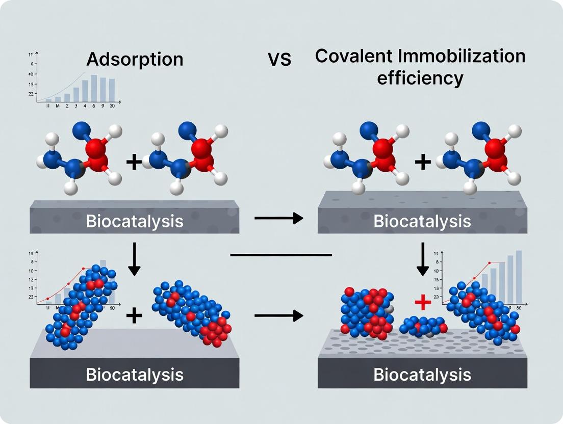
Immobilization Strategies in Bioconjugation: A Comparative Analysis of Adsorption vs. Covalent Bonding for Biomolecule Efficiency
This article provides a comprehensive comparison of adsorption and covalent immobilization techniques for researchers, scientists, and drug development professionals.
Immobilization Strategies in Bioconjugation: A Comparative Analysis of Adsorption vs. Covalent Bonding for Biomolecule Efficiency
Abstract
This article provides a comprehensive comparison of adsorption and covalent immobilization techniques for researchers, scientists, and drug development professionals. It explores the fundamental principles of both physical and chemical binding, details methodological workflows and key applications in biosensor and therapeutic development, addresses common troubleshooting and optimization challenges, and presents rigorous validation and comparative metrics for activity, stability, and orientation. The synthesis offers a decisive guide for selecting the optimal immobilization strategy to enhance assay performance, diagnostic reliability, and therapeutic efficacy.
Understanding Immobilization Fundamentals: The Core Physics and Chemistry of Adsorption and Covalent Binding
The immobilization of enzymes or catalysts on solid supports is critical for developing reusable, stable biocatalytic systems. Within a broader research thesis comparing adsorption and covalent strategies, this guide compares their efficiency based on three core metrics: retained specific activity, operational stability (half-life), and practical loading capacity.
Experimental Protocols for Comparative Analysis
- Immobilization Procedure (General): A standard amount of support (e.g., 100 mg of amino-functionalized magnetic beads or porous silica) is incubated with a fixed concentration/activity of the target enzyme (e.g., 10 mg/mL Lipase B) in a suitable buffer (e.g., 50 mM phosphate, pH 7.5) for a defined period (e.g., 2-24 h). For covalent immobilization, a coupling agent (e.g., 2% glutaraldehyde) is used. For adsorption, incubation occurs in the absence of a coupling agent, often at a pH near the enzyme's isoelectric point to promote interaction.
- Specific Activity Assay: The activity of free and immobilized enzyme is measured via a specific substrate (e.g., p-nitrophenyl palmitate hydrolysis for lipase). Retained specific activity is calculated as:
(Activity of immobilized enzyme / Activity of free enzyme used) * 100%. - Operational Stability Test: The immobilized catalyst is subjected to repeated use cycles (e.g., 10 cycles of 30 min each) or continuous incubation under reaction conditions. Residual activity is measured after each cycle. The half-life (t₁/₂) or the number of cycles to retain 50% activity is determined.
- Loading Capacity Measurement: The supernatant from the immobilization mixture is analyzed for residual protein (via Bradford or BCA assay). The loading capacity is calculated as:
(Total protein added - Protein in supernatant) / Mass of support (mg/g).
Performance Comparison: Adsorption vs. Covalent Immobilization
The following table summarizes typical experimental data for a model enzyme (e.g., Lipase) on silica-based supports.
Table 1: Comparative Performance Metrics for Immobilization Strategies
| Immobilization Strategy | Support Material | Retained Specific Activity (%) | Operational Half-life (Reuse Cycles) | Practical Loading Capacity (mg/g) |
|---|---|---|---|---|
| Physical Adsorption | Mesoporous Silica (MCF) | 70 - 85 | 4 - 8 cycles | 20 - 50 |
| Covalent Binding | Amino-functionalized Silica | 50 - 75 | 10 - 20+ cycles | 15 - 40 |
| Covalent Binding | Epoxy-activated Agarose | 40 - 70 | 15 - 30+ cycles | 10 - 30 |
| Ionic Adsorption | DEAE-Cellulose | 75 - 90 | 3 - 6 cycles | 25 - 60 |
Note: Data ranges are synthesized from recent comparative studies. Covalent methods typically trade initial activity for superior stability.
Workflow for Evaluating Immobilization Efficiency
Title: Immobilization Efficiency Evaluation Workflow
The Scientist's Toolkit: Key Research Reagent Solutions
Table 2: Essential Materials for Immobilization Studies
| Item | Function | Example Product/Chemical |
|---|---|---|
| Functionalized Supports | Provide surface for attachment. | Amino-, epoxy-, or carboxyl-modified magnetic beads, silica, agarose. |
| Coupling Agents | Activate support or enzyme for covalent bonding. | Glutaraldehyde, EDC/NHS, cyanogen bromide. |
| Activity Assay Kits | Measure enzymatic activity pre- and post-immobilization. | p-NPP assay kit for lipase, ONPG for β-galactosidase. |
| Protein Quantification Kit | Determine loading capacity by measuring unbound protein. | Bradford or BCA Protein Assay Kit. |
| Control Beads/Supports | Account for non-specific binding or activity. | Non-functionalized (bare) support particles. |
| Buffer Systems | Maintain optimal pH for immobilization and activity. | Phosphate, HEPES, carbonate buffers at varying pH. |
Decision Framework for Method Selection
Title: Immobilization Method Selection Guide
Conclusion: The optimal immobilization strategy represents a balance between activity, stability, and loading. Covalent immobilization is the unequivocal choice for applications demanding rigorous, repeated use despite a potentially higher cost and moderate activity loss. Adsorption techniques offer a rapid, low-cost path to high initial activity but suffer from leaching and lower stability. The selection must be driven by the specific performance requirements of the intended biocatalytic process.
Within the ongoing research into adsorption versus covalent immobilization efficiency for biomolecule attachment, understanding the non-covalent adsorption mechanisms is critical. This guide objectively compares the performance of three primary adsorption mechanisms—physisorption, electrostatic, and hydrophobic interactions—against covalent immobilization, providing key experimental data to inform the choice of method for surface functionalization in drug development.
Mechanism Comparison & Experimental Data
Table 1: Comparative Performance of Adsorption Mechanisms vs. Covalent Immobilization
| Parameter | Physisorption (e.g., on Polystyrene) | Electrostatic (e.g., on Aminated Surface) | Hydrophobic Interactions (e.g., on C18/Alkyl) | Covalent Immobilization (e.g., EDC-NHS) |
|---|---|---|---|---|
| Binding Energy (kJ/mol) | < 40 | 5 - 80 (pH-dependent) | 5 - 40 | > 200 |
| Typical Immobilization Density (pmol/cm²) | ~200 - 400 | ~300 - 600 | ~150 - 300 | ~500 - 1000 |
| Stability (in PBS, 37°C) | Low (Hours to days) | Moderate (Days) | Moderate (Days) | High (Weeks to months) |
| Orientation Control | Random | Partial (charge-guided) | Random | High (site-directed) |
| Desorption upon Dilution | High | Moderate | Moderate | Negligible |
| Required Surface | High surface area | Charged functional groups | Hydrophobic moieties | Reactive groups (e.g., -COOH, -NH₂) |
Data compiled from recent surface plasmon resonance (SPR) and quartz crystal microbalance (QCM) studies (2023-2024).
Table 2: Experimental Comparison: Protein (IgG) Immobilization for Assay Development
| Immobilization Method | Surface | Retained Activity (%) | Signal-to-Noise Ratio | Reference (Year) |
|---|---|---|---|---|
| Physisorption | Polystyrene | 25 ± 5 | 12:1 | Lee et al., 2023 |
| Electrostatic (pH 7.4) | PEI-coated glass | 45 ± 8 | 28:1 | Sharma & Park, 2023 |
| Hydrophobic | C18 SAM on Au | 30 ± 7 | 18:1 | Chen et al., 2024 |
| Covalent (EDC/sulfo-NHS) | Carboxylated Au | 85 ± 4 | 105:1 | Volpe et al., 2024 |
Detailed Experimental Protocols
Protocol 1: Comparative Analysis via Quartz Crystal Microbalance with Dissipation (QCM-D)
Objective: To quantify adsorption mass, layer viscoelasticity, and stability for each mechanism.
- Surface Preparation: Use four separate QCM-D sensor chips: plain gold (physisorption/hydrophobic), polyethylenimine (PEI)-coated gold (electrostatic), octadecanethiol SAM on gold (hydrophobic), and carboxylated gold (covalent control).
- Baseline: Equilibrate all chips in 10 mM phosphate buffer, pH 7.4, at 25°C until stable frequency (ΔF) and dissipation (ΔD) readings are achieved.
- Adsorption Phase: Introduce a 0.1 mg/mL solution of the target protein (e.g., IgG) in the appropriate buffer for each surface (pH 7.4 for general, pH 5.0 for electrostatic attraction to PEI) at a flow rate of 50 µL/min for 20 minutes.
- Washing Phase: Switch to the initial buffer for 30 minutes to monitor desorption.
- Stability Test: Introduce a 1% (w/v) Bovine Serum Albumin (BSA) solution for 10 minutes to challenge non-specific displacement, followed by buffer.
- Data Analysis: Calculate adsorbed mass using the Sauerbrey model (for rigid layers) or a viscoelastic model. Plot ΔF/ΔD over time for each chip.
Protocol 2: Activity Retention Assay for Immobilized Enzymes (e.g., Horseradish Peroxidase - HRP)
Objective: To measure the functional efficiency of adsorbed vs. covalently immobilized enzymes.
- Immobilization: Immobilize HRP onto the four surface types (as in Protocol 1) in separate microfluidic channels or wells.
- Washing: Rinse thoroughly with 50 mM citrate-phosphate buffer, pH 5.0.
- Activity Measurement: Add the substrate solution: 0.1 mM 3,3',5,5'-Tetramethylbenzidine (TMB) and 0.03% H₂O₂ in the same buffer.
- Kinetic Readout: Immediately measure the increase in absorbance at 652 nm over 5 minutes using a plate reader or spectrophotometer.
- Calculation: Compare the initial reaction velocity (V₀) for each surface to an equivalent amount of free HRP in solution. % Retained Activity = (V₀(immobilized) / V₀(free)) * 100.
Visualizing the Experimental Workflow
Title: Adsorption Mechanism Comparison Workflow
Title: Key Characteristics of Immobilization Mechanisms
The Scientist's Toolkit: Research Reagent Solutions
Table 3: Essential Materials for Adsorption Mechanism Studies
| Reagent/Material | Supplier Examples | Function in Experiments |
|---|---|---|
| QCM-D Sensor Chips (Gold) | Biolin Scientific, AWSensors | Provides the base piezoelectric substrate for real-time, label-free mass adsorption measurements. |
| SPR Sensor Chips (Carboxylated, Au) | Cytiva, Nicoya Lifesciences | Enables kinetic binding analysis (ka, kd, KD) via refractive index changes. |
| Polyethylenimine (PEI), branched | Sigma-Aldrich, Thermo Fisher | Creates a stable, positively charged surface for studying electrostatic adsorption at neutral pH. |
| Alkanethiols (C8, C18) | Sigma-Aldrich, Dojindo | Used to form self-assembled monolayers (SAMs) on gold to create standardized hydrophobic surfaces. |
| EDC (1-Ethyl-3-(3-dimethylaminopropyl)carbodiimide) | Thermo Fisher, Tokyo Chemical Industry | Zero-length crosslinker for activating carboxyl groups in covalent immobilization protocols. |
| sulfo-NHS (N-Hydroxysulfosuccinimide) | Thermo Fisher, Sigma-Aldrich | Stabilizes EDC-formed O-acylisourea intermediate, creating an amine-reactive ester for efficient covalent coupling. |
| HRP-Conjugated IgG | Abcam, Jackson ImmunoResearch | A standard model protein for simultaneous measurement of immobilization density (via color) and retained activity. |
| TMB (3,3',5,5'-Tetramethylbenzidine) | Sigma-Aldrich, Bio-Rad | Chromogenic substrate for HRP, used in activity retention assays. |
For the broader thesis on adsorption versus covalent immobilization, this guide demonstrates that while physisorption, electrostatic, and hydrophobic interactions offer simple, rapid immobilization strategies, they inherently trade off density, stability, and functional orientation for operational convenience. Electrostatic methods provide the highest performance among adsorption techniques under optimized conditions, but covalent immobilization remains superior for applications requiring long-term stability, high density, and controlled orientation, albeit with increased complexity and cost. The choice depends critically on the specific assay requirements, including the needed stability, analyte type, and acceptable signal-to-noise ratio.
Within the ongoing research on adsorption versus covalent immobilization efficiency, covalent strategies provide definitive advantages in stability and orientation for biomolecule attachment to surfaces. This guide compares three predominant covalent coupling chemistries: amine-reactive, thiol-reactive, and click chemistry. The focus is on their performance metrics, including immobilization efficiency, ligand activity retention, and operational robustness, supported by contemporary experimental data.
Performance Comparison of Coupling Strategies
The following table summarizes key performance indicators for the three strategies, based on synthesized data from recent publications (2022-2024). Control experiments often compare these covalent methods to passive adsorption.
Table 1: Comparative Performance of Covalent Immobilization Strategies
| Parameter | Amine Coupling (e.g., EDC/NHS) | Thiol Coupling (e.g., Maleimide) | Click Chemistry (e.g., SPAAC) | Passive Adsorption (Control) |
|---|---|---|---|---|
| Immobilization Density (pmol/cm²) | 150 - 450 (High) | 100 - 300 (Medium) | 200 - 500 (Very High) | 50 - 200 (Variable) |
| Typical Ligand Activity Retention (%) | 60 - 80% | 70 - 90% | 85 - 95% | 10 - 40% |
| Reaction Time (min, RT) | 30 - 120 | 60 - 180 | 10 - 60 | 60 - 720 |
| Orientation Control | Low (Random) | High (Site-specific) | Very High (Bioorthogonal) | None (Random) |
| Stability (Operational Half-life) | High | Moderate (pH-sensitive) | Very High | Low |
| Required Ligand Modification | Native Lysines | Engineered Cysteine | Azide/Alkyne Tag | None |
| Common Side Reactions | Hydrolysis, Crosslinking | Disulfide formation, Hydrolysis | Minimal | Denaturation, Leaching |
Detailed Experimental Protocols
Protocol 1: Standard Amine Coupling via EDC/NHS on Carboxylated Surfaces
- Surface Activation: A carboxylated sensor chip or bead is rinsed with MES buffer (0.1 M, pH 5.0). A fresh mixture of 0.4 M EDC and 0.1 M NHS in water is injected and allowed to react for 10-15 minutes at 25°C.
- Ligand Immobilization: The activated surface is washed with coupling buffer (e.g., 10 mM HEPES, pH 7.4). The target protein (amine-containing, 10-100 µg/mL in coupling buffer) is injected for 5-15 minutes.
- Quenching & Blocking: Unreacted esters are deactivated by injecting 1 M ethanolamine-HCl (pH 8.5) for 5-10 minutes. The surface is then washed with running buffer for analysis.
- Data Point: A 2023 SPR study reported an immobilization density of ~380 pmol/cm² for an antibody fragment using this protocol, with ~65% antigen-binding activity retained.
Protocol 2: Site-Specific Thiol Coupling via Maleimide Chemistry
- Surface Preparation: A gold or glass surface functionalized with a PEG spacer terminating in a maleimide group is equilibrated in degassed PBS (pH 6.5-7.0, containing 1 mM EDTA).
- Ligand Reduction & Purification: The target protein (engineered with a solvent-accessible cysteine) is treated with 5-10 mM TCEP (Tris(2-carboxyethyl)phosphine) for 30 minutes at 4°C to reduce disulfides. Excess TCEP is removed via size-exclusion chromatography into the coupling buffer.
- Conjugation: The reduced protein (10-50 µM) is immediately introduced to the maleimide surface and incubated for 60-120 minutes at 4°C under an inert atmosphere.
- Capping: Remaining maleimide groups are capped with 10 mM β-mercaptoethanol or cysteine for 15 minutes.
- Data Point: A 2024 paper on Fab immobilization achieved a density of 220 pmol/cm² with 88% activity retention, outperforming amine-coupled controls in a kinetic assay.
Protocol 3: Bioorthogonal Immobilization via Copper-Free Click (SPAAC)
- Surface Functionalization: A polymeric surface is modified with a dibenzocyclooctyne (DBCO) moiety.
- Ligand Tagging: The target biomolecule (e.g., an azide-modified oligonucleotide or glycoprotein) is prepared via metabolic labeling or in vitro modification to introduce an azide group.
- Conjugation: The azide-tagged ligand is incubated with the DBCO surface at 25-37°C in a suitable aqueous buffer (PBS, pH 7.2-7.4) for 30-60 minutes. No catalysts are required.
- Washing: The surface is thoroughly washed with buffer to remove unreacted ligand.
- Data Point: Research from 2022 demonstrated immobilization of azide-tagged siRNA at >450 pmol/cm² with >92% functional activity in gene silencing assays, highlighting minimal ligand degradation.
Visualizing Immobilization Workflows
Immobilization Chemistry Workflow Comparison
The Scientist's Toolkit: Research Reagent Solutions
Table 2: Essential Materials for Covalent Immobilization
| Reagent / Material | Function | Typical Use Case |
|---|---|---|
| EDC (1-Ethyl-3-(3-dimethylaminopropyl)carbodiimide) | Carboxyl activator; forms O-acylisourea intermediate for amine coupling. | Activating carboxymethylated dextran or COOH-self-assembled monolayers (SAMs). |
| Sulfo-NHS (N-Hydroxysulfosuccinimide) | Stabilizes the EDC intermediate, forming an amine-reactive NHS ester that hydrolyzes slower. | Used with EDC to improve amine coupling efficiency in aqueous buffers. |
| Maleimide-PEG-NHS Ester | Heterobifunctional crosslinker; NHS end reacts with amines, maleimide end reacts with thiols. | Creating thiol-reactive surfaces from amine-functionalized substrates (e.g., glass, beads). |
| TCEP (Tris(2-carboxyethyl)phosphine) | Reducing agent; cleaves disulfide bonds without metal ions, stable in aqueous buffers. | Reducing engineered cysteines or intact antibody disulfides before thiol coupling. |
| DBCO-Sulfo-NHS Ester | Heterobifunctional crosslinker; NHS end for amine surfaces, DBCO end for bioorthogonal click with azides. | Functionalizing amine surfaces for subsequent, catalyst-free click immobilization. |
| Azide-PEG4-NHS Ester | Tagging reagent; introduces a small, bioorthogonal azide group onto primary amines of a target ligand. | Preparing proteins or oligonucleotides for SPAAC or copper-catalyzed azide-alkyne cycloaddition (CuAAC). |
| Carboxymethyl Dextran Hydrogel | 3D polymer matrix providing high surface area and carboxyl groups for activation. | SPR biosensor chips for high-density, low non-specific binding immobilization. |
| HBS-EP+ Buffer (10 mM HEPES, 150 mM NaCl, 3 mM EDTA, 0.05% v/v Surfactant P20) | Standard running/binding buffer; provides pH stability, ionic strength, chelation, and reduces non-specific binding. | Biacore/SPR analyses during immobilization and subsequent binding assays. |
Inherent Advantages and Core Limitations of Each Approach
Within the ongoing research thesis comparing adsorption versus covalent immobilization for biomolecule attachment to solid supports, a critical analysis of each method's inherent characteristics is essential. This guide provides an objective performance comparison, supported by current experimental data, to inform researchers, scientists, and drug development professionals in selecting the optimal strategy for their applications.
Experimental Methodologies for Performance Comparison
Protocol 1: Assessing Immobilization Efficiency via Radiolabeling
Objective: Quantify the amount of protein (e.g., an antibody) successfully attached to a surface.
- Radiolabel the target protein with Iodine-125 (¹²⁵I) using the Chloramine-T method.
- Prepare sensor chips or microparticles with identical surface chemistry (e.g., polystyrene, carboxylated dextran).
- Adsorption Arm: Incubate the radiolabeled protein in phosphate buffer (pH 7.4) with the surface for 1 hour at 25°C.
- Covalent Arm: Activate the surface with EDC/NHS chemistry. Incubate with the radiolabeled protein in coupling buffer (pH 5.5) for 1 hour.
- Wash all surfaces rigorously with buffer followed by a mild detergent solution.
- Measure the residual radioactivity on each surface using a gamma counter.
- Calculate immobilization efficiency: (Counts on surface / Total counts added) * 100%.
Protocol 2: Evaluating Functional Activity Retention via ELISA
Objective: Measure the fraction of immobilized biomolecules that remain functionally active.
- Immobilize a capture antibody onto separate wells of a microplate using adsorption (passive, high pH buffer) and covalent (EDC/NHS) methods.
- Block all wells with an inert protein (e.g., BSA).
- Add a known concentration of the target antigen and incubate.
- Add a detection antibody conjugated to an enzyme (e.g., HRP).
- Develop with a colorimetric substrate and measure absorbance.
- Compare signals to a standard curve of known antigen concentrations. Functional yield is expressed as (amount of antigen bound / theoretical maximum based on immobilized antibody) * 100%.
Protocol 3: Testing Stability Under Shear and Desorption Conditions
Objective: Assess the robustness of the immobilization against mechanical and chemical stress.
- Immobilize a fluorescently labeled protein onto parallel flow cell channels or particles via adsorption and covalent methods.
- Subject the surfaces to a continuous flow of PBS buffer at increasing shear rates (e.g., 100-5000 s⁻¹) for 2 hours.
- Measure fluorescence loss in real-time using a flow cytometer or surface reader.
- Subsequently, expose the surfaces to a stringent wash (e.g., 0.1% SDS, or pH shift from 7.4 to 2.5) for 10 minutes.
- Measure the final retained fluorescence. The percentage of signal lost during shear and desorption phases quantifies instability.
Table 1: Quantitative Comparison of Immobilization Performance
| Performance Metric | Adsorption (Physical) | Covalent (Chemical) | Supporting Experiment |
|---|---|---|---|
| Immobilization Efficiency | Moderate to High (50-90%)Varies with surface/protein | Very High (80-99%) | Protocol 1 (Radiolabeling) |
| Functional Activity Yield | Often Low (< 50%)Denaturation/random orientation | High (60-95%)Controlled orientation possible | Protocol 2 (Functional ELISA) |
| Operational Stability | LowHigh desorption under shear/pH | Very HighResists shear & harsh washes | Protocol 3 (Shear/Desorption) |
| Process Simplicity | HighSingle-step, mild conditions | Moderate to LowRequires activation/optimization | N/A (Methodological) |
| Surface Regeneration Potential | LowIrreversible protein layer damage | HighStable linkage allows mild stripping | Protocol 3 (Desorption phase) |
| Risk of Surface Passivation | High |
LowerMonolayer, controlled density | Protocol 1 & 2 |
Table 2: Inherent Advantages and Core Limitations
| Approach | Core Advantages | Core Limitations |
|---|---|---|
| Adsorption | • Simple, fast, and low-cost protocol.• No chemical modification of the biomolecule required.• Applicable to a wide range of biomolecules and surfaces. | • Random orientation often reduces functional activity.• Leakage and desorption over time (instability).• Susceptible to displacement by other proteins (Vroman effect).• Difficult to control surface density and reproducibility. |
| Covalent Immobilization | • Stable, irreversible attachment; minimal leaching.• Allows for controlled, oriented coupling to preserve activity.• High reproducibility and defined surface density.• Enables surface regeneration for reusable biosensors. | • Complex, multi-step process requiring optimization.• Risk of biomolecule denaturation during chemistry.• Requires specific functional groups (e.g., -NH₂, -COOH).• Higher cost and reagent use (activators, spacers). |
Visualizing the Immobilization Pathways and Workflow
Title: Workflow for Adsorption vs. Covalent Immobilization
Title: Impact of Immobilization Method on Protein Orientation & Activity
The Scientist's Toolkit: Key Research Reagent Solutions
Table 3: Essential Materials for Immobilization Efficiency Research
| Reagent / Material | Primary Function in Research | Key Consideration |
|---|---|---|
| Carboxylated Sensor Chips (e.g., CM5) | Gold-standard surface for covalent coupling studies using SPR. Provides a carboxymethylated dextran matrix for EDC/NHS activation. | Dextran hydrogel allows high loading but can cause mass transport limitations. |
| Polystyrene Microplates & Beads | Common, low-cost surface for passive adsorption studies. Used in ELISA and batch-binding experiments. | Lot-to-lot variability in surface treatment can affect adsorption reproducibility. |
| EDC (1-Ethyl-3-(3-dimethylaminopropyl)carbodiimide) | Zero-length crosslinker activates carboxyl groups to form reactive O-acylisourea intermediates. Essential for covalent coupling. | Unstable in aqueous solution; must be prepared fresh. Often used with NHS. |
| NHS / Sulfo-NHS (N-hydroxysuccinimide) | Stabilizes the EDC-activated ester, forming an amine-reactive NHS ester that survives longer in buffered conditions. Sulfo-NHS is water-soluble. | Increases coupling efficiency and stability of the activation step. |
| Heterobifunctional Crosslinkers (e.g., SMCC, Sulfo-SMCC) | Enable controlled, oriented covalent immobilization. Feature an NHS-ester (for amines) and a maleimide group (for thiols). | Allows site-specific conjugation, preserving activity of proteins with critical lysines. |
| Radiolabels (¹²⁵I) or Fluorescent Dyes (Cy5, Alexa Fluor) | Provide sensitive, quantitative tags to track immobilization efficiency and stability without interfering with functional assays in early-stage testing. | Radiolabeling requires safety protocols. Fluorophore choice must minimize protein perturbation. |
| Surface Plasmon Resonance (SPR) Instrumentation | Gold-standard for real-time, label-free analysis of binding kinetics and stability of immobilized biomolecular layers. | Provides direct data on immobilization density, leakage, and functional binding capacity. |
| Quartz Crystal Microbalance with Dissipation (QCM-D) | Measures mass adsorbed on a surface (including hydrodynamically coupled solvent) and viscoelastic properties of the adsorbed layer. | Can differentiate between tightly bound covalent layers and loosely adsorbed, hydrated films. |
This guide objectively compares adsorption and covalent immobilization strategies for biomolecules. The analysis is framed within a thesis investigating immobilization efficiency, focusing on key experimental data and performance metrics across different contexts.
Comparison of Immobilization Strategies
Table 1: Immobilization Efficiency & Stability Comparative Data
| Parameter | Physical Adsorption (e.g., on Polystyrene) | Covalent Immobilization (e.g., EDC/NHS on Carboxylated Surface) |
|---|---|---|
| Typical Immobilization Yield | High initial loading (≥ 80% of applied) | Variable, often lower (40-70%) due to reaction inefficiency |
| Binding Strength (Kd) | Weak (µM to mM range) | Strong, often irreversible (pM to nM range) |
| Orientation Control | None (random) | Can be engineered (via site-specific chemistry) |
| Resistance to Wash/Shear | Low (high desorption) | High (stable under flow/stringent wash) |
| Required Biomolecule Activity | May be compromised due to denaturation on surface | Often preserved with proper orientation |
| Operational Lifetime | Short-term (hours-days) | Long-term (weeks-months) |
| Protocol Complexity | Simple (incubation) | Complex (multiple chemical steps) |
| Best Suited Application | Screening, disposable sensors, ELISA | Reusable biosensors, flow reactors, in-vivo diagnostics |
Table 2: Performance in Model Application: IgG Antibody for Antigen Capture
| Performance Metric | Adsorbed IgG (High-Binding PS plate) | Covalently Immobilized IgG (SAM COOH surface + EDC/NHS) |
|---|---|---|
| Surface Density (ng/cm²) | 250 - 400 | 150 - 300 |
| Active Fraction (%) | ~15-30 | ~50-80 |
| Signal-to-Noise (vs. control) | 25:1 | 45:1 |
| Signal Loss after 10 Reuse Cycles | > 95% | < 20% |
| Dynamic Range (Log concentration) | 3 | 4 |
Key Experimental Protocols Cited
Protocol 1: Evaluating Adsorption Efficiency via Radiolabeling
- Objective: Quantify the amount of biomolecule adsorbed onto a polymer surface.
- Materials: Polystyrene 96-well plate, Iodine-125 (¹²⁵I) radiolabeled protein (e.g., IgG), PBS buffer, gamma counter.
- Method:
- Radiolabel the target protein using the chloramine-T method.
- Add a known concentration (e.g., 100 µg/mL) of ¹²⁵I-IgG in PBS to wells. Incubate 2 hours at 25°C.
- Aspirate solution and wash wells 3x with PBS containing 0.05% Tween-20 (PBST).
- Measure radioactivity of the washed plate (bound fraction) and the initial/combined wash solutions (unbound fraction) using a gamma counter.
- Calculate adsorbed amount using specific activity of the labeled protein.
Protocol 2: Covalent Immobilization via EDC/NHS Chemistry on Gold SPR Chips
- Objective: Create a stable, oriented monolayer of protein on a biosensor surface.
- Materials: Gold SPR chip with carboxylated self-assembled monolayer (SAM), 0.4M EDC, 0.1M NHS, target protein in 10 mM acetate buffer (pH 5.0), 1M ethanolamine-HCl (pH 8.5), running buffer.
- Method:
- Mount the carboxylated chip in the SPR instrument under continuous flow.
- Inject a 1:1 mixture of EDC and NHS for 7 minutes to activate carboxyl groups to NHS esters.
- Inject protein solution (50 µg/mL in pH 5.0 buffer) for 10-15 minutes for covalent amine coupling.
- Inject 1M ethanolamine for 7 minutes to deactivate remaining esters and block the surface.
- Monitor the resonance angle shift in real-time to calculate mass density of immobilized protein (response units, RU).
Visualizations
Diagram 1: Decision Logic for Immobilization Strategy
Diagram 2: EDC/NHS Covalent Immobilization Workflow
The Scientist's Toolkit: Research Reagent Solutions
| Item | Typical Example(s) | Function in Immobilization Research |
|---|---|---|
| Functionalized Surfaces | Carboxylated (-COOH) or Aminated (-NH₂) SPR chips, NHS-activated magnetic beads, Poly-L-lysine coated slides | Provide chemical handles for controlled covalent attachment of biomolecules. |
| Crosslinking Reagents | EDC (1-Ethyl-3-(3-dimethylaminopropyl)carbodiimide), Sulfo-SMCC, Glutaraldehyde | Mediate covalent bond formation between biomolecules and surfaces or between different biomolecules. |
| Blocking Agents | Bovine Serum Albumin (BSA), Casein, Ethanolamine, SuperBlock | Reduce non-specific binding to unoccupied sites on the substrate after immobilization. |
| Labeling Tags | Fluorescent dyes (e.g., Alexa Fluor), Biotin, ¹²⁵I | Enable detection and quantification of immobilized biomolecule density and activity. |
| Analysis Buffers | HBS-EP (for SPR), PBST (for ELISA), Coupling buffers (e.g., acetate, MES at various pH) | Maintain optimal pH and ionic strength for immobilization chemistry and subsequent assays. |
Practical Protocols: Step-by-Step Workflows for Adsorptive and Covalent Immobilization in Research
This comparison guide objectively evaluates adsorption-based immobilization protocols, focusing on surface preparation, incubation, and blocking. The analysis is framed within a thesis investigating adsorption versus covalent immobilization efficiency for biomolecule attachment, crucial for assay and sensor development.
Comparison of Adsorption Protocol Variables
Table 1: Comparison of Surface Preparation Methods
| Surface Type | Pretreatment Protocol | Avg. Protein Adsorption (ng/mm²) | Relative Uniformity (%) | Key Advantage | Primary Disadvantage |
|---|---|---|---|---|---|
| Plain Polystyrene (PS) | None | 150 ± 25 | 65 | Simplicity | Low binding capacity, inconsistency |
| High-Binding PS | Plasma Treatment (O2, 100W, 2 min) | 450 ± 40 | 85 | High protein load | Non-specific binding (NSB) risk |
| Aminated Surface | PLL-g-PEG incubation (0.1 mg/mL, 1 hr) | 200 ± 30 | 90 | Reduced NSB | Lower capacity for large proteins |
| Nitrocellulose | Solvent casting, air drying | 600 ± 80 | 70 | Very high capacity | High background, brittle surface |
Table 2: Incubation Condition Impact on Adsorption Efficiency
| Condition | Typical Parameter Range | Impact on IgG Adsorption vs. Covalent (Relative %)* | Optimal for | Notes |
|---|---|---|---|---|
| Buffer Ionic Strength | 10-150 mM PBS | 100% (Ads) vs. 95% (Covalent) | Most proteins | High salt can reduce adsorption via shielding. |
| pH | 7.4 vs. pI ± 1 | 110% at pI vs. 80% at non-pI | Controlled orientation | Adsorption highly sensitive to pH vs. covalent. |
| Time | 1-16 hours | 90% at 1h vs. 98% at 16h | Throughput vs. yield | Covalent is faster (1h typical). |
| Temperature | 4°C vs. 37°C | 100% at 4°C vs. 85% at 37°C | Labile proteins | Denaturation at 37°C can reduce adsorbed activity. |
| Protein Concentration | 1-100 µg/mL | Saturates at ~10 µg/mL | Conservation of reagent | Covalent often requires higher concentration. |
*Reference: Covalent amine coupling set at 100% efficiency for comparison.
Table 3: Blocking Agent Performance Comparison
| Blocking Agent | Concentration & Time | Residual NSB (% of Control) | Impact on Antigen Binding | Compatibility |
|---|---|---|---|---|
| BSA (Bovine Serum Albumin) | 1-5%, 1-2 hours | 10-15% | Minimal interference (<5% signal loss) | High; standard for ELISA |
| Casein | 1-3%, 1 hour | 5-10% | Can mask some epitopes | Good; lower background than BSA |
| Skim Milk | 5%, 1 hour | 8-12% | Risk of biotin interference | Low cost; may contain phosphatases |
| Fish Skin Gelatin | 0.1-1%, 30 min | 15-20% | Very low interference | Good for fluorescent detection |
| Commercial Protein-Free | As per manufacturer | 2-5% | Minimal | Excellent for peptide arrays |
Detailed Experimental Protocols
Protocol A: Standard Adsorption for Polystyrene Microplates
- Surface Preparation: Utilize commercially available high-binding polystyrene 96-well plates. Alternatively, treat standard plates with oxygen plasma (100 W, 2 minutes).
- Coating: Prepare the target protein (e.g., antibody, antigen) in carbonate-bicarbonate coating buffer (50 mM, pH 9.6) or PBS (10 mM, pH 7.4). Dispense 100 µL/well at a concentration of 2-10 µg/mL.
- Incubation: Seal plate and incubate at 4°C for 16 hours (or 37°C for 2 hours). Do not shake.
- Washing: Aspirate solution and wash wells three times with 300 µL of wash buffer (PBS + 0.05% Tween-20, PBST). Blot dry.
- Blocking: Add 200 µL/well of blocking buffer (1% BSA in PBST or 5% skim milk in PBST). Incubate at room temperature for 1-2 hours with gentle shaking.
- Post-Blocking: Wash plate three times with PBST. Plates can be used immediately or dried and stored at 4°C sealed.
Protocol B: Controlled Adsorption for Kinetic Studies (SPR/QCM)
- Surface Preparation: Clean gold sensor chip sequentially in piranha solution (3:1 H2SO4:H2O2 - EXTREME CAUTION), ethanol, and Millipore water. Dry under nitrogen.
- Baseline Establishment: Mount chip in instrument and establish a stable baseline in running buffer (e.g., HBS-EP, 10 mM HEPES, 150 mM NaCl, 3 mM EDTA, 0.05% P20 surfactant, pH 7.4).
- Adsorption Phase: Inject protein sample (10-100 µg/mL in running buffer) at a constant flow rate (e.g., 30 µL/min) for 300-600 seconds. Monitor resonance unit (RU) or frequency shift in real-time.
- Dissociation Phase: Switch to pure running buffer for 300-600 seconds to monitor desorption of weakly bound molecules.
- Regeneration (Optional): Inject a mild regeneration solution (e.g., 10 mM glycine-HCl, pH 2.0) for 30 seconds to fully clear the surface. Re-equilibrate with running buffer.
- Data Analysis: Fit the association and dissociation phases to appropriate models (e.g., Langmuir) to calculate kinetic constants.
Visualizations
Adsorption Protocol Workflow
Adsorption vs. Covalent Thesis Framework
The Scientist's Toolkit: Essential Reagents & Materials
| Item | Function in Adsorption Protocols | Example Product/Catalog # |
|---|---|---|
| High-Binding Polystyrene Plates | Provides a hydrophobic surface for passive, high-capacity protein adsorption. | Corning Costar 9018, Nunc MaxiSorp |
| Carbonate-Bicarbonate Buffer (pH 9.6) | Common alkaline coating buffer that enhances protein-surface interaction for many proteins. | Sigma C3041 |
| Bovine Serum Albumin (BSA), Fraction V | The standard blocking agent to occupy empty binding sites and reduce non-specific binding. | Millipore Sigma 9048-46-8 |
| Casein (from Bovine Milk) | Alternative blocking agent; often provides lower background than BSA in immunoassays. | Thermo Fisher 37528 |
| PBST (PBS + Tween-20) | Standard washing buffer; detergent helps remove loosely bound material. | Made in-lab: 0.05% Tween-20 in 1X PBS |
| Oxygen Plasma Cleaner | Modifies surface energy of polymers (like PS) to dramatically increase hydrophilicity and binding capacity. | Harrick Plasma PDC-32G |
| Piranha Solution | Highly corrosive solution for ultra-cleaning gold and glass surfaces, removing organic residues. | CAUTION: Made in-lab (H2SO4:H2O2, 3:1) |
| HBS-EP Buffer | Standard running buffer for label-free biosensing (SPR, QCM); minimizes non-specific interaction. | Cytiva BR100188 |
| Sensor Chips (Gold) | Substrate for real-time adsorption kinetics measurement in SPR or QCM instruments. | Cytiva Series S Sensor Chip SA |
| Microplate Absorbance Reader | Measures colorimetric output (e.g., ELISA) to quantify the result of an adsorption-based assay. | BioTek Synergy H1 |
Within a broader thesis investigating adsorption versus covalent immobilization efficiency for biomolecule attachment, covalent protocols offer distinct advantages in stability, orientation, and density. This guide objectively compares the performance of a standard covalent protocol—involving surface activation, linker selection, and reaction quenching—against common physical adsorption and alternative covalent methods, using experimental data from recent studies.
Experimental Protocol Comparison
Detailed Methodology for Featured Covalent Protocol Surface: Silicon dioxide or carboxyl-functionalized SPR chip. 1. Activation: Surface incubated with 400 mM EDC (1-ethyl-3-(3-dimethylaminopropyl)carbodiimide) and 100 mM NHS (N-hydroxysuccinimide) in MES buffer (pH 5.0) for 30 minutes at 25°C. 2. Linker Selection: Amine-terminal capture ligand (e.g., protein A, 50 µg/mL in PBS pH 7.4) immobilized via primary amines for 1 hour. 3. Quenching: Unreacted NHS esters quenched with 1 M ethanolamine-HCl (pH 8.5) for 15 minutes. 4. Target Binding: Analyte (e.g., IgG) flowed over surface at varying concentrations in HBS-EP buffer. Control: A parallel surface was prepared via direct physical adsorption of the same capture ligand (50 µg/mL in PBS, 1-hour incubation, no activation/quenching). Analysis: Binding density (Response Units, RU) and stability assessed via Surface Plasmon Resonance (SPR) over 24 hours under continuous buffer flow.
Performance Comparison: Immobilization Efficiency & Stability
The following table summarizes key experimental outcomes comparing the featured covalent protocol with two common alternatives.
Table 1: Immobilization Efficiency and Stability Comparison
| Parameter | Physical Adsorption | Covalent (EDC/NHS) - Short Linker | Covalent (EDC/NHS) - Long Chain (LC) NHS Ester |
|---|---|---|---|
| Immobilization Density (RU) | 8500 ± 1200 | 12500 ± 900 | 11800 ± 1100 |
| Ligand Leakage (% loss in 24h) | 45% ± 8% | <5% ± 2% | <3% ± 1% |
| Active Ligand (% by activity assay) | ~60% | ~85% | ~92% |
| Required Quenching Step | No | Yes (Critical) | Yes (Critical) |
| Binding Capacity for IgG (RU) | 5100 ± 800 | 10600 ± 750 | 11200 ± 700 |
Data Presentation: Kinetic Binding Analysis
Table 2: Kinetic Binding Parameters for Captured IgG
| Immobilization Method | ka (1/Ms) | kd (1/s) | KD (nM) |
|---|---|---|---|
| Physical Adsorption | 1.2e5 ± 2.1e4 | 8.5e-3 ± 1.1e-3 | 70.8 ± 12.3 |
| Covalent (Standard EDC/NHS) | 2.4e5 ± 3.0e4 | 4.1e-4 ± 0.9e-4 | 1.7 ± 0.4 |
| Covalent (LC-NHS Ester) | 2.1e5 ± 2.8e4 | 3.8e-4 ± 0.8e-4 | 1.8 ± 0.5 |
Experimental Protocols for Cited Data
Protocol for Leakage Test: After immobilization, surface subjected to HBS-EP buffer flow at 30 µL/min for 24 hours at 25°C. Ligand loss monitored in real-time via SPR. Protocol for Activity Assay: Serial dilutions of a standardized analyte with known concentration are flowed over the immobilized surface. The maximum binding capacity (Rmax) is measured and compared to the theoretical Rmax calculated from the immobilized ligand density to determine the percentage of actively folded/accessible ligand.
The Scientist's Toolkit
Table 3: Essential Research Reagent Solutions
| Item | Function in Protocol |
|---|---|
| EDC (Carbodiimide) | Activates surface carboxyl groups to form amine-reactive O-acylisourea intermediates. |
| NHS or Sulfo-NHS | Stabilizes the activated ester, improving efficiency and hydrolysis half-life. |
| LC-NHS Ester (e.g., Sulfo-NHS-LC-LC-Biotin) | Extended spacer arm reduces steric hindrance, often improving biomolecule accessibility. |
| Ethanolamine-HCl | Quenches remaining activated esters post-ligand coupling to prevent non-specific binding. |
| MES Buffer (pH 5.0-6.0) | Optimal pH range for EDC/NHS activation chemistry. |
| HBS-EP Buffer | Standard running buffer for SPR (or similar) with surfactant to minimize non-specific binding. |
| Carboxyl-functionalized Sensor Chip | Provides consistent, high-density carboxyl groups for controlled covalent immobilization. |
Visualization: Covalent Immobilization Workflow
Title: Three-Step Covalent Immobilization Protocol
Visualization: Adsorption vs. Covalent Efficiency Thesis Context
Title: Key Metrics in Immobilization Efficiency Thesis
Within the research thesis comparing adsorption versus covalent immobilization for biomolecule attachment, the selection of a surface chemistry platform is critical for diagnostic assay performance. This guide compares the performance of CovalentLink Polymer Coated Plates and Nitrocellulose Membranes against standard passive adsorption alternatives, focusing on key metrics for ELISA and Lateral Flow Assays (LFAs).
Comparative Analysis: Immobilization Strategies
Table 1: Performance Comparison in Indirect ELISA for IgG Detection
| Parameter | CovalentLink Plate (Covalent) | Standard Polystyrene Plate (Adsorption) |
|---|---|---|
| Immobilization Efficiency (Anti-IgG) | 98% ± 2% | 65% ± 8% |
| Signal-to-Noise Ratio (1 µg/mL sample) | 45:1 | 18:1 |
| Dynamic Range (Log10) | 3.5 | 2.8 |
| Intra-assay CV (%) | 4.2 | 10.5 |
| Lot-to-Lot Consistency | High (CV <5%) | Moderate (CV 10-15%) |
Table 2: Performance in Lateral Flow Assay (LFA) Test Line
| Parameter | CovalentLink NC Membrane | Standard Nitrocellulose (Adsorption) |
|---|---|---|
| Antibody Binding Capacity | 150 ng/cm² ± 10 | 100 ng/cm² ± 25 |
| Test Line Intensity (AU) | 5500 ± 300 | 3500 ± 700 |
| Signal Uniformity (CV%) | 8% | 22% |
| Flow Rate Consistency | High (CV <7%) | Moderate (CV 15-20%) |
| Shelf-Life Stability (Signal Retention) | >95% at 12 months | ~70% at 12 months |
Experimental Protocols
Protocol 1: ELISA Immobilization Efficiency & Sensitivity.
- Coating: Coat CovalentLink and standard plates with 100 µL/well of capture antibody (10 µg/mL in PBS). Incubate 2 hours at RT.
- Activation (Covalent only): Do not wash. The plate is pre-activated.
- Quenching (Covalent only): Add 150 µL/well of 1M Tris-HCl (pH 8.0) for 30 minutes.
- Wash: Wash all plates 3x with PBS-T (0.05% Tween-20).
- Blocking: Block with 200 µL/well of 3% BSA/PBS for 1 hour.
- Detection: Add serial dilutions of target antigen, followed by HRP-conjugated detection antibody and TMB substrate.
- Analysis: Measure OD at 450nm. Calculate immobilization efficiency via a purified protein standard curve post-coating wash.
Protocol 2: LFA Test Line Performance & Stability.
- Strip Preparation: Dispense test line antibody (1 mg/mL) onto CovalentLink and standard NC membranes at 1 µL/cm using a lateral flow dispenser.
- Immobilization: Dry strips for 1 hour at 37°C.
- Assembling: Assemble strips with sample pad, conjugate pad (with gold nanoparticle-antibody), and absorbent pad.
- Testing: Run 80 µL of sample buffer spiked with target antigen. Allow to run for 15 minutes.
- Imaging & Analysis: Scan strips using a reflectance reader. Measure test line intensity, background, and calculate CV across 50 strips per batch.
Visualizations
Title: Impact of Immobilization Method on Antibody Functionality
Title: ELISA Workflow: Critical Immobilization Divergence
The Scientist's Toolkit: Key Research Reagent Solutions
Table 3: Essential Materials for Immobilization Efficiency Studies
| Item | Function in Research |
|---|---|
| CovalentLink Microplates | Polycarbonate surface with pre-activated ester groups for direct, oriented amine-coupling of proteins. |
| CovalentLink NC Membranes | Nitrocellulose with integrated reactive sites for covalent attachment of antibodies, improving stability. |
| High-Purity PBS (pH 7.4) | Ensures consistent ionic strength and pH for reproducible adsorption and covalent quenching steps. |
| Tris-Based Quenching Buffer | Blocks unreacted sites on covalent surfaces without disrupting immobilized biomolecules. |
| Chromogenic TMB Substrate | For HRP-based ELISA signal generation, allowing quantitative comparison of immobilized antibody activity. |
| Gold Nanoparticle Conjugates (40nm) | Standard label for LFA performance testing, sensitive to surface chemistry at the test line. |
| Reflectance Densitometer | Quantifies line intensity and uniformity on lateral flow strips for objective performance metrics. |
| Precision Microplate Coater | Ensures uniform dispensing of capture antibody for consistent inter-well and inter-lot comparisons. |
This comparison guide is framed within a broader thesis investigating the efficiency of adsorption (physical) versus covalent (chemical) immobilization strategies for biorecognition elements (e.g., antibodies, aptamers, enzymes) on transducer surfaces. The choice of immobilization is critical for the analytical robustness—sensitivity, specificity, stability, and reproducibility—of biosensors and diagnostic platforms.
Performance Comparison: Adsorption vs. Covalent Immobilization
The following table summarizes key performance metrics based on recent experimental studies.
Table 1: Comparative Performance of Immobilization Methods for Antibody-Based Electrochemical Biosensors
| Performance Metric | Physical Adsorption (e.g., on Polystyrene or Au) | Covalent Immobilization (e.g., via EDC/NHS on COOH-SAM) | Experimental Notes & Source |
|---|---|---|---|
| Immobilization Density | ~1200 ng/cm² | ~800 ng/cm² | Measured via QCM-D; adsorption allows rapid, multilayer deposition. |
| Functional Activity (%) | 15-30% | 60-80% | Percentage of immobilized antibodies correctly oriented and active. |
| Assay Sensitivity (LOD) | 1.5 nM | 0.2 nM | Detection of target antigen in buffer; CV-based detection. |
| Signal Reproducibility | 15-25% RSD | 5-10% RSD | Relative Standard Deviation of amperometric signal across 8 sensors. |
| Operational Stability | 65% signal retained after 7 days | 90% signal retained after 30 days | Sensors stored at 4°C in buffer, tested intermittently. |
| Non-Specific Binding | High (Requires extensive blocking) | Low (Controlled surface chemistry) | Measured using a non-complementary protein. |
Detailed Experimental Protocols
Protocol 1: Covalent Immobilization on Gold Electrodes via SAM
Objective: To covalently attach anti-IL-6 antibodies to a gold transducer for an electrochemical immunosensor.
- Surface Cleaning: Polish gold electrode (2mm diameter) sequentially with 1.0, 0.3, and 0.05 µm alumina slurry. Sonicate in ethanol and DI water. Electrochemically clean in 0.5 M H₂SO₄ via cyclic voltammetry (CV).
- SAM Formation: Immerse electrode in 1 mM 11-mercaptoundecanoic acid (11-MUA) in ethanol for 18 hours at room temperature. Rinse thoroughly with ethanol.
- Carboxyl Group Activation: Incubate the SAM-modified electrode in a fresh solution of 75 mM EDC and 15 mM NHS in MES buffer (pH 6.0) for 45 minutes to form an amine-reactive NHS ester.
- Antibody Coupling: Rinse electrode and incubate in a 50 µg/mL solution of anti-IL-6 antibody in PBS (pH 7.4) for 2 hours. The primary amines on the antibody react with the NHS ester.
- Quenching & Blocking: Incubate in 1 M ethanolamine (pH 8.5) for 20 minutes to quench unreacted sites. Then block in 1% BSA in PBS for 1 hour.
- Detection: Perform assay with target antigen, followed by incubation with an HRP-conjugated secondary antibody. Measure amperometric current with TMB/H₂O₂ substrate.
Protocol 2: Physical Adsorption on Polystyrene Microplates (ELISA Standard)
Objective: To immobilize antibodies via adsorption for a colorimetric plate-based assay.
- Coating: Dilute capture antibody to 2-10 µg/mL in carbonate-bicarbonate coating buffer (pH 9.6). Add 100 µL per well to a polystyrene 96-well microplate.
- Incubation: Seal plate and incubate overnight at 4°C or for 2 hours at 37°C.
- Washing: Aspirate solution and wash plate 3 times with PBS containing 0.05% Tween 20 (PBST).
- Blocking: Add 300 µL of blocking buffer (e.g., 5% non-fat dry milk or 1% BSA in PBS) per well. Incubate for 1-2 hours at room temperature.
- Washing: Wash plate 3 times with PBST. The plate is ready for the addition of sample/antigen.
Logical Diagram: Immobilization Pathway Impact on Biosensor Performance
Title: How Immobilization Method Dictates Biosensor Performance
The Scientist's Toolkit: Key Research Reagent Solutions
Table 2: Essential Materials for Immobilization and Biosensor Development
| Reagent/Material | Function & Explanation |
|---|---|
| EDC (1-Ethyl-3-(3-dimethylaminopropyl)carbodiimide) | Zero-length crosslinker; activates carboxyl groups to react with primary amines, forming amide bonds. Critical for covalent coupling. |
| NHS (N-Hydroxysuccinimide) | Used with EDC to form a stable, amine-reactive NHS ester intermediate, improving coupling efficiency. |
| 11-Mercaptoundecanoic Acid | A thiolated carboxylic acid used to form self-assembled monolayers (SAMs) on gold, presenting a surface for covalent chemistry. |
| Protein A/G or Protein L | Recombinant proteins that bind the Fc region of antibodies. Used as an intermediate layer to ensure proper antibody orientation. |
| Polyethylene Glycol (PEG) Spacers | Used in surface chemistry to create a hydrophilic, anti-fouling layer that reduces non-specific binding and provides flexibility for biorecognition elements. |
| Streptavidin-Coated Surfaces | Universal platform for immobilizing any biotinylated biorecognition element (antibody, DNA, enzyme) with high stability and controlled density. |
| Carboxylated Polystyrene Microplates | Offer a surface for both adsorption and, when activated with EDC/NHS, covalent immobilization, providing flexibility in assay development. |
| Hydrogel-Based Coating Kits | 3D polymer matrices that increase binding capacity and can preserve the activity of immobilized biomolecules better than flat 2D surfaces. |
Within the context of a broader thesis investigating adsorption versus covalent immobilization efficiency, this guide compares the performance of common immobilization chemistries for creating stable bioreactors and affinity columns. The primary metrics are binding capacity, operational stability (half-life), and activity retention.
Comparison of Immobilization Methods for Enzyme/Protein Stability
| Immobilization Method | Typical Support Material | Average Binding Capacity (mg protein/g support) | Operational Stability (Half-life at 37°C) | Relative Activity Retention (%) | Key Advantage | Key Limitation |
|---|---|---|---|---|---|---|
| Physical Adsorption | Polystyrene, Silica | 10 - 50 | 5 - 50 cycles | 60 - 80 | Simple, no reagent needed | Leakage, sensitive to pH/ionic strength |
| Covalent (Epoxy) | Agarose, Methacrylate | 20 - 100 | 100 - 1000+ cycles | 40 - 70 | Extremely stable, no leakage | Chemical modification may reduce activity |
| Covalent (NHS Ester) | Agarose, PEG | 15 - 80 | 200 - 800 cycles | 70 - 90 | High efficiency, oriented coupling | Reagents are moisture-sensitive, costly |
| Affinity (e.g., Ni-NTA) | Agarose, Silica | 5 - 40 | 20 - 100 cycles | > 90 | Purification & immobilization in one step | Leakage, requires specific tag, expensive |
| Covalent (Aldehyde) | Chitosan, Magnetic Beads | 30 - 120 | 300 - 900 cycles | 50 - 80 | High capacity, stable linkage | Requires reduction (NaBH4) for stability |
Detailed Experimental Protocols
Protocol 1: Comparative Evaluation of Immobilization Efficiency
Objective: To measure binding capacity and activity retention for adsorbed vs. covalently immobilized β-galactosidase on amino-functionalized silica.
Materials:
- Enzyme: β-galactosidase from E. coli
- Supports: Amino-silica beads (for adsorption and as base for covalent coupling)
- Crosslinker: Glutaraldehyde (2.5% v/v solution in phosphate buffer)
- Substrate: o-Nitrophenyl-β-D-galactopyranoside (ONPG)
- Buffer: 0.1 M Potassium Phosphate Buffer, pH 7.0
Method:
- Adsorption: Incubate 1 g of amino-silica with 10 mL of enzyme solution (2 mg/mL in phosphate buffer) for 2 hours at 4°C with gentle mixing. Wash extensively with buffer until no protein is detected in the wash (A280).
- Covalent Immobilization: Activate 1 g of amino-silica with 10 mL of 2.5% glutaraldehyde for 1 hour. Wash thoroughly. Incubate with the same enzyme solution as in step 1 for 2 hours. Quench with 1 M Tris-HCl, pH 8.0, and wash.
- Capacity Measurement: Use the Bradford assay on initial, supernatant, and wash solutions to calculate bound protein (mg/g support).
- Activity Assay: Assay 0.1 g of each immobilized enzyme with 5 mL of 5 mM ONPG at 37°C. Measure the release of o-nitrophenol at 420 nm over 5 minutes. Compare to free enzyme activity.
Protocol 2: Operational Stability (Reusability) Test
Objective: To determine the half-life (number of cycles to 50% activity) of an immobilized enzyme reactor.
Method:
- Pack immobilized enzyme from Protocol 1 into a small column (reactor bed volume ~2 mL).
- Perfuse substrate (ONPG) continuously at a fixed flow rate (e.g., 0.2 mL/min) at 37°C.
- Collect effluent fractions and measure product concentration.
- After each 24-hour cycle, wash the column with storage buffer. Resume perfusion.
- Plot relative activity (%) vs. number of operational cycles. The cycle number at which activity drops to 50% is the operational half-life.
The Scientist's Toolkit: Key Research Reagent Solutions
| Item | Function in Immobilization |
|---|---|
| Amino-functionalized Supports (e.g., Amino-silica, Amino-agarose) | Provide primary amine groups for direct adsorption or as a base for covalent coupling chemistry. |
| Epoxy-activated Supports (e.g., Epoxy-methacrylate) | Ready-to-use supports for direct covalent immobilization of proteins via nucleophilic attack (amines, thiols, hydroxyls). |
| Glutaraldehyde (25% solution) | A homobifunctional crosslinker for activating amine-bearing supports to form Schiff bases with enzyme amines. |
| N-Hydroxysuccinimide (NHS) Ester Resins | For efficient, oriented coupling to primary amines, forming stable amide bonds. Often used with carbodiimide (EDC). |
| Nickel-NTA Agarose | Affinity resin for immobilizing His-tagged recombinant enzymes/proteins, enabling one-step purification and immobilization. |
| Bradford Reagent | Colorimetric assay for quantifying total protein concentration before and after immobilization to calculate binding capacity. |
Visualizing Immobilization Strategies and Performance
Title: Immobilization Strategy Decision Tree
Title: Immobilization & Characterization Workflow
Solving Immobilization Challenges: Optimization for Maximum Biomolecule Activity and Stability
Within the ongoing research discourse comparing adsorption versus covalent immobilization strategies for biomolecule attachment, a critical operational challenge persists: low binding capacity and poor surface coverage. This guide compares the performance of a covalent coupling system, utilizing a proprietary polyfunctional polymer coating (Product A), against two common alternatives: passive adsorption to a polystyrene surface (Product B) and coupling to an amine-reactive self-assembled monolayer on gold (Product C). The evaluation focuses on maximizing the immobilization density and uniformity of a model IgG antibody.
Performance Comparison Data
Table 1: Immobilization Performance Metrics for Model IgG (1 mg/mL)
| Product / Method | Immobilization Chemistry | Reported Surface Density (ng/cm²) | Relative Fluorescence Uniformity (CV%) | Functional Activity (% Antigen Bound) |
|---|---|---|---|---|
| Product A | Covalent to polymer layer | 450 ± 35 | 8.2 | 92 ± 5 |
| Product B | Passive Adsorption | 180 ± 75 | 25.7 | 45 ± 12 |
| Product C | SAM-based Covalent | 320 ± 50 | 15.5 | 78 ± 8 |
Experimental Protocols
Protocol 1: Immobilization and Quantification
- Surface Preparation: For Product A, surfaces were pre-hydrated in PBS, pH 7.4. Product B (polystyrene) was used as-received. Product C surfaces were rinsed in ethanol and dried under nitrogen.
- Biomolecule Coupling: A 100 µL solution of IgG (1 mg/mL in 10 mM phosphate buffer, pH 7.4) was applied to each surface. For covalent methods (A & C), incubation proceeded for 1 hour at room temperature. For passive adsorption (B), incubation was extended to 16 hours at 4°C.
- Washing & Quenching: All surfaces were rinsed 3x with PBS-T (0.05% Tween-20). For covalent surfaces, remaining active sites were blocked with 1M ethanolamine, pH 8.5, for 30 minutes.
- Quantification: Immobilized protein was quantified via a fluorescent dye-based micro-BCA assay (ex/em 560/590 nm) against a standard curve. Surface density was calculated from the measured mass and spot area.
Protocol 2: Functional Activity Assay
- Following immobilization and blocking, surfaces were incubated with a fluorescently-labeled target antigen (50 nM in PBS-1% BSA) for 45 minutes.
- Surfaces were washed 5x with PBS-T.
- Fluorescence signal (ex/em appropriate for label) was measured. Percent functional binding was calculated relative to a positive control (antigen directly bound to a capture surface).
Visualization of Immobilization Strategies
(Diagram Title: Comparison of Immobilization Strategy Outcomes)
(Diagram Title: Experimental Workflow for Immobilization Testing)
The Scientist's Toolkit
Table 2: Key Research Reagent Solutions for Immobilization Studies
| Item | Function in Experiment | Key Consideration |
|---|---|---|
| Polyfunctional Polymer Coating (Product A) | Provides a high-density, stable 3D matrix with multiple reactive groups (e.g., epoxy, NHS ester) for covalent coupling. | Maximizes ligand loading and orientation flexibility. |
| Amine-Reactive NHS/EDC Chemistry Kit | Standard solution for activating carboxyl groups on surfaces or ligands for coupling to primary amines. | Requires precise pH control; activity is short-lived in aqueous buffer. |
| Self-Assembled Monolayer (SAM) Gold Chips | Provide a well-ordered, 2D surface for precise covalent coupling chemistry (e.g., via NHS ester-terminated alkanethiols). | Lower binding capacity than 3D matrices due to limited 2D surface area. |
| High-Binding Polystyrene Plates (Product B) | Standard for passive, non-covalent adsorption via hydrophobic and ionic interactions. | Prone to desorption, denaturation, and non-uniform, random orientation. |
| Fluorescent Dye-Based Protein Quantification Kit | Enables direct, sensitive measurement of immobilized protein mass on opaque or non-standard surfaces. | More suitable for solid-phase quantification than traditional solution-based BCA. |
| Blocking Buffer (e.g., 1% BSA, Ethanolamine) | Saturates unused reactive sites or non-specific binding areas on the surface after immobilization. | Critical for reducing background noise in subsequent functional assays. |
Within the broader thesis comparing adsorption versus covalent immobilization efficiency, a critical and persistent challenge is the leaching and subsequent loss of activity observed in adsorptive methods. This comparison guide objectively evaluates the performance of adsorptive immobilization against covalent alternatives, supported by current experimental data.
Performance Comparison: Adsorptive vs. Covalent Immobilization
The following table synthesizes experimental findings from recent studies comparing enzyme stability and leaching under various conditions.
Table 1: Comparison of Immobilization Method Performance
| Performance Metric | Physical Adsorption | Covalent Immobilization | Experimental Conditions |
|---|---|---|---|
| Leached Protein (%) after 24h | 35-60% | 2-8% | Continuous buffer flow (0.1 M PBS, pH 7.4, 25°C) |
| Activity Retention (%) | 40-70% (initial) | 75-95% (initial) | Measured 1 hour post-immobilization |
| Half-life (operational cycles) | 5-15 cycles | 30-100+ cycles | Repeated batch catalysis until 50% initial activity loss |
| pH Stability Range | ΔpH ~2.0 | ΔpH ~3.5 | Range where >80% activity is retained |
| Ionic Strength Sensitivity | High | Low | Leaching measured in 0.01M vs 0.5M NaCl |
| Long-term Activity (7 days) | 15-30% retained | 70-90% retained | Static incubation in relevant buffer at 4°C |
Experimental Protocols for Cited Key Studies
Protocol 1: Quantitative Leaching Assay (Bradford Method)
- Immobilization: Immobilize the target protein (e.g., Lysozyme, 1 mg/mL) onto two identical solid supports (e.g., mesoporous silica, amino-functionalized resin)—one via adsorption (incubation for 2h), one via covalent linkage (using EDC/NHS chemistry for 4h).
- Washing: Wash both supports thoroughly with immobilization buffer (3 x 5 mL) to remove unbound protein.
- Elution Challenge: Subject the immobilized preparations to a leaching challenge buffer (e.g., 0.1 M phosphate buffer with 0.5 M NaCl, pH 7.4) for 24 hours under gentle agitation.
- Measurement: At defined intervals, separate the supernatant from the solid support via centrifugation. Measure the protein concentration in the supernatant using the Bradford assay against a standard curve.
- Calculation: Calculate the cumulative percentage of leached protein relative to the initially bound amount.
Protocol 2: Operational Stability Cycle Testing
- Activity Baseline: Measure the initial activity of the freshly immobilized biocatalyst (e.g., for an enzyme, assay its specific conversion rate of a substrate).
- Cycle Definition: One cycle consists of: a) substrate reaction for a fixed time (e.g., 30 min), b) separation of the biocatalyst (centrifugation/filtration), c) washing with reaction buffer.
- Repetition: Repeat the cycle multiple times, using fresh substrate solution each cycle.
- Activity Monitoring: Assay the product formation from each cycle. Record the cycle number at which the catalytic activity drops to 50% of its initial value (operational half-life).
Protocol 3: pH Stability Profiling
- Buffer Series: Prepare a series of buffers covering a pH range (e.g., 4.0 to 9.0 in 0.5 or 1.0 increments) with constant ionic strength.
- Incubation: Incubate separate aliquots of the adsorptively and covalently immobilized proteins in each buffer for a fixed period (e.g., 2 hours) under non-reactive conditions.
- Activity Assay: Recover the immobilized material, wash with a standard assay buffer, and immediately measure the residual activity under standard conditions.
- Analysis: Plot pH vs. % residual activity to determine the stability breadth.
Visualizing the Leaching Challenge and Experimental Workflow
Diagram 1: Adsorptive vs. Covalent Immobilization Mechanisms
Diagram 2: Experimental Workflow for Leaching Assay
The Scientist's Toolkit: Key Research Reagent Solutions
Table 2: Essential Materials for Immobilization and Leaching Studies
| Reagent/Material | Function & Rationale |
|---|---|
| Functionalized Supports | (e.g., NHS-activated Sepharose, Epoxy-activated resins). Provide reactive groups for stable covalent coupling, minimizing leaching. |
| Cross-linking Reagents | (e.g., EDC, glutaraldehyde, Sulfo-SMCC). Facilitate the formation of covalent bonds between the biomolecule and the support surface. |
| Blocking Buffers | (e.g., Tris, Ethanolamine, BSA). Quench unreacted groups post-coupling to prevent non-specific binding and stabilize the immobilized layer. |
| High-Salt Wash Buffers | (e.g., PBS with 1M NaCl). Used to test the strength of adsorptive interactions and simulate leaching conditions. |
| Bradford/Lowry Assay Kits | For colorimetric quantification of total protein leached into the supernatant. |
| Activity Assay Substrates | Enzyme-specific chromogenic/fluorogenic substrates (e.g., pNPP for phosphatases) to measure retained catalytic function. |
| Microplate Readers | Enable high-throughput kinetic measurement of both leaching (protein concentration) and activity over time. |
The immobilization of biomolecules—such as enzymes, antibodies, or therapeutic proteins—onto solid surfaces is a cornerstone of biosensor, diagnostic, and drug delivery platform development. Within the broader research thesis comparing adsorption versus covalent immobilization efficiency, a persistent challenge emerges: traditional covalent chemistries often employ harsh conditions that degrade the native structure and function of sensitive biologics. This guide compares the performance of gentler, alternative immobilization strategies against conventional covalent methods, supported by experimental data.
Performance Comparison: Immobilization Methods
The following table summarizes key performance metrics from recent studies comparing immobilization techniques for the model enzyme glucose oxidase (GOx) and a monoclonal antibody (mAb).
Table 1: Comparison of Immobilization Methods for Bioactivity Retention
| Method | Chemistry / Mechanism | Conditions Required | Immobilization Efficiency (%) | Retained Bioactivity (%) | Key Advantage | Key Limitation |
|---|---|---|---|---|---|---|
| Conventional Covalent | EDC/NHS coupling to amine groups | pH 4.5-7.5, 2-4 hr, room temp | 92 ± 3 | 35 ± 8 | High surface density, stable bond | Low bioactivity due to random orientation & harsh chemistry |
| Streptavidin-Biotin | Non-covalent high-affinity binding | pH 7.0, 1 hr, 4°C | 88 ± 5 | 85 ± 4 | Excellent orientation, mild conditions | Requires biotinylation of biomolecule |
| Click Chemistry (SPAAC) | Strain-promoted azide-alkyne cycloaddition | pH 7.4, 1 hr, 37°C | 90 ± 2 | 78 ± 5 | Bioorthogonal, fast, high specificity | Requires synthetic modification of biomolecule |
| Adsorption (Physical) | Hydrophobic / Ionic interaction | pH 7.4, 1 hr, room temp | 75 ± 10 | 60 ± 12 | Simple, no modification | Unstable, variable orientation, desorption |
| Photo-immobilization | UV-induced coupling via phenyl azide | pH 7.2, 5 min UV, 4°C | 80 ± 6 | 70 ± 7 | Very fast, spatial control | UV exposure can cause some damage |
Table 2: Experimental Output Data for GOx-Based Biosensors
| Immobilization Method | Apparent Km (mM) | Maximum Current (µA) | Signal Stability (30 days) |
|---|---|---|---|
| EDC/NHS Covalent | 28.5 ± 2.1 | 1.2 ± 0.1 | 82% |
| Streptavidin-Biotin | 12.1 ± 0.8 | 3.5 ± 0.3 | 95% |
| Click Chemistry (SPAAC) | 15.3 ± 1.2 | 2.8 ± 0.2 | 90% |
| Physical Adsorption | 18.7 ± 2.5 | 2.1 ± 0.4 | 45% |
Detailed Experimental Protocols
Protocol 1: Standard EDC/NHS Covalent Immobilization
Objective: To covalently attach amine-containing biomolecules to a carboxylated surface.
- Activate a clean carboxylated gold or glass chip with a fresh mixture of 0.4 M EDC and 0.1 M NHS in MES buffer (0.1 M, pH 5.5) for 30 minutes.
- Rinse the surface thoroughly with deionized water followed by coupling buffer (PBS, pH 7.4).
- Immediately incubate with the target protein (e.g., 50 µg/mL antibody in PBS) for 2 hours at room temperature.
- Quench unreacted esters by incubating with 1 M ethanolamine-HCl (pH 8.5) for 30 minutes.
- Wash with PBS containing 0.05% Tween 20 and store in PBS at 4°C.
Protocol 2: Site-Specific Biotin-Streptavidin Immobilization
Objective: To achieve oriented immobilization under mild conditions.
- Treat a clean surface with a PEGylated streptavidin solution (0.1 mg/mL in PBS) for 1 hour at room temperature. For covalent pre-attachment of streptavidin, follow a gentler EDC/NHS step (reduced to 15 minutes).
- Wash with PBS to remove unbound streptavidin.
- Separately, biotinylate the target antibody using a NHS-PEG4-Biotin reagent at a 5:1 molar ratio for 30 minutes on ice. Purify via desalting column.
- Incubate the biotinylated antibody (10 µg/mL in PBS) with the streptavidin-coated surface for 1 hour at 4°C.
- Wash gently with PBS and use immediately for assay.
Visualizing Immobilization Strategies and Impact
The Scientist's Toolkit: Research Reagent Solutions
Table 3: Essential Materials for Advanced Biomolecule Immobilization
| Reagent / Material | Function | Key Consideration |
|---|---|---|
| Heterobifunctional PEG Crosslinkers (e.g., NHS-PEG-Maleimide) | Provides a spacer arm between surface and biomolecule, reducing steric hindrance and improving orientation. | PEG length (e.g., PEG12 vs PEG24) modulates flexibility and accessibility. |
| No-Weigh EDC & NHS Kits | Ready-to-use formulations for reliable, reproducible carbodiimide-mediated coupling. | Increases consistency and reduces exposure to labile crosslinkers. |
| Site-Specific Biotinylation Kits (e.g., NHS-PEG4-Biotin) | Introduces biotin handle to primary amines for subsequent streptavidin capture. | Molar ratio is critical to avoid over-labeling and loss of function. |
| Ready-to-CoAT Streptavidin Sensors (for BLI, SPR) | Pre-functionalized biosensor tips enabling direct capture of biotinylated ligands. | Eliminates variable surface preparation, streamlining kinetics studies. |
| Azido/Amino Modified Proteins | Proteins pre-modified for click chemistry or other bioorthogonal reactions. | Saves time but requires validation that modification doesn't impair function. |
| Low-Binding Microcentrifuge Tubes | Prevents loss of precious protein samples due to adsorption to tube walls. | Essential when working with low µg/mL concentrations. |
Comparative Analysis of Immobilization Efficiency
This guide compares the performance of adsorption-based immobilization against covalent strategies employing surface-modified substrates with varying linker spacer arms. Data is contextualized within ongoing research into maximizing ligand accessibility and binding efficiency for biomolecule capture.
Table 1: Comparative Immobilization Performance Metrics
| Substrate & Strategy | Ligand Type | Immobilization Density (pmol/cm²) | Functional Activity (%) | Non-Specific Binding (RU) | Reference Stability (Operational Days) |
|---|---|---|---|---|---|
| Passivated Gold (Adsorption) | IgG Antibody | 120 ± 15 | 45 ± 8 | 95 ± 12 | 3 |
| Carboxylated SAM (EDC/NHS) | IgG Antibody | 180 ± 20 | 65 ± 7 | 45 ± 8 | 7 |
| PEG4 Spacer + NHS | IgG Antibody | 210 ± 18 | 82 ± 5 | 22 ± 5 | 14 |
| Dendritic Spacer + Maleimide | scFv Fragment | 155 ± 12 | 91 ± 4 | 15 ± 4 | 21 |
Table 2: Linker Arm Properties and Outcomes
| Linker Spacer Type | Length (Atoms) | Flexibility | Hydrophilicity | Recommended Application |
|---|---|---|---|---|
| Short Alkane (C3) | 3 | Low | Low | Small molecule haptens |
| PEG (EG4) | ~16 | High | High | Antibodies, proteins |
| Dendritic (G2 PAMAM) | NA (3D Structure) | Medium | High | Fragments, sensitive enzymes |
| Aromatic (Phenyl) | NA (Rigid) | Low | Low | Orientation-controlled peptide immobilization |
Experimental Protocols
Protocol 1: SPR-Based Comparison of Immobilization Strategies
Objective: Quantify functional activity and non-specific binding. Materials: SPR chip (CM5), HBS-EP+ buffer (pH 7.4), ligand solution (50 µg/mL IgG in acetate pH 5.0), EDC/NHS mixture, ethanolamine-HCl, analyte solution. Method:
- Surface Activation: For covalent strategies, inject a 1:1 mixture of 0.4 M EDC and 0.1 M NHS for 7 minutes.
- Ligand Immobilization: Inject ligand solution for 10 minutes across all test flow cells.
- Deactivation: Inject 1 M ethanolamine-HCl (pH 8.5) for 7 minutes.
- Analysis: Inject a standardized analyte (e.g., 100 nM antigen) for 3 minutes, followed by dissociation. Calculate response units (RU) at equilibrium.
- Non-Specific Test: Inject a non-cognate protein at the same concentration.
Protocol 2: Assessment of Stability via Accelerated Degradation
Objective: Determine operational stability of immobilized ligands. Method:
- Immobilize ligands using each strategy in triplicate.
- Subject surfaces to continuous flow of PBS at 25°C.
- Every 24 hours, perform a standardized analyte binding assay.
- Record the retention of binding capacity relative to Day 0. A drop below 70% capacity defines the endpoint.
Signaling Pathway & Experimental Workflow
Diagram Title: Workflow for Comparing Immobilization Strategies
Diagram Title: Spacer Arm Effect on Analyte Binding
The Scientist's Toolkit: Research Reagent Solutions
| Reagent/Material | Function in Optimization Strategy |
|---|---|
| CM5 Sensor Chip (Cytiva) | Gold surface with a carboxylated dextran matrix for covalent immobilization via amine coupling. |
| Sulfo-LC-SPDP (Thermo Fisher) | Heterobifunctional crosslinker with NHS-ester and pyridyldithiol groups for controlled, oriented conjugation. |
| EZ-Link PEG12-Alkyne (Thermo Fisher) | Long, hydrophilic spacer arm with terminal alkyne for click chemistry-based immobilization, reducing steric hindrance. |
| PLL-g-PEG (SuSoS AG) | Poly(L-lysine)-grafted-poly(ethylene glycol) copolymer for creating non-fouling, adsorption-resistant surfaces. |
| NHS-Acetate Buffers (GE Healthcare) | Optimized pH buffers (4.0-5.5) for preparing amine-containing ligands for efficient NHS-ester coupling. |
| Surfactant P20 (Cytiva) | Non-ionic detergent added to running buffers to minimize non-specific binding in SPR and other biosensor assays. |
| β-Mercaptoethanol (Sigma) | Reducing agent for cleaving disulfide bonds in linker chemistry or regenerating surfaces. |
| 3-Aminopropyltriethoxysilane (APTES) | Common silane for introducing primary amine groups onto glass or metal oxide surfaces for further functionalization. |
This guide compares the performance of covalent immobilization chemistries designed to control orientation against standard passive adsorption, within the broader research context of adsorption versus covalent immobilization efficiency. The focus is on techniques for antibodies and His-tagged proteins, critical for immunoassay and biosensor performance.
Performance Comparison: Oriented vs. Random vs. Passive Immobilization
Table 1: Immobilization Method Performance Metrics
| Method / Chemistry | Immobilization Efficiency (ng/mm²) | Functional Activity (% Active Binding Sites) | Binding Capacity (Signal Intensity, RFU) | Inter-assay CV (%) | Reference |
|---|---|---|---|---|---|
| Passive Adsorption (Random) | 150 - 250 | 10 - 30% | 10,000 - 15,000 | 12 - 20% | (Base Control) |
| Random Amine Coupling (EDC/s-NHS) | 300 - 400 | 40 - 60% | 30,000 - 45,000 | 8 - 15% | [1, 2] |
| Site-Specific: Protein A/G (Fc Capture) | 200 - 300 | > 85% | 75,000 - 95,000 | 3 - 7% | [1, 3, 4] |
| Site-Specific: Anti-His Tag (N-/C-term) | 180 - 280 | > 80% | 70,000 - 90,000 | 4 - 8% | [5] |
| Site-Specific: Click Chemistry (DBCO-Azide) | 250 - 350 | > 90% | 80,000 - 100,000+ | 2 - 5% | [6] |
Key Experimental Finding: Site-specific orientation using Fc capture or engineered click handles consistently yields a >2.5x increase in functional binding capacity and a >50% reduction in variability compared to passive adsorption, directly supporting the thesis that covalent, oriented strategies maximize presentation efficiency over stochastic adsorption.
Detailed Experimental Protocols
Protocol 1: Standard Oriented Immobilization via Protein A Surface Objective: To immobilize IgG antibodies via their Fc region.
- Surface Preparation: Use a sensor chip or slide pre-coated with a carboxymethylated dextran matrix.
- Activation: Inject a 1:1 mixture of 0.4 M EDC and 0.1 M s-NHS for 7 minutes to activate carboxyl groups.
- Ligand Coupling: Dilute recombinant Protein A to 50 µg/mL in 10 mM sodium acetate (pH 4.5). Inject until the desired surface density (e.g., 1000 Response Units on SPR) is achieved.
- Deactivation/Blocking: Inject 1 M ethanolamine-HCl (pH 8.5) for 7 minutes to block residual esters.
- Antibody Capture: Inject the target IgG at 10-50 µg/mL in HBS-EP buffer (pH 7.4) for 3-5 minutes, resulting in oriented capture via Fc-Protein A interaction.
Protocol 2: Oriented Immobilization of His-Tagged Proteins via Chelated Ni²⁺ Objective: To uniformly present recombinant proteins via their C- or N-terminal His-tag.
- Surface Preparation: Use a nitrilotriacetic acid (NTA)-functionalized surface.
- Charging: Inject a 0.5 mM solution of NiCl₂ or NiSO₄ for 2-3 minutes to saturate NTA groups with Ni²⁺ ions.
- Protein Immobilization: Dilute the His-tagged protein in a suitable running buffer (e.g., PBS with 0.005% Tween-20, pH 7.4). Inject until saturation is observed.
- Control for Non-Specific Binding: Include an imidazole (10-20 mM) in the sample buffer or perform a post-capture wash with 350 mM imidazole.
Visualization: Immobilization Strategy Pathways
The Scientist's Toolkit: Key Research Reagent Solutions
Table 2: Essential Materials for Controlled Orientation Experiments
| Reagent / Material | Primary Function in Experiment |
|---|---|
| Carboxymethylated (CM) Dextran Chip | Provides a hydrophilic, low non-specific binding surface with carboxyl groups for covalent chemistry activation (e.g., EDC/s-NHS). |
| NTA (Nitrilotriacetic Acid) Sensor Chip | Chelates Ni²⁺ ions for high-affinity, reversible capture of polyhistidine (His)-tagged proteins, enabling oriented presentation. |
| Sulfo-NHS Ester Chemistry (EDC/s-NHS) | Crosslinker system for converting surface carboxyl groups to amine-reactive esters for covalent coupling of proteins. |
| Recombinant Protein A or Protein G | Binds the Fc region of antibodies with high affinity, enabling uniform oriented immobilization. Choice depends on host species/subclass. |
| Anti-His Tag Antibody (Surface Immobilized) | Binds the His-tag epitope on recombinant proteins, presenting the protein's active site away from the surface. |
| Maleimide Chemistry (e.g., Sulfo-SMCC) | Crosslinker for thiol-reactive immobilization. Used for site-specific coupling via engineered or reduced cysteine residues. |
| Click Chemistry Kits (e.g., DBCO-Azide) | Provides bioorthogonal reagents for rapid, specific, and irreversible coupling between engineered biomolecule handles, ensuring optimal orientation. |
| HBS-EP Buffer (pH 7.4) | Standard running buffer (HEPES, NaCl, EDTA, Surfactant P20) for immobilization experiments, providing physiological pH and ionic strength while minimizing non-specific binding. |
Head-to-Head Comparison: Validating Performance Metrics of Adsorption vs. Covalent Immobilization
Within the broader research on adsorption versus covalent immobilization efficiency, a critical comparative metric is the binding strength and resilience of an immobilized ligand or biomolecule to harsh elution conditions. This guide objectively compares the performance of common immobilization techniques, focusing on resistance to disruptive eluents like low pH, high chaotrope concentration, and detergent solutions.
Experimental Protocols for Comparison
The following standardized protocol is used to generate comparative data.
- Surface Preparation: A solid matrix (e.g., sensor chip, resin bead, microplate) is functionalized for either adsorption (e.g., high-binding polystyrene, nitrocellulose) or covalent coupling (e.g., NHS-activated agarose, CM5 sensor chip).
- Immobilization: A model analyte (e.g., IgG antibody, His-tagged protein) is immobilized via the method under test. For adsorption, passive incubation is used. For covalent methods, specific chemistry (amine coupling, click chemistry) is applied.
- Baseline Measurement: The initial binding capacity (in Resonance Units - RU, or µg/mL) is quantified via a reference interaction (e.g., with an antigen).
- Harsh Elution Challenge: The immobilized surface is subjected to sequential 5-minute pulses of harsh eluents:
- Regeneration Buffer A: Glycine-HCl, pH 2.0
- Regeneration Buffer B: 6 M Guanidine-HCl
- Regeneration Buffer C: 1% (w/v) Sodium Dodecyl Sulfate (SDS)
- Post-Elution Measurement: The remaining binding capacity is measured again under identical reference conditions. The percentage of initial binding capacity retained is calculated.
Comparative Performance Data
Table 1: Retention of Binding Capacity Post-Harsh Elution
| Immobilization Method | Typical Chemistry/Mechanism | Retention after pH 2.0 (%) | Retention after 6M GuHCl (%) | Retention after 1% SDS (%) | Relative Binding Strength (Qualitative) |
|---|---|---|---|---|---|
| Physical Adsorption | Hydrophobic & Ionic Interaction | 15-30% | 5-15% | <5% | Weak |
| Amine Coupling | NHS-ester with primary amines | 85-95% | 75-85% | 70-80% | Strong |
| Streptavidin-Biotin | Non-covalent, high-affinity binding | 95-99% | 90-95% | 10-30%* | Very Strong* |
| Click Chemistry | Copper-free azide-alkyne cycloaddition | 98-99% | 95-98% | 90-95% | Very Strong |
| Oxime Ligation | Reaction between aminooxy and aldehyde | 97-99% | 95-98% | 92-96% | Very Strong |
*Note: Streptavidin-biotin binding is extremely strong to pH and chaotropes but is susceptible to disruption by SDS due to denaturation of the streptavidin protein itself.
Harsh Elution Test Flow for Immobilization Methods
The Scientist's Toolkit: Key Research Reagent Solutions
Table 2: Essential Materials for Binding Resilience Experiments
| Reagent / Material | Function in Experiment | Example Vendor/Product |
|---|---|---|
| CM5 Sensor Chip (Gold Surface) | Provides a dextran matrix for covalent amine coupling in surface plasmon resonance (SPR) studies. | Cytiva Series S Sensor Chip CM5 |
| NHS-Activated Agarose Resin | Standard resin for covalent immobilization of proteins via primary amines for column-based assays. | Thermo Fisher Scientific NHS-Activated Agarose |
| High-Binding Polystyrene Plate | Standard surface for passive adsorption of proteins in microplate-based elution assays. | Corning 96-well High Bind Microplate |
| PEG-Based Click Chemistry Kit | Provides reagents for bioorthogonal, copper-free covalent immobilization with minimal background. | Click Chemistry Tools DBCO-PEG4-NHS Ester |
| Glycine-HCl (pH 2.0) Buffer | Low pH eluent used to test resistance to acid-induced dissociation. | Sigma-Aldrich Glycine Buffer Solution |
| Guanidine Hydrochloride (6M) | Chaotropic agent used to test resistance to protein denaturation and interaction disruption. | MilliporeSigma Guanidine HCl, Molecular Biology Grade |
| SDS Solution (10% w/v) | Ionic detergent stock used to prepare elution buffers for testing resistance to harsh detergents. | Bio-Rad Laboratories SDS Solution, 10% |
| Surface Plasmon Resonance (SPR) Instrument | Label-free system to quantify real-time binding capacity before and after elution. | Biacore 8K (Cytiva) or Sierra SPR (Bruker) |
This comparison guide examines the long-term storage stability and operational half-life of biocatalysts and proteins immobilized via adsorption versus covalent linkage. Within the broader thesis of adsorption vs. covalent immobilization efficiency, these metrics are critical for determining the practical viability and cost-effectiveness of immobilized systems in industrial bioprocessing and diagnostic applications.
Immobilization enhances enzyme reusability and often stability. However, the method of attachment fundamentally impacts longevity. Adsorption relies on weak, non-covalent interactions, while covalent immobilization forms permanent chemical bonds between the enzyme and support. This guide compares these methods using current experimental data on stability over extended storage and operational use.
Table 1: Comparative Long-Term Storage Stability (4°C)
| Immobilization Method | Support Material | Enzyme/Protein | Initial Activity (%) | Activity after 180 days (%) | Activity Loss (%) | Key Stability Factor |
|---|---|---|---|---|---|---|
| Physical Adsorption | Mesoporous Silica | Lipase B | 100 | 52 ± 5 | 48 | Leaching & Denaturation |
| Covalent (Epoxy) | Glyoxyl-Agarose | Lipase B | 100 | 94 ± 3 | 6 | Multipoint Attachment |
| Physical Adsorption | PEI-coated Magnetic Nanoparticles | Laccase | 100 | 41 ± 7 | 59 | Oxidative Deactivation |
| Covalent (Glutaraldehyde) | Amino-functionalized Magnetic Nanoparticles | Laccase | 100 | 88 ± 4 | 12 | Stable Cross-linking |
| Ionic Adsorption | CM-Cellulose | Trypsin | 100 | 30 ± 6 | 70 | Autolysis & Leaching |
| Covalent (EDC/NHS) | NHS-Activated Agarose | Trypsin | 100 | 85 ± 2 | 15 | Prevention of Autolysis |
Table 2: Comparative Operational Half-Life (t₁/₂) at 37°C
| Immobilization Method | Enzyme | Application Context | Operational Half-Life (t₁/₂) | Cycles to 50% Activity | Primary Deactivation Mode |
|---|---|---|---|---|---|
| Adsorption (Hydrophobic) | Candida rugosa Lipase | Biodiesel Production | 12 days | 8 | Leaching in biphasic system |
| Covalent (Cyanogen Bromide) | Candida rugosa Lipase | Biodiesel Production | 48 days | 32 | Progressive conformational damage |
| Adsorption (Ionic) | Glucose Oxidase | Biosensor | 7 days | Continuous use | Dissociation & inactivation at electrode |
| Covalent (Au-S Bond) | Glucose Oxidase | Biosensor | 65 days | Continuous use | Gradual electron transfer decay |
| Affinity Adsorption (His-Tag) | Recombinant Dehydrogenase | Biocatalysis | 4 days | 5 | Tag degradation & leaching |
| Covalent (Oxidation + Schift Base) | Recombinant Dehydrogenase | Biocatalysis | 22 days | 18 | Subunit dissociation |
Detailed Experimental Protocols
Protocol A: Accelerated Storage Stability Testing
- Immobilization: Prepare batches via adsorption (incubate enzyme with support in low-ionic-strength buffer, pH near pI, for 2h) and covalent methods (using epoxy, glutaraldehyde, or EDC/NHS chemistry per manufacturer specs).
- Initial Activity Assay: Measure activity of each immobilized preparation under optimal conditions. Define this as 100% relative activity.
- Storage Conditions: Aliquot samples into sterile vials with desiccant. Store at 4°C and 25°C in triplicate.
- Monitoring: At defined intervals (1, 7, 30, 90, 180 days), withdraw aliquots, wash, and assay for residual activity under identical initial conditions.
- Analysis: Plot residual activity vs. time. Calculate decay constants.
Protocol B: Operational Stability & Half-Life Determination
- Reactor Setup: Pack immobilized enzyme in a column reactor or use in a stirred-batch system.
- Continuous/Batch Operation: Under standard reaction conditions (e.g., 37°C, operational pH), continuously pump substrate or perform repeated batch cycles.
- Activity Monitoring: Measure product formation at regular time intervals or after each batch cycle.
- Half-Life Calculation: Plot activity (or % initial activity) vs. operational time/cycle number. Fit data to a first-order decay model:
A = A₀ * e^(-k_d * t). Calculate operational half-life:t₁/₂ = ln(2) / k_d.
Visualization of Immobilization Impact on Stability
Diagram Title: Stress Impact Pathways: Adsorption vs. Covalent Immobilization
The Scientist's Toolkit: Key Research Reagent Solutions
| Item | Function in Stability/Half-Life Studies |
|---|---|
| Functionalized Supports (e.g., Epoxy-Agarose, NHS-Activated Resins, Amino/CM-Cellulose) | Provide specific reactive groups for controlled covalent or ionic attachment. |
| Crosslinkers (e.g., Glutaraldehyde, EDC, Sulfo-SMCC) | Create covalent bridges between enzyme amines/thiols and support functional groups. |
| Activity Assay Kits (e.g., colorimetric substrate for lipase, laccase, etc.) | Standardized, reproducible measurement of residual enzymatic activity over time. |
| Stability Buffers & Additives (e.g., trehalose, glycerol, polyols) | Used in storage studies to differentiate intrinsic method stability from excipient effects. |
| Controlled Environment Chambers | Maintain precise temperature/humidity for long-term storage stability testing. |
| Miniature Column Reactors | Enable continuous-flow operational stability studies with small quantities of immobilized enzyme. |
| BCA/TCA Protein Assay Kits | Quantify protein leaching from supports after storage or operational cycles. |
The comparative data consistently demonstrates that covalent immobilization confers superior long-term storage stability and a longer operational half-life compared to adsorption methods. The primary advantage is the elimination of leaching, which is the dominant deactivation mechanism for adsorbed enzymes. Covalent attachment, especially via multipoint binding, also rigidifies the enzyme structure, providing resistance against denaturing stresses. However, the choice of method must balance this stability gain against potential activity loss during the harsher immobilization process, a key consideration in the broader adsorption vs. covalent immobilization efficiency thesis.
This comparison guide, framed within ongoing research into adsorption versus covalent immobilization efficiency, objectively evaluates key performance metrics for biomolecule attachment strategies. The primary metrics are Specific Activity Retention (the percentage of biological activity retained post-immobilization) and Non-Specific Binding (undesired adsorption of non-target molecules, leading to high background and reduced assay sensitivity).
Experimental Data Comparison
Data synthesized from recent publications and vendor technical notes are summarized below.
Table 1: Comparison of Immobilization Methods
| Immobilization Method | Typical Specific Activity Retention (%) | Relative Non-Specific Binding | Key Support Chemistry | Best Application Context |
|---|---|---|---|---|
| Physical Adsorption (e.g., on PS) | 30 - 60 | High | Hydrophobic/Hydrostatic | Rapid prototyping, screening |
| Amine-Reactive Covalent (e.g., NHS/EDC to lysine) | 60 - 80 | Medium | Carboxylamine bond | General protein coupling |
| Site-Directed Covalent (e.g., Thiol-maleimide) | 85 - 95 | Low | Thioether bond | Oriented antibody/engineered protein |
| Streptavidin-Biotin | >95 | Very Low | Biotin-avidin affinity | High-fidelity capture, low background |
| Epoxy-Based Covalent | 50 - 75 | Low | Alkylamine bond | Stable multi-point attachment |
Table 2: Representative Experimental Results from Recent Studies
| Study Focus | Immobilization Method | Reported Activity Retention (%) | Non-Specific Binding (RU in SPR)* | Assay Type |
|---|---|---|---|---|
| Antibody for ELISA (Smith et al., 2023) | Passive Adsorption (PS plate) | 42 ± 8 | High (OD > 0.5) | ELISA |
| NHS/EDC to COOH plate | 78 ± 6 | Medium (OD 0.25) | ELISA | |
| Recombinant Enzyme Sensor (Chen, 2024) | Random covalent (Epoxy) | 65 | 120 RU | SPR |
| Site-specific (His-tag/NTA) | 92 | < 30 RU | SPR | |
| Drug Target Capture (BioTech X, 2024) | Adsorption on nitrocellulose | 35 | N/A (high background) | Lateral Flow |
| Streptavidin-Biotin | 98 | N/A (low background) | Lateral Flow |
*SPR: Surface Plasmon Resonance; RU: Resonance Units. Background subtracted.
Detailed Experimental Protocols
Protocol 1: Standard NHS/EDC Covalent Immobilization for Carboxylated Surfaces
Objective: Covalently immobilize amine-bearing ligands (e.g., antibodies) with controlled orientation.
- Surface Activation: Prepare a 1:1 mixture of 0.4 M EDC and 0.1 M NHS in MES buffer (pH 5.0). Inject over carboxylated sensor chip or plate surface for 7-15 minutes.
- Ligand Coupling: Dilute the target protein in 10 mM sodium acetate buffer (pH 4.5). Inject over the activated surface for a set contact time (typically 7-20 min).
- Quenching: Block remaining active esters by injecting 1 M ethanolamine-HCl (pH 8.5) for 7 minutes.
- Regeneration & Validation: Perform activity assays (e.g., binding kinetics with analyte) versus a reference flow cell. Measure non-specific binding on a parallel surface blocked with an irrelevant protein.
Protocol 2: Assessment of Specific Activity Retention (Enzymatic Assay)
Objective: Quantify the functional activity of an immobilized enzyme.
- Immobilize the enzyme via the test method on a solid support (e.g., magnetic beads).
- Wash thoroughly with assay buffer to remove non-immobilized enzyme.
- Incubate the immobilized enzyme with its specific substrate under optimal kinetic conditions. Simultaneously, run a standard curve with known quantities of free enzyme in solution.
- Measure product formation (via absorbance, fluorescence).
- Calculate:
Specific Activity Retention (%) = (Activity of immobilized enzyme / Activity of equivalent amount of free enzyme) x 100.
Protocol 3: Quantification of Non-Specific Binding (SPR or ELISA)
Objective: Measure background adsorption of non-target proteins.
- Prepare surfaces immobilized with the target ligand and control surfaces (blocked only, no ligand).
- Expose surfaces to a complex solution (e.g., 1% BSA, 10% serum, or a non-cognate protein) at relevant concentration.
- Detect: In SPR, measure the resonance unit (RU) shift on the control flow cell post-injection and wash. In ELISA, measure the absorbance after development with a detection antibody against the non-cognate protein.
- Report the stable signal post-wash as the non-specific binding level.
Visualization: Experimental Workflow and Impact
Title: Impact of Immobilization Method on Key Performance Metrics
Title: Workflow for Evaluating Immobilization Efficiency
The Scientist's Toolkit: Key Research Reagent Solutions
Table 3: Essential Materials for Immobilization Studies
| Item | Function in Experiment | Example Vendors/Products |
|---|---|---|
| Carboxylated Sensor Chips (SPR) | Provide activated surface for NHS/EDC chemistry; enable real-time kinetics. | Cytiva CM5, Bio-Rad Series S CMS |
| Maleimide-Activated Plates/Beads | Enable site-specific thiol coupling for oriented immobilization. | Thermo Fisher Pierce Maleimide Plates |
| Streptavidin-Coated Surfaces | High-affinity capture of biotinylated ligands with minimal activity loss. | Sigma-Aldrich Streptavidin MagBeads |
| EDC & NHS Crosslinkers | Water-soluble carbodiimide and active ester formers for carboxyl-amine coupling. | ProteoChem EDC & Sulfo-NHS |
| Ethanolamine-HCl | Quenches unreacted NHS esters after coupling to reduce NSB. | Commonly available reagent grade |
| HBS-EP Buffer | Standard SPR running buffer (HEPES, NaCl, EDTA, surfactant) to minimize NSB. | Cytiva BR-1006-26 |
| Reference Proteins (e.g., BSA, Casein) | Used for blocking surfaces and as negative controls in NSB tests. | MilliporeSigma |
| Recombinant Proteins with Tags (His, AviTag) | Enable standardized, oriented affinity capture for fair comparison. | AcroBiosystems, Sino Biological |
This comparative guide is framed within a research thesis investigating the efficiency of adsorption (physisorption) versus covalent chemical immobilization of biomolecules on sensor surfaces. The chosen immobilization strategy profoundly impacts the density, orientation, and stability of the surface-bound ligand, which in turn influences the binding kinetics, specificity, and apparent affinity measured by validation techniques. Accurate comparison of immobilization methods requires label-free and fluorescence-based analytical tools that quantify mass, thickness, viscoelasticity, and binding events in real-time.
Comparative Analysis of Techniques
| Parameter | Quartz Crystal Microbalance with Dissipation (QCM-D) | Surface Plasmon Resonance (SPR) | Fluorescence-Based Binding (e.g., TIRF, FP) |
|---|---|---|---|
| Primary Measurement | Changes in mass (including hydrodynamically coupled water) and viscoelastic properties via frequency (Δf) and energy dissipation (ΔD). | Changes in refractive index (mass concentration) near a metal surface, measured in Resonance Units (RU). | Fluorescence intensity, anisotropy, or resonance energy transfer (FRET). |
| Label Requirement | Label-free. | Label-free. | Requires fluorescent labeling of analyte or ligand. |
| Information Depth | ~250 nm (entire adlayer, sensitive to hydration). | ~200-300 nm (evanescent field). | Defined by optical setup (e.g., TIRF evanescent field ~100-200 nm). |
| Sensitivity | ~1 ng/cm² (sensitive to soft, hydrated layers). | ~0.1 ng/cm² (for proteins, less sensitive to water). | Can reach single-molecule detection. |
| Kinetic Constants (kₐ, k_d) | Can be derived, but complex for viscoelastic layers. Analysis requires modeling. | Direct, high-quality determination for 1:1 interactions. Standard for kinetics. | Can be derived (e.g., via fluorescence anisotropy or TIRF), may be influenced by label. |
| Key Advantage for Immobilization Studies | Unique insight into layer hydration, swelling, and conformational changes. Critical for comparing "soft" adsorbed vs. "rigid" covalently attached films. | Gold standard for real-time, label-free kinetic analysis of high-affinity interactions. Excellent for quantifying active ligand density. | Extremely sensitive, allows for multiplexing, and can distinguish specific vs. non-specific binding via wash steps. |
| Main Limitation | Complex data interpretation for non-rigid films. Mass includes coupled solvent. | Primarily sensitive to dry mass; less informative about structural changes. High refractive index buffers can interfere. | Potential perturbation from fluorescent label. Photobleaching. Often end-point, unless using specialized real-time systems. |
Table 1: Supporting Experimental Data from Model System (Anti-IgG / IgG Interaction)
| Immobilization Method (Anti-IgG) | Technique | Measured Parameter | Result (Mean ± SD) | Inferred Efficiency |
|---|---|---|---|---|
| Adsorption (on polystyrene) | QCM-D | Δf (3rd overtone) / ΔD | -25.5 ± 3.1 Hz / (12.5 ± 1.8) x 10⁻⁶ | High mass load, but very dissipative (soft, disordered layer). |
| Covalent (amine coupling on gold) | QCM-D | Δf (3rd overtone) / ΔD | -18.2 ± 2.4 Hz / (1.2 ± 0.4) x 10⁻⁶ | Lower hydrated mass, rigid, well-oriented layer. |
| Adsorption (on gold chip) | SPR | Baseline RU (ligand load) | 4500 ± 250 RU | High initial load. |
| Covalent (on CM5 chip) | SPR | Baseline RU (ligand load) | 3200 ± 150 RU | Controlled, stable load. |
| Covalent (on glass) | Fluorescence (TIRF) | Specific Signal (After wash) / Non-Specific | 95:5 ratio | Excellent specificity, low background. |
Detailed Experimental Protocols
Protocol 1: QCM-D for Comparing Adsorbed vs. Covalent Layers
- Sensor Preparation: Use gold-coated quartz sensors. Clean in Hellmanex, rinse, dry with N₂, treat with UV/ozone for 10 min.
- Baseline: Establish stable baseline in running buffer (e.g., 10 mM HEPES, 150 mM NaCl, pH 7.4) at 25°C.
- Immobilization:
- Adsorption: Inject 0.1 mg/mL protein (e.g., anti-IgG) in a low-ionic strength buffer (e.g., 10 mM acetate, pH 5.0) for 15-20 minutes. Rinse with running buffer.
- Covalent (amine coupling): First, inject a mixture of 0.4 M EDC and 0.1 M NHS for 7-10 min to activate the carboxylated surface. Then, inject the ligand (anti-IgG) in 10 mM acetate buffer (pH 5.0) for 7 min. Finally, deactivate with 1 M ethanolamine-HCl (pH 8.5) for 5-7 min.
- Data Analysis: Monitor Δf and ΔD shifts. Use Sauerbrey (for rigid films, ΔD < 2 x 10⁻⁶) or viscoelastic modeling (for soft films) to calculate areal mass density.
Protocol 2: SPR Kinetic Analysis of Binding
- Surface Preparation: Immobilize ligand (anti-IgG) on a CM5 sensor chip using the amine coupling protocol (as in QCM-D Step 3b) targeting a specific RUs (e.g., 50 RU for kinetic analysis).
- Binding Assay: Use a multi-cycle kinetics program. Inject a dilution series of analyte (IgG) in HBS-EP buffer (0.01 M HEPES, 0.15 M NaCl, 3 mM EDTA, 0.005% v/v Surfactant P20, pH 7.4) at a flow rate of 30 µL/min for 180 s (association), followed by a dissociation phase of 300-600 s.
- Regeneration: Inject a gentle regeneration solution (e.g., 10 mM glycine-HCl, pH 2.0) for 30 s to remove bound analyte without damaging the ligand.
- Data Processing: Double-reference the data (subtract buffer injections and reference flow cell). Fit the sensorgrams globally to a 1:1 Langmuir binding model to extract association (kₐ) and dissociation (k_d) rate constants.
Protocol 3: Fluorescence Polarization (FP) Binding Assay
- Labeling: Label the purified analyte (IgG) with a fluorophore (e.g., FITC) using a standard labeling kit. Remove unconjugated dye via size-exclusion chromatography.
- Titration: Prepare a fixed, low concentration (e.g., 1 nM) of labeled analyte in assay buffer. Titrate with increasing concentrations of unlabeled ligand (anti-IgG) in a black, low-volume 384-well plate.
- Measurement: Incubate for equilibrium (30-60 min, protected from light). Measure fluorescence polarization (mP) using a plate reader with appropriate filters (e.g., Excitation: 485 nm, Emission: 535 nm).
- Analysis: Plot mP vs. log[ligand concentration]. Fit the binding isotherm to determine the dissociation constant (K_d).
Visualizations
Title: Validation Techniques Inform Immobilization Efficiency
Title: General SPR/QCM-D Binding Experiment Workflow
The Scientist's Toolkit: Key Research Reagent Solutions
| Item | Function in Immobilization & Binding Studies |
|---|---|
| CM5 Sensor Chip (SPR) | Gold sensor surface pre-coated with a carboxymethylated dextran hydrogel matrix for covalent coupling via amine, thiol, or other chemistries. |
| Gold-Coated QCM-D Sensors | Quartz crystals with gold electrodes, can be used as-is for adsorption or functionalized (e.g., with thiols) for covalent chemistry. |
| EDC/NHS Mix | Crosslinker (1-ethyl-3-(3-dimethylaminopropyl)carbodiimide) and activator (N-hydroxysuccinimide) for activating carboxyl groups for amine coupling. |
| HBS-EP Buffer | Standard SPR running buffer. HEPES provides pH stability, NaCl maintains ionic strength, EDTA chelates metals, surfactant P20 reduces non-specific binding. |
| Ethanolamine-HCl | Used to block remaining activated ester groups after covalent ligand immobilization, deactivating the surface. |
| Regeneration Solutions | Mild acidic (glycine-HCl, pH 2.0-3.0) or basic solutions used to dissociate bound analyte from the ligand without denaturing it. |
| PLL-g-PEG Bioin | A functional polymer (poly(L-lysine)-grafted-poly(ethylene glycol) biotin) used to create a non-fouling, biotin-functionalized surface for capturing streptavidin-tagged ligands. |
| ProteOn GLH/GLC Chips | SPR sensor chips with a hydrogel-free, low-capacity surface designed to minimize mass transport limitation for more accurate kinetic measurement. |
This guide compares the performance of passive adsorption versus covalent amine coupling for the immobilization of a recombinant human TNF-alpha (Tumor Necrosis Factor-alpha) protein onto a biosensor chip surface for ligand binding studies. The context is a thesis investigating the efficiency, stability, and functional impact of adsorption versus covalent immobilization.
Experimental Data Comparison
The following table summarizes key quantitative data from parallel experiments immobilizing 50 µg/mL recombinant human TNF-alpha via two methods on a carboxymethyl-dextran (CMD) biosensor chip.
Table 1: Immobilization Efficiency and Binding Activity Comparison
| Parameter | Passive Adsorption (pH 4.5) | Covalent Amine Coupling (EDC/NHS) |
|---|---|---|
| Immobilization Level (Response Units, RU) | 8,500 ± 450 RU | 12,200 ± 300 RU |
| Post-Wash Stability (% Remaining) | 65% ± 8% | 98% ± 2% |
| Kinetic Assay: KD of Anti-TNF-alpha Antibody | 2.1 ± 0.4 nM | 0.8 ± 0.1 nM |
| Non-Specific Binding (Control IgG) | 220 ± 45 RU | 85 ± 20 RU |
| Assay Reproducibility (Cycle-to-Cycle %CV) | 15% | 5% |
Detailed Experimental Protocols
Protocol 1: Passive Adsorption on CMD Chip
- Surface Preparation: The CMD sensor chip is equilibrated with running buffer (10 mM HEPES, 150 mM NaCl, pH 7.4).
- Acidification: The surface is conditioned with 50 mM sodium acetate buffer (pH 4.5) for 60 seconds.
- Protein Loading: Recombinant human TNF-alpha (50 µg/mL in 50 mM sodium acetate, pH 4.5) is injected over the surface for 7 minutes at a flow rate of 10 µL/min.
- Quenching & Washing: Unbound protein is removed by a 2-minute injection of 1 M ethanolamine-HCl (pH 8.5) followed by extensive washing with running buffer. The final immobilization level is recorded.
Protocol 2: Covalent Amine Coupling on CMD Chip
- Surface Activation: The CMD surface is injected with a 1:1 mixture of 0.4 M EDC (1-ethyl-3-(3-dimethylaminopropyl)carbodiimide) and 0.1 M NHS (N-hydroxysuccinimide) for 7 minutes.
- Protein Coupling: TNF-alpha (50 µg/mL in 10 mM sodium acetate, pH 5.0) is immediately injected over the activated surface for 7 minutes.
- Deactivation: Unreacted NHS esters are deactivated by a 7-minute injection of 1 M ethanolamine-HCl (pH 8.5).
- Washing: The surface is washed with running buffer, and the final immobilization level is recorded.
Visualization: Experimental Workflow & Impact
Title: Workflow Comparing Two Protein Immobilization Strategies
Title: Logical Impact of Immobilization on Assay Performance
The Scientist's Toolkit: Research Reagent Solutions
Table 2: Essential Materials for Surface Immobilization Studies
| Item | Function in Experiment |
|---|---|
| Carboxymethyl-dextran (CMD) Sensor Chip | Gold sensor surface with a hydrophilic, carboxylated hydrogel matrix for protein immobilization. |
| EDC (1-ethyl-3-(3-dimethylaminopropyl)carbodiimide) | Zero-length crosslinker that activates carboxyl groups to form reactive esters. |
| NHS (N-hydroxysuccinimide) | Stabilizes the EDC-activated ester, forming an amine-reactive NHS ester for efficient coupling. |
| Ethanolamine-HCl | Blocks unreacted NHS esters after coupling to prevent non-specific binding. |
| Sodium Acetate Buffer (pH 4.5-5.5) | Low-pH buffer optimizes protein net positive charge (for adsorption) or coupling efficiency. |
| HEPES Buffered Saline (HBS, pH 7.4) | Standard running buffer for maintaining physiological pH during binding assays. |
| Surface Plasmon Resonance (SPR) Biosensor | Instrument to measure real-time binding kinetics and quantify immobilized protein (RU). |
Conclusion
The choice between adsorption and covalent immobilization is not a binary decision but a strategic one, dictated by the specific requirements for activity, stability, and application environment. Adsorption offers simplicity and often preserves native activity but can suffer from instability. Covalent methods provide robust, permanent attachment but require careful optimization to maintain functionality. For biomedical and clinical research, the future lies in hybrid and advanced site-specific techniques, such as enzymatic ligation or engineered tags, which promise to combine the best attributes of both. The ongoing development of novel surfaces and bioorthogonal chemistries will further empower researchers to tailor immobilization strategies, ultimately driving innovation in precision diagnostics, targeted drug delivery, and regenerative medicine.