Unlocking Novel Biocatalysts: A Comprehensive Guide to Metagenomic Library Screening for Carboxylesterase Activity
This article provides a detailed roadmap for researchers and biotechnologists seeking to discover novel carboxylesterases from uncultured microbial communities.
Unlocking Novel Biocatalysts: A Comprehensive Guide to Metagenomic Library Screening for Carboxylesterase Activity
Abstract
This article provides a detailed roadmap for researchers and biotechnologists seeking to discover novel carboxylesterases from uncultured microbial communities. We cover the foundational principles of metagenomic library construction and the ecological role of carboxylesterases, followed by a step-by-step guide to current high-throughput screening methodologies using chromogenic/fluorogenic substrates. The guide delves into practical troubleshooting for common pitfalls in library expression and host compatibility, and establishes robust validation protocols for hit confirmation and characterization. Finally, we present comparative frameworks to assess novel enzyme performance against known benchmarks, highlighting applications in pharmaceutical synthesis, bioremediation, and diagnostic development.
Why Mine Metagenomes for Carboxylesterases? Unveiling a Universe of Hidden Enzymes
Application Notes
Carboxylesterases (CEs; EC 3.1.1.1) are ubiquitous serine hydrolases that catalyze the hydrolysis of a wide range of ester- and amide-containing compounds. Within the context of metagenomic library screening, identifying novel CEs offers immense potential for understanding microbial ecology and accessing biocatalysts with unique properties for industrial and pharmaceutical applications.
1. Metabolic and Detoxification Roles CEs are critical in phase I metabolism, converting prodrugs to active forms or facilitating the detoxification of xenobiotics by introducing polar groups for phase II conjugation. In humans, variations in CE activity (e.g., CES1 and CES2 isoforms) directly impact drug efficacy and toxicity profiles.
2. Industrial Biocatalysis Their broad substrate specificity, high enantioselectivity, and stability under non-aqueous conditions make CEs indispensable in biotechnology. Key applications include:
- Chiral Resolution: Synthesis of pure enantiomers for pharmaceuticals.
- Biopolymer Modification: Synthesis and degradation of polyesters like polylactic acid (PLA).
- Flavor and Fragrance Ester Synthesis: Green chemistry alternative to traditional chemical synthesis.
Table 1: Key Industrial Applications of Carboxylesterases
| Application Sector | Example Reaction | Typical Yield/Enantiomeric Excess (e.e.) Range |
|---|---|---|
| Chiral Pharma Intermediate | Hydrolysis of racemic ethyl 3-hydroxybutyrate to (S)-3-hydroxybutyric acid. | 90-99% e.e. |
| Polymer Degradation | Hydrolytic cleavage of polylactic acid (PLA) to lactic acid monomers. | >80% monomer recovery. |
| Biodiesel Production | Transesterification of triglycerides to fatty acid methyl esters (FAME). | 85-98% conversion. |
| Food & Fragrance | Synthesis of geranyl acetate from geraniol and vinyl acetate. | 70-95% conversion. |
Protocol 1: High-Throughput Screening of Metagenomic Libraries for CE Activity Using α-Naphthyl Acetate
Objective: To identify clones expressing carboxylesterase activity from a metagenomic fosmid or cosmid library.
Materials (The Scientist's Toolkit):
- Metagenomic Library: Fosmid/cosmid library prepared from environmental DNA in E. coli.
- Substrate Solution: 1 mM α-naphthyl acetate (in acetone or DMSO). Serves as a chromogenic esterase substrate.
- Coupling Agent: 1 mg/mL Fast Blue RR Salt (in water, prepared fresh). Couples with released α-naphthyl to form a brownish-red azo dye.
- Agar Plates: LB agar containing appropriate antibiotic (e.g., chloramphenicol for fosmids).
- Overlay Agar: 0.7% soft agar in LB, maintained at 45°C.
- Positive Control: Clone with known CE activity.
- Negative Control: E. coli host without insert.
Procedure:
- Plate the metagenomic library to obtain ~300-500 colonies per standard Petri dish. Incubate at 37°C until colonies are 0.5-1 mm in diameter.
- Gently overlay each plate with 5 mL of soft agar containing 100 µL of substrate solution (α-naphthyl acetate) and 500 µL of Fast Blue RR Salt solution. Mix by swirling.
- Allow the overlay to solidify and incubate plates at room temperature or 37°C for 15-60 minutes.
- Activity Detection: Positive clones expressing CE activity will hydrolyze the substrate, producing α-naphthyl, which couples with Fast Blue RR to form an insoluble brownish-red precipitate around the colony.
- Pick positive colonies, restreak for purity, and confirm activity. Prepare fosmid/cosmid DNA for sequencing and downstream characterization.
Protocol 2: Quantitative CE Activity Assay Using p-Nitrophenyl Acetate (pNPA)
Objective: To quantify the hydrolytic activity and determine basic kinetic parameters of a purified CE.
Materials (The Scientist's Toolkit):
- Purified CE Enzyme: In suitable buffer (e.g., 50 mM Tris-HCl, pH 7.5).
- Substrate Stock: 100 mM p-nitrophenyl acetate (pNPA) in acetonitrile. A chromogenic substrate releasing yellow p-nitrophenolate upon hydrolysis.
- Assay Buffer: 50 mM Potassium Phosphate Buffer, pH 7.0.
- Microplate Reader or Spectrophotometer: Capable of measuring absorbance at 405 nm.
Procedure:
- Prepare a reaction mixture in a microcuvette or 96-well plate containing 980 µL of assay buffer and 10 µL of appropriately diluted enzyme.
- Initiate the reaction by adding 10 µL of pNPA stock solution (final concentration typically 1 mM). Mix rapidly.
- Immediately measure the increase in absorbance at 405 nm (A405) for 2-5 minutes at 25-30°C.
- Calculate the initial reaction velocity (V0). Use the molar extinction coefficient for p-nitrophenolate (ε405 ≈ 16,800 M⁻¹cm⁻¹ for pH >7.0) to convert ΔA405/min to µmol/min/mL.
- Kinetic Analysis: Repeat steps 1-3 with varying pNPA concentrations (e.g., 0.05 to 5 mM). Plot V0 vs. [S] and fit data to the Michaelis-Menten equation to determine Km and Vmax.
Table 2: Example Kinetic Parameters of CEs from Diverse Sources
| Enzyme Source | Substrate | Km (mM) | kcat (s⁻¹) | Temperature Optimum |
|---|---|---|---|---|
| Human CES1 | p-Nitrophenyl acetate | 0.12 - 0.25 | 450 - 580 | 37°C |
| Thermophilic Metagenome Clone | p-Nitrophenyl butyrate | 0.8 | 1200 | 65°C |
| Bacterial (Pseudomonas) | Ethyl butyrate | 5.2 | 320 | 40°C |
HT Screeng Metagenomic Library
CE Catalytic Detox Pathway
The vast majority of microbial life (>99%) resists cultivation under standard laboratory conditions, constituting the "Microbial Dark Matter" (MDM). This uncultured majority represents an immense reservoir of novel biochemical functions, including hydrolytic enzymes like carboxylesterases. These enzymes are crucial in drug metabolism (activating prodrugs, detoxifying agents) and industrial biocatalysis. Direct metagenomic sequencing identifies potential genes but provides no direct evidence of functional activity or a means for protein expression and characterization. Therefore, functional metagenomic library construction is a critical, indispensable step. It captures environmental DNA (eDNA) in a cultivable host, creating a stable, screenable resource that links function to genetic identity, enabling the discovery and subsequent engineering of novel carboxylesterases from MDM.
Application Notes: Key Considerations for Library Construction Targeting Carboxylesterases
2.1. Sample Selection & eDNA Extraction Target environments with high microbial diversity and presumed esterolytic activity.
- Quantitative Data Summary:
| Sample Type | Rationale for Carboxylesterase Discovery | Key eDNA Yield Metric (Typical Range) | Critical Quality Metric (A260/A280) |
|---|---|---|---|
| Soil (e.g., forest, agricultural) | High organic matter decomposition; diverse ester-containing compounds. | 1–40 µg/g of soil | >1.8 (High purity) |
| Marine Sediment | Cold-adapted & salt-tolerant enzymes; unique lipid substrates. | 0.5–10 µg/g of sediment | >1.7 |
| Animal Gut (e.g., termite, ruminant) | Specialized in digesting complex plant polyesters (e.g., lignocellulose). | 5–60 µg/g of content | 1.7–2.0 |
| Activated Sludge | Microbial adaptation to degrade anthropogenic esters (e.g., plastics, pollutants). | 10–100 µg/mL of sample | ~1.8 |
2.2. Vector & Host Selection Choice depends on desired insert size and expression efficiency.
- Quantitative Data Summary:
| Vector Type | Typical Insert Size | Primary Host | Advantage for Carboxylesterase Screening |
|---|---|---|---|
| Plasmid (e.g., pUC19) | < 10 kbp | E. coli | High copy number; ideal for single genes/small operons; rapid screening. |
| Cosmid | 30–45 kbp | E. coli | Clones larger operons; maintains gene clusters; moderate throughput. |
| Fosmid/BAC | 30–200 kbp | E. coli | Very stable; minimizes host bias; essential for complex gene clusters from MDM. |
| Shuttle Vector | Varies | E. coli + alternative (e.g., Pseudomonas) | Broadens host expression capability; improves odds for correct folding/activity. |
2.3. Library Quality Assessment Adequate size and diversity are prerequisites for successful screening.
- Quantitative Data Summary:
| Assessment Parameter | Target Minimum for Screening | Calculation Method |
|---|---|---|
| Library Size (in clones) | 10⁶ – 10⁷ independent clones | Colony count on selective plates x total volume. |
| Average Insert Size | > 5 kbp | PCR or restriction digest of random clones. |
| Functional Diversity | N/A | Restriction Fragment Length Polymorphism (RFLP) analysis of random clones. |
| Coverage (Gb of DNA) | 1–5 Gb | (Library size x Avg. insert size) / 10⁹. |
Detailed Protocols
3.1. Protocol: High-Purity eDNA Extraction from Complex Soil for Fosmid Library Construction
- Materials: See "The Scientist's Toolkit" (Table 1).
- Procedure:
- Cell Lysis: Homogenize 5 g of soil in 10 mL Lysis Buffer A. Incubate at 65°C for 30 min with gentle inversion.
- Inhibitor Removal: Add 2 mL of 2M Potassium Acetate (pH 5.0). Incubate on ice for 30 min. Centrifuge at 10,000 x g for 15 min at 4°C.
- eDNA Precipitation: Transfer supernatant. Add 0.7 volumes of isopropanol. Pellet DNA by centrifugation at 12,000 x g for 20 min. Wash pellet with 70% ethanol.
- Purification: Dissolve pellet in TE buffer. Perform gel electrophoresis on 0.8% low-melting-point agarose. Excise high molecular weight (>20 kbp) DNA. Purify using GELase enzyme per manufacturer's instructions.
- Final Concentration: Use drop dialysis on MF-Millipore membranes against TE buffer for 1 hr. Quantify using Qubit fluorometer.
3.2. Protocol: Construction of a Fosmid Metagenomic Library in E. coli
- Materials: CopyControl Fosmid Library Production Kit (Lucigen), End-Repair Enzyme Mix, T4 DNA Ligase.
- Procedure:
- eDNA End-Repair: Incubate 1–2 µg of size-selected eDNA with End-Repair Mix for 1 hr at room temperature. Purify using column purification.
- Ligation: Ligate repaired eDNA into pre-linearized, dephosphorylated pCC2FOS vector at a 3:1 (insert:vector) molar ratio using T4 DNA Ligase overnight at 16°C.
- Packaging & Transduction: Perform in vitro packaging of ligation mix using MaxPlax Lambda Packaging Extracts. Transduce packaged fosmids into EPI300-T1R E. coli cells.
- Library Plating & Arraying: Plate transduced cells on LB agar + Chloramphenicol (12.5 µg/mL). Incubate at 37°C for 24 hr. Pick individual colonies into 384-well plates containing LB freezing medium. Store at -80°C as the permanent library resource.
3.3. Protocol: Primary Activity Screening for Carboxylesterase on Agar Plates
- Materials: LB Agar + Chloramphenicol, 1 mM IPTG, α-naphthyl acetate substrate solution (10 mg/mL in acetone), Fast Blue RR salt (1 mg/mL in H₂O).
- Procedure:
- Replica Plating: Using the 384-well library stock, replicate clones onto fresh LB/Chloramphenicol agar plates. Incubate at 37°C for 16-24 hr.
- Induction & Overlay: Induce fosmid copy number and gene expression by overlaying the colonies with a soft agar mix (0.7% agar) containing 1 mM IPTG. Let solidify.
- Reaction & Detection: Spray or overlay with a freshly prepared detection mix: 1 mL α-naphthyl acetate solution + 5 mL Fast Blue RR solution. Positive clones expressing active carboxylesterase hydrolyze the substrate, forming a brownish-red precipitate (α-naphthol coupled with Fast Blue RR) within 5-30 minutes.
Visualization
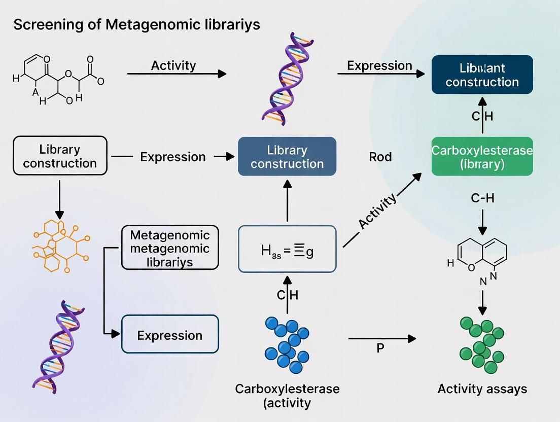
Functional Metagenomic Library Construction & Screening Workflow
Carboxylesterase Activity & Detection Principle
The Scientist's Toolkit
Table 1: Key Research Reagent Solutions for Library Construction & Screening
| Item/Reagent | Function/Benefit |
|---|---|
| Lysis Buffer A (100 mM Tris-HCl, 100 mM EDTA, 1.5 M NaCl, 1% CTAB) | Efficient disruption of diverse environmental cells and inhibition of nucleases. |
| GELase Enzyme | Purifies large DNA fragments from agarose without shearing; critical for HMW eDNA. |
| CopyControl pCC2FOS Vector | Fosmid vector with inducible copy number; increases DNA yield for sequencing & expression. |
| EPI300-T1R E. coli Strain | Optimized host for fosmid propagation; T1 phage resistance improves library stability. |
| MaxPlax Lambda Packaging Extracts | High-efficiency, in vitro packaging system for fosmid transduction into E. coli. |
| α-Naphthyl Acetate | Chromogenic esterase substrate; hydrolysis yields α-naphthol for colorimetric detection. |
| Fast Blue RR Salt | Diazonium salt dye coupler; reacts with α-naphthol to form an insoluble, colored azo dye. |
| LB Freezing Medium (LB broth + 36 mM K₂HPO₄, 13.2 mM KH₂PO₄, 1.7 mM citrate, 0.4 mM MgSO₄, 6.8 mM (NH₄)₂SO₄, 4.4% v/v glycerol) | Long-term storage of library clones at -80°C without significant loss of viability. |
Application Notes
Within a thesis focused on activity-based screening of metagenomic libraries for novel carboxylesterases, the construction of high-quality, large-insert libraries is paramount. This protocol outlines core principles for converting complex environmental samples into fosmid/cosmid libraries, maximizing the probability of capturing large, intact operons and novel enzyme-encoding genes. The use of fosmid (pCC1FOS or equivalent) or cosmid vectors, which accommodate ~30-45 kb inserts, reduces the number of clones needed for sufficient coverage and maintains gene cluster integrity. Success hinges on obtaining high-molecular-weight (HMW), pure environmental DNA (eDNA), its careful size selection, and efficient packaging/cloning to create a stable, representative resource for downstream functional screening in E. coli.
Key Quantitative Benchmarks
Table 1: Critical Quantitative Benchmarks for Library Construction
| Parameter | Target Specification | Rationale |
|---|---|---|
| eDNA Purity (A260/A280) | 1.8 - 2.0 | Indicates protein contamination if outside range. |
| eDNA Size Pre-Repair | >100 kb, ideally >200 kb | Essential for efficient end-repair and large-insert cloning. |
| Size-Selected DNA Fragments | 35 - 50 kb (for fosmids) | Matches vector capacity and optimizes packaging efficiency. |
| Vector:Insert Molar Ratio | 1:1 to 1:3 | Critical for minimizing empty vector or concatemer background. |
| Packaging Efficiency (Test) | >10^8 pfu/µg control DNA | Validates commercial packaging extract performance. |
| Primary Library Titer | >10^5 CFU/mL | Ensures sufficient clone diversity for screening. |
| Average Insert Size (QC) | >35 kb | Confirms successful cloning of large fragments. |
| Library Representation | >1 Gb of metagenomic DNA | Reduces screening bias against rare genes. |
Detailed Protocols
Protocol 1: HMW Environmental DNA Extraction from Soil/Sediment
Principle: Gently lyse microbial cells while minimizing physical shearing and co-extraction of humic acids that inhibit downstream enzymes.
Reagents: PowerSoil Pro Kit (QIAGEN) or modified CTAB-based buffers, Polyvinylpolypyrrolidone (PVPP), Sodium Phosphate Buffer (120 mM, pH 8.0), SDS Lysis Buffer (2% SDS, 200 mM NaCl, 100 mM Tris-HCl pH 8.0), Precipitation Solution (3M sodium acetate, pH 5.2), Isopropanol, 70% Ethanol, TE Buffer.
Procedure:
- Homogenization: Suspend 5 g of soil/sediment in 10 mL Sodium Phosphate Buffer and 2 g PVPP. Vortex vigorously for 10 minutes.
- Centrifugation: Centrifuge at 700 x g for 5 min at 4°C. Transfer supernatant to a new tube.
- Secondary Clearing: Centrifuge supernatant at 10,000 x g for 15 min at 4°C. Collect supernatant.
- Concentration: Concentrate microbial cells by centrifuging at 15,000 x g for 30 min at 4°C. Discard supernatant.
- Chemical Lysis: Resuspend pellet in 1 mL SDS Lysis Buffer. Incubate at 65°C for 1-2 hours with gentle inversion every 15 min.
- Precipitation: Add 1/10 volume Precipitation Solution, mix, then add 0.7 volumes isopropanol. Mix by gentle inversion. Spool out DNA using a sterile glass rod or hook.
- Wash & Elute: Wash DNA hook in 70% ethanol, air-dry briefly, and dissolve in 100 µL TE Buffer overnight at 4°C.
- QC: Analyze 2 µL by pulsed-field gel electrophoresis (PFGE) or on a 0.6% agarose gel alongside a lambda ladder to confirm size >100 kb. Measure purity via nanodrop.
Protocol 2: End-Repair and Size Selection of eDNA
Principle: Blunt-end fragmented HMW DNA and selectively isolate fragments in the 35-50 kb range.
Reagents: NEBNext Ultra II FS DNA Module, Agarose (Low Melt), GELase Enzyme, Size Selection Buffer (20 mM Tris-HCl, 1 mM EDTA, pH 8.0).
Procedure:
- End-Repair: Set up reaction with 1-5 µg HMW eDNA using NEBNext End Repair enzyme mix per manufacturer's instructions. Incubate at 20°C for 30 min, then 65°C for 30 min.
- Agarose Embed: Cast the entire reaction in a 1% low-melt agarose plug. Solidify at 4°C for 30 min.
- Size Fractionation: Perform PFGE (6 V/cm, 14°C, 120° inclusion angle, 5-50 s switch time, 16 hours) alongside a PFGE marker (e.g., BioRad's CHEF DNA Size Standard).
- Excise & Digest Agarose: Excise gel slice corresponding to 35-50 kb. Melt slice at 68°C, then treat with GELase per instructions to digest agarose.
- Concentration: Concentrate DNA using isopropanol precipitation. Resuspend in 10-20 µL TE buffer.
Protocol 3: Fosmid Vector Ligation, Packaging, and Library Assembly
Principle: Ligate size-selected eDNA into a linearized, dephosphorylated fosmid vector, package into phage particles in vitro, and transfer into E. coli for propagation.
Reagents: CopyControl Fosmid Library Production Kit (Lucigen), T4 DNA Ligase, ATP, PEG 8000, E. coli EPI300 plating strain, LB Agar with Chloramphenicol (12.5 µg/mL).
Procedure:
- Ligation: In a 10 µL reaction, combine 100 ng pCC1FOS vector, a 1:2 molar ratio of size-selected insert, 1X T4 DNA Ligase buffer, and 5 Weiss units T4 DNA Ligase. Incubate at 16°C overnight.
- Packaging: Add the entire ligation to a tube of MaxPlax Lambda Packaging Extracts. Follow kit protocol (typically 90 min incubation at 30°C).
- Phage Infection: Add packaged mix to 1 mL of EPI300 culture (OD600 ~0.5-0.8). Incubate at 37°C for 45 min with gentle shaking.
- Titering & Library Assembly: Plate serial dilutions on LB/Chloramphenicol plates to determine titer (CFU/mL). Plate the remaining infection mixture on large, square bioassay dishes to generate the primary library (aim for >50,000 colonies).
- Harvesting: Scrape colonies into 10 mL LB/15% glycerol using a sterile spreader. Mix thoroughly, aliquot, and store at -80°C as the library stock.
Visualizations
Diagram 1: Fosmid Library Construction Workflow
Diagram 2: Screening Logic for Carboxylesterase Thesis
The Scientist's Toolkit
Table 2: Essential Research Reagent Solutions
| Item | Function & Relevance |
|---|---|
| PowerSoil Pro Kit | Optimized for inhibitor removal; critical for obtaining PCR & enzyme-inhibitor-free HMW eDNA. |
| Polyvinylpolypyrrolidone (PVPP) | Binds polyphenolic compounds (humics) during extraction, improving DNA purity. |
| Pulsed-Field Gel Electrophoresis (PFGE) System | Essential for accurate size verification of HMW eDNA (>100 kb) and precise size selection. |
| CopyControl pCC1FOS Vector | Fosmid vector with inducible high-copy number replication; enables easy DNA isolation for sequencing or subcloning. |
| MaxPlax Lambda Packaging Extracts | High-efficiency in vitro packaging system for converting ligated fosmids into infectious particles. |
| EPI300 E. coli Strain | T7 RNAP-deficient, recA- end-host for stable fosmid maintenance; allows induction for higher copy number. |
| Tributyrin Agar Plates | Primary activity screen substrate; carboxylesterase activity produces clear halos around expressing colonies. |
| NEBNext Ultra II FS Module | Provides optimized enzymes for blunt-ending and polishing sheared DNA ends for efficient ligation. |
| GELase Enzyme | Digests agarose in the presence of EDTA, allowing recovery of size-selected DNA without damage or inhibition. |
This application note supports the thesis that environmental niche selection is a primary determinant of success in activity-based screening of metagenomic libraries for novel carboxylesterases. The following table summarizes key quantitative and qualitative characteristics of four high-potential niches.
Table 1: Comparative Analysis of Environmental Niches for Carboxylesterase Discovery
| Niche Parameter | Soil | Marine | Gut (Mammalian) | Extreme (e.g., Hot Spring) |
|---|---|---|---|---|
| Estimated Microbial Richness | 10^8 - 10^9 species per gram | 10^5 - 10^6 cells per mL | 10^10 - 10^11 cells per gram (colon) | Variable, often low diversity |
| Dominant Phyla | Proteobacteria, Actinobacteria, Acidobacteria | Proteobacteria, Bacteroidetes, Cyanobacteria | Bacteroidetes, Firmicutes | Crenarchaeota, Aquificae, Thermotogae |
| Key Selection Pressure | Complex polymer degradation, toxin deactivation | High salinity/pressure, oligotrophy | Bile salts, host-derived glycans, low oxygen | High temperature (>80°C), extreme pH, high salinity |
| Predicted Esterase Adaptations | Broad substrate range, humic acid tolerance | Halotolerance, cold-activity, pressure stability | Bile salt resistance, mucin degradation | Thermostability, pH stability, solvent tolerance |
| Avg. DNA Yield (per g/mL sample) | 1-10 µg (high humics) | 0.1-1 µg | 5-20 µg (inhibitor-sensitive) | 0.01-1 µg |
| Metagenomic Library Complexity | Very High | High | High, host DNA contamination | Moderate, highly novel sequences |
| Primary Screening Challenge | Inhibitor (humic acid) removal | Biomass concentration, salt removal | Host DNA depletion, anaerobic handling | Biased lysis, DNA fragmentation |
Detailed Protocols for Niche-Specific Metagenomic Library Construction
Protocol 2.1: Marine Biomass Concentration and DNA Extraction (Modified from ISC Protocol) Objective: Obtain high-molecular-weight DNA from planktonic microbial communities.
- Sample: Collect 1-10L seawater via Niskin bottle. Pre-filter through 3µm membrane to remove eukaryotes.
- Concentration: Filter remaining water through a 0.22µm polyethersulfone (PES) membrane under gentle vacuum (<5 psi).
- Cell Lysis: Cut membrane into strips, place in 2mL tube with 0.5mL lysis buffer (500mM NaCl, 50mM Tris-HCl pH 8.0, 50mM EDTA, 1% SDS). Add 0.5mg/mL Proteinase K. Incubate at 56°C for 2h with rotation.
- Humic/Salt Removal: Add an equal volume of chloroform:isoamyl alcohol (24:1), mix, centrifuge. Transfer aqueous phase to a new tube. Add 0.1x volume of 7.5M ammonium acetate and 0.7x volume of isopropanol. Incubate at -20°C for 1h.
- DNA Precipitation: Pellet DNA at 16,000 x g for 30 min at 4°C. Wash pellet twice with cold 70% ethanol.
- Final Resuspension: Air-dry pellet and resuspend in 50µL TE buffer (10mM Tris, 0.1mM EDTA, pH 8.0). Assess quality via Nanodrop (260/280 ~1.8) and gel electrophoresis.
Protocol 2.2: Functional Screening for Carboxylesterase Activity in Fosmid Libraries Objective: Identify clones expressing esterase activity on indicator plates.
- Library Construction: Use the CopyControl Fosmid Library Production Kit with size-selected (~40kb) environmental DNA.
- Host Strain: Transform into E. coli EPI300-T1R.
- Activity Screening: Plate transformed cells on LB agar + CopyControl Induction Solution + 12.5µg/mL chloramphenicol + 1% tributyrin (or 0.01% α-naphthyl acetate with Fast Blue RR). Incubate at 37°C for 48h.
- Positive Clone Identification: Tributyrin plates: look for clear halo zones around colonies. α-Naphthyl acetate plates: look for brown/red precipitate formation.
- Secondary Screening: Pick positive clones, re-streak for purity, and assay with chromogenic p-nitrophenyl esters (pNPC2-C16) in microtiter plates to determine substrate specificity range.
Visualizing the Screening Workflow and Enzyme Discovery Logic
Title: Activity-Based Screening Workflow for Metagenomic Esterases
Title: Logic of Niche-Driven Enzyme Discovery
The Scientist's Toolkit: Key Research Reagent Solutions
Table 2: Essential Reagents for Metagenomic Carboxylesterase Screening
| Reagent/Material | Supplier Examples | Function in Protocol |
|---|---|---|
| CopyControl Fosmid Library Kit | Lucigen | Construction of large-insert (40kb) metagenomic libraries with inducible copy number for improved expression. |
| Tributyrin (Glyceryl tributyrate) | Sigma-Aldrich | Lipid substrate for primary esterase screening. Hydrolysis produces a clear halo in opaque agar. |
| p-Nitrophenyl ester series (C2-C16) | Sigma-Aldrich, Cayman Chemical | Chromogenic substrates for quantitative, spectrophotometric determination of esterase substrate specificity and kinetics. |
| Fast Blue RR Salt | Thermo Fisher | Coupling agent used with α/β-naphthyl acetate substrates to form an insoluble colored precipitate for plate screening. |
| Polyethersulfone (PES) Membrane Filters (0.22µm) | Millipore | For gentle concentration of microbial cells from marine or aqueous samples with minimal DNA binding. |
| Humic Acid Removal Solution (e.g., PVPP) | Sigma-Aldrich | Binds polyphenolic compounds (humics) from soil extracts that inhibit downstream enzymatic reactions. |
| EPI300-T1R E. coli Strain | Lucigen | Optimized fosmid host with high transformation efficiency, inducible copy control, and reduced recombinant bias. |
| Proteinase K, Molecular Biology Grade | Roche, Qiagen | Broad-spectrum serine protease for efficient digestion of proteins during cell lysis, freeing DNA. |
Within the broader thesis on activity screening of metagenomic libraries for novel carboxylesterases, bioinformatic pre-screening is a critical first step. Carboxylesterases (CEs; EC 3.1.1.1) are key enzymes in drug metabolism and synthesis. Direct functional screening of vast metagenomic libraries is resource-intensive. Targeting conserved sequence motifs (e.g., the catalytic triad GXSXG, HGGG, and GX sequences) through PCR and probe-based methods enables the enrichment of clones harboring putative esterase genes before expression screening, dramatically increasing hit rates and efficiency.
Key Conserved Motifs in Carboxylesterases
Carboxylesterases share conserved motifs critical for catalysis and structural integrity. The table below summarizes the primary motifs targeted for primer/probe design.
Table 1: Conserved Motifs in α/β-Hydrolase Fold Carboxylesterases
| Motif Name | Consensus Sequence (Amino Acids) | Functional Role | Variability in Metagenomes |
|---|---|---|---|
| Catalytic Nucleophile | G-E-S/A-G (GXSXG) | Contains the nucleophilic serine residue. Highly conserved. | Moderate (2nd & 4th positions). |
| Oxyanion Hole | H-G-G-G | Stabilizes the tetrahedral transition state. | Low (Gly residues highly conserved). |
| Catalytic Acid | G-X (typically downstream) | Often contains the catalytic glutamate/aspartate. | High. Requires degenerate design. |
| N-terminal Nucleophile Cap | Sm-X-Nu (where Nu is nucleophile) | Structural motif capping the nucleophile. | Moderate. |
Experimental Protocol: Primer Design and Enrichment PCR
Protocol 3.1: Degenerate Primer Design from Aligned Motifs
Objective: To design degenerate primers amplifying internal fragments of putative carboxylesterase genes from metagenomic DNA.
Materials & Reagents:
- Multiple sequence alignment (MSA) of known carboxylesterases (e.g., from Pfam family PF00135).
- Metagenomic DNA extract (environmental sample).
- Primer design software (e.g., Primer3, CODEHOP).
Methodology:
- Generate MSA: Curate a reference set of diverse carboxylesterase protein sequences from public databases (NCBI, UniProt). Perform alignment using Clustal Omega or MUSCLE.
- Identify Conserved Blocks: Visually or algorithmically identify blocks containing the GXSXG and HGGG motifs and their flanking 5-10 amino acids.
- Translate to Nucleotide: Back-translate the amino acid blocks to nucleotide sequence, considering codon usage bias from the source environment (if known).
- Assign Degeneracy: Introduce degenerate bases (IUPAC codes) at variable codon positions. Critical: Limit total degeneracy to <1024-fold to maintain PCR efficiency.
- Design Primer Pairs: Design forward primer from region upstream of GXSXG. Design reverse primer from region downstream of HGGG, ensuring amplicon size of 300-700 bp.
- Add Adapters: Add defined 5' sequences (e.g., for subsequent TA-cloning or sequencing) to the degenerate cores.
Example Primer Sequences (Theoretical):
- CE_Fwd: 5'-TAATACGACTCACTATAGGG-GCIWSITGYGGIWSITTYGG-3' (T7 promoter + GXSXG region)
- CE_Rev: 5'-CAGCTATGACCATG-CCRTAIARTCICCRCA-3' (Adapter + HGGG region)
Protocol 3.2: Touchdown PCR for Metagenomic Amplification
Objective: To amplify target fragments from complex metagenomic DNA using degenerate primers.
PCR Master Mix:
| Component | Volume (50 µL rxn) | Final Concentration |
|---|---|---|
| Metagenomic DNA (10-100 ng) | 2 µL | Variable |
| 10X High-Fidelity Buffer | 5 µL | 1X |
| dNTP Mix (10 mM each) | 1 µL | 200 µM each |
| Degenerate Forward Primer (10 µM) | 2.5 µL | 0.5 µM |
| Degenerate Reverse Primer (10 µM) | 2.5 µL | 0.5 µM |
| High-Fidelity DNA Polymerase | 0.5 µL | 1-2.5 U |
| Nuclease-free H₂O | to 50 µL | - |
Thermocycling Profile (Touchdown):
- Initial Denaturation: 98°C for 30 sec.
- 10x Cycles: Denature 98°C, 10 sec; Anneal 65-55°C (-1°C/cycle), 20 sec; Extend 72°C, 45 sec/kb.
- 25x Cycles: Denature 98°C, 10 sec; Anneal 55°C, 20 sec; Extend 72°C, 45 sec/kb.
- Final Extension: 72°C, 2 min.
- Hold: 4°C.
Analysis: Purify PCR product, clone into a vector, and sequence individual clones. Perform BLASTX analysis to confirm hits are related to carboxylesterases.
The Scientist's Toolkit: Research Reagent Solutions
Table 2: Essential Reagents for Bioinformatic Pre-Screening of Carboxylesterases
| Item / Reagent | Function / Role | Example Product / Note |
|---|---|---|
| High-Fidelity DNA Polymerase | Accurate amplification from complex templates with low error rate. | Phusion U Green, KAPA HiFi. |
| Degenerate Primer Mix | Synthesized oligonucleotide pool to target variable motif sequences. | HPLC-purified, resuspended in TE buffer. |
| Metagenomic DNA Kit | Isolation of high-molecular-weight, inhibitor-free DNA from soil/water/gut samples. | DNeasy PowerSoil Pro Kit. |
| Gel Extraction Kit | Purification of correctly sized amplicons from agarose gels. | Zymoclean Gel DNA Recovery Kit. |
| TA/Blunt-End Cloning Kit | Ligation of degenerate PCR products into sequencing vector. | pGEM-T Easy Vector System. |
| Sanger Sequencing Service | Verification of insert sequence and identity. | Mix of vector and insert-specific primers. |
| Multiple Sequence Alignment Tool | Identification of conserved blocks for primer design. | Clustal Omega, MEGA XI. |
| CODEHOP Software | Hybrid primer design combining conserved cores with degenerate 3' ends. | Public web tool or standalone. |
Workflow Visualization
Diagram 1: Workflow for primer/probe-based pre-screening of metagenomic libraries.
Data Integration and Validation
Table 3: Expected Outcomes from a Typical Pre-Screening Experiment
| Metric | Untreated Metagenomic Library | Library Post-Motif Enrichment | Notes |
|---|---|---|---|
| Clones to Screen | 10⁵ - 10⁶ | 10³ - 10⁴ | Drastic reduction in screening burden. |
| Putative Esterase Hit Rate | 0.01% - 0.1% | 5% - 20% | Based on published studies. |
| Amplicon Diversity (OTUs) | N/A | 10 - 50 unique sequences | Indicative of novelty captured. |
| Time to First Validated Hit | Weeks - Months | 1 - 2 Weeks | Includes sequencing and validation time. |
Advanced Protocol: Probe Design for Hybridization Screening
Protocol 7.1: Design of Conserved Motif Probes for Array/Membrane Screening
Objective: To design and apply labeled oligonucleotide probes for colony or plaque hybridization to identify carboxylesterase-positive clones.
- Probe Design: From the MSA, select the most conserved 18-25 nucleotide region spanning part of the GXSXG motif. Avoid high degeneracy (>32-fold). Synthesize as a mixture or use inosine at highly variable positions.
- Probe Labeling: Label probe 5' end with digoxigenin (DIG) or biotin using terminal transferase.
- Library Plating: Plate metagenomic fosmid/cosmid library at medium density (~3000 colonies per membrane).
- Hybridization: Use low-stringency hybridization (Tm -20°C) overnight, followed by medium-stringency washes.
- Detection: Use chemiluminescent (for DIG) or colorimetric detection to identify positive signals. Isplicate corresponding clones for downstream analysis.
This bioinformatic pre-screening pipeline, embedded within the carboxylesterase discovery thesis, creates a focused, sequence-informed sub-library, ensuring downstream functional screening efforts are concentrated on the most promising genetic material.
High-Throughput Screening in Action: Step-by-Step Protocols for Activity-Based Discovery
Within the context of activity screening of metagenomic libraries for carboxylesterase (CE) research, substrate selection is a critical first step. Carboxylesterases (EC 3.1.1.1) hydrolyze ester bonds, and their activity can be detected using specific chromogenic or fluorogenic ester substrates. This guide details the properties, applications, and protocols for these substrates, enabling researchers to efficiently identify novel esterases from complex environmental DNA libraries.
Substrate Characteristics and Quantitative Comparison
Table 1: Comparison of Common Chromogenic and Fluorogenic Ester Substrates
| Substrate Name | Type (Core) | Detection Method | λex / λem (nm) | Product (Color/Fluorescence) | Relative Sensitivity | Typical Working Conc. | Key Advantage for Metagenomics |
|---|---|---|---|---|---|---|---|
| α-Naphthyl acetate | Chromogenic | Colorimetric (Azo-dye coupling) | N/A | Red-purple precipitate | Moderate | 0.1 - 1.0 mM | Low cost, direct visual screening on plates. |
| β-Naphthyl acetate | Chromogenic | Colorimetric (Azo-dye coupling) | N/A | Red precipitate | Moderate | 0.1 - 1.0 mM | Faster coupling reaction than α-naphthyl. |
| p-Nitrophenyl acetate (pNPA) | Chromogenic | Direct spectrophotometric | 405 (product) | Yellow (p-nitrophenolate) | Good | 0.05 - 2.0 mM | Quantitative, continuous assay; no coupling step. |
| 4-Methylumbelliferyl acetate (4-MUA) | Fluorogenic | Fluorescence | 355 / 460 | Blue fluorescence (4-MU) | High | 10 - 200 µM | Very high sensitivity for low-activity clones. |
| Fluorescein diacetate (FDA) | Fluorogenic | Fluorescence | 490 / 514 | Green fluorescence (fluorescein) | High | 10 - 100 µM | Cell-permeable;可用于活细胞/viability staining in situ. |
| Resorufin acetate | Fluorogenic | Fluorescence (or colorimetric) | 571 / 585 | Pink/red fluorescence (resorufin) | Very High | 5 - 50 µM | Extremely sensitive; dual detection modes. |
Research Reagent Solutions Toolkit
Table 2: Essential Materials for Esterase Activity Screening
| Item | Function in Screening | Example/Notes |
|---|---|---|
| Agar plates with substrate | Primary library screening | LB-agar with 0.1-0.5 mM chromogenic ester (e.g., α-naphthyl acetate). |
| Fast Blue RR / Fast Red TR Salt | Diazo coupling reagent for naphthyl esters | Forms insoluble azo dye with released α/β-naphthol. Fast Red TR gives red precipitate. |
| Lysis Buffer (e.g., BugBuster) | Cell lysis for intracellular enzyme assays | For screening lysates from E. coli expression clones. |
| Assay Buffer (Tris or Phosphate, pH 7.0-8.5) | Optimal enzymatic activity | Carboxylesterases typically have neutral to alkaline pH optima. |
| Positive Control Enzyme (e.g., Porcine liver esterase) | Assay validation and standardization | Verifies substrate performance and assay conditions. |
| Microplate Reader (UV-Vis & Fluorescence) | High-throughput quantitative screening | Essential for kinetic analysis of lysates from 96/384-well formats. |
| Overlay Agar Solution (soft agar with substrate) | In situ activity detection on plates | 0.7% agar containing substrate and coupling agent poured over grown colonies. |
Detailed Experimental Protocols
Protocol 1: Primary Plate Screening with α/β-Naphthyl Acetate
Objective: Visually identify esterase-active clones from a metagenomic library plated on agar.
Materials:
- LB-agar plates with appropriate antibiotic (e.g., chloramphenicol for fosmid libraries).
- α- or β-Naphthyl acetate stock solution (100 mM in acetone or DMSO).
- Fast Blue RR Salt stock solution (20 mg/mL in DMSO, prepared fresh).
- 0.1 M Sodium Phosphate Buffer, pH 7.0.
Procedure:
- Plate Library: Spread or replica-plate metagenomic library clones onto LB-agar plates. Incubate at 37°C until colonies are 0.5-1.0 mm in diameter (16-24 h).
- Prepare Overlay: Gently warm 10 mL of 0.7% agarose in 0.1 M phosphate buffer (pH 7.0). Cool to ~50°C.
- Add Substrate & Dye: Quickly add α/β-naphthyl acetate (from stock) to a final concentration of 0.2 mM and Fast Blue RR to 0.1 mg/mL to the melted agarose. Mix gently to avoid bubbles.
- Overlay Plates: Pour the mixture evenly over the surface of the plate containing grown colonies. Allow to solidify (5-10 min).
- Incubate & Score: Incubate the plates at room temperature or 37°C. Positive colonies will be surrounded by a red-purple (α-naphthyl) or red (β-naphthyl) halo within minutes to a few hours.
- Pick Positives: Mark and pick active colonies for further cultivation and secondary screening.
Protocol 2: Quantitative Microplate Assay with Fluorogenic Substrate (4-MUA)
Objective: Quantitatively measure esterase activity in cell lysates from hit clones in a 96-well format.
Materials:
- Clonal cultures in 96-deep well blocks.
- Lysis buffer (e.g., 50 mM Tris-HCl pH 8.0, 0.1% Triton X-100, 1 mg/mL lysozyme).
- Assay buffer: 50 mM Tris-HCl, pH 8.0, 150 mM NaCl, 0.1% BSA.
- 4-Methylumbelliferyl acetate (4-MUA) stock: 10 mM in DMSO (store at -20°C, protect from light).
- Fluorescence microplate reader.
Procedure:
- Prepare Lysates: Grow hits in 1 mL LB medium for 24-48 h. Pellet cells by centrifugation (3000 x g, 10 min). Resuspend in 200 µL lysis buffer. Incubate 30 min on ice with occasional shaking. Clarify by centrifugation (4000 x g, 20 min, 4°C). Transfer supernatant (crude lysate) to a new plate.
- Prepare Assay Plate: In a black 96-well plate, add 80 µL of assay buffer per well.
- Initiate Reaction: Add 10 µL of clarified lysate (or assay buffer for blank) to each well. Start the reaction by adding 10 µL of 4-MUA stock diluted in assay buffer to a final well concentration of 100 µM (final volume = 100 µL).
- Measure Kinetics: Immediately place the plate in a pre-warmed (30°C) microplate reader. Measure fluorescence (λex = 355 nm, λem = 460 nm) every 30-60 seconds for 10-20 minutes.
- Analyze Data: Subtract the blank rate. Calculate activity from the linear portion of the progress curve using a 4-methylumbelliferone standard curve (0-10 µM in assay buffer).
Visualization of Screening Workflows
Primary & Secondary Screening Workflow
Esterase Activity Detection Principle
Within the context of a thesis focused on activity-based screening of metagenomic libraries for novel carboxylesterases, the selection of an initial primary screening method is critical. This protocol compares two established plate-based methods: the Agar Overlay Assay and the Liquid Culture Microplate Screening method. Both are designed for high-throughput functional screening of clone libraries using chromogenic or fluorogenic ester substrates (e.g., α- or β-naphthyl acetate). The choice impacts throughput, sensitivity, false positive/negative rates, and subsequent recovery of active clones.
Table 1: Quantitative Comparison of Screening Methods
| Parameter | Agar Overlay Assay | Liquid Culture Microplate Screening |
|---|---|---|
| Throughput | Moderate (colony picking required) | High (direct culture in microplate) |
| Time to Result | 24-48 hours (including colony growth) | 6-24 hours (from pre-grown culture) |
| Sensitivity | Lower (substrate diffusion barrier) | Higher (homogeneous substrate mixing) |
| Quantification | Semi-quantitative (zone size) | Quantitative (kinetic fluorescence/absorbance) |
| Clone Recovery | Direct from master plate | Requires replica plating or glycerol stock |
| Reagent Cost per Clone | Low | Moderate to High |
| False Positives | Low (activity is visually confirmed) | Moderate (can arise from cell lysis) |
| Primary Readout | Insoluble colored precipitate (halo) | Soluble fluorescent/colored product |
Table 2: Key Decision Factors for Method Selection
| Research Goal | Recommended Method | Rationale |
|---|---|---|
| Initial library sweep (>10^4 clones) | Agar Overlay | Cost-effective, simple, preserves spatial clone mapping. |
| Quantitative activity ranking | Liquid Culture | Enables kinetic measurements and dose-response. |
| Screening for thermostability/pH optimum | Liquid Culture | Easy control of assay conditions in liquid phase. |
| Detection of weak esterase activity | Liquid Culture | Superior sensitivity due to lack of diffusion limit. |
| Minimal equipment availability | Agar Overlay | Requires only plates, substrate, and incubator. |
Detailed Protocols
Protocol 1: Agar Overlay Assay for Carboxylesterase Activity
Principle: Colonies expressing esterase activity hydrolyze a chromogenic substrate (e.g., α-naphthyl acetate) within an agar overlay. The released α-naphthyl couples with a diazo dye (e.g., Fast Blue RR salt) to form an insoluble, colored precipitate around active colonies.
Materials (Research Reagent Toolkit):
- LB Agar Plates with Inducer: Contains antibiotic and IPTG for gene expression from metagenomic inserts in vector (e.g., pET, pBAD).
- Soft Agar Overlay: 0.7-1% agar in suitable buffer (e.g., 50 mM Tris-HCl, pH 7.5).
- Ester Substrate Stock: 50 mM α-naphthyl acetate in acetone or DMSO.
- Coupling Dye Stock: 30 mg/mL Fast Blue RR salt in DMSO (prepare fresh).
- Assay Buffer: 50 mM phosphate buffer, pH 7.0, with 0.1% Triton X-100.
Procedure:
- Library Plating: Plate metagenomic library clones on LB agar containing appropriate antibiotic and induce with IPTG (or appropriate inducer) until pinpoint colonies appear (12-16 h, 30°C).
- Overlay Preparation: Melt soft agar and cool to 55°C. Quickly add α-naphthyl acetate (final 0.1 mM) and Fast Blue RR salt (final 0.2 mg/mL). Mix gently to avoid bubbles.
- Assay: Pour the mixture evenly over the colonies to form a thin overlay (~5 mL per 90 mm plate). Swirl gently for even distribution.
- Incubation & Detection: Incubate plates at the desired screening temperature (e.g., 30°C or 37°C). Positive clones are identified by the formation of a reddish-brown halo within 10 minutes to 2 hours.
- Clone Recovery: Mark positive colonies on the back of the plate. Pick corresponding clones from the master plate for secondary screening and sequencing.
Protocol 2: Liquid Culture Microplate Screening for Esterase Activity
Principle: Clones are grown in liquid culture in 96- or 384-well microplates. Esterase activity is measured in a homogeneous assay by adding a fluorogenic substrate (e.g., 4-methylumbelliferyl acetate). Hydrolysis releases the fluorescent product (4-methylumbelliferone), which is quantified kinetically.
Materials (Research Reagent Toolkit):
- Deep-Well Culture Plates: 96-well 2 mL plates for aerobic culture.
- Rich Media: TB or LB with antibiotic and inducer.
- Lysis Solution: BugBuster Master Mix or Lysozyme (0.2 mg/mL) in assay buffer.
- Fluorogenic Substrate: 10 mM 4-methylumbelliferyl acetate (4-MUA) in DMSO.
- Assay Buffer: 50 mM Tris-HCl, pH 8.0, 150 mM NaCl.
- Microplate Reader: Capable of fluorescence measurement (Ex: 355 nm, Em: 460 nm).
Procedure:
- Inoculation & Growth: Inoculate single clones into deep-well plates containing 1 mL of media with antibiotic. Grow with shaking (900 rpm) at 30°C for 18-24 hours. Induce expression at mid-log phase.
- Cell Harvest & Lysis: Centrifuge plates at 3000 x g for 10 min. Discard supernatant. Resuspend cell pellets in 200 µL of lysis solution per well. Incubate with shaking for 30 min at room temperature.
- Clarification: Centrifuge plates at 4000 x g for 20 min to pellet debris.
- Activity Assay: Transfer 50 µL of supernatant (cell lysate) to a black, clear-bottom 384-well assay plate. Add 150 µL of assay buffer containing 4-MUA (final concentration 100 µM). Mix immediately.
- Measurement: Immediately place plate in a pre-warmed (e.g., 30°C) microplate reader. Measure fluorescence every 30 seconds for 10 minutes. Use wells with empty vector lysates as negative controls.
- Data Analysis: Calculate initial reaction velocities (RFU/min). Clones exhibiting activity >3 standard deviations above the negative control mean are considered positive. Correlate well position with the original culture plate for recovery from parallel glycerol stocks.
Visualizations
Agar Overlay Screening Workflow
Liquid Culture Microplate Screening Workflow
Method Selection Decision Tree
Application Notes: Integrating Automation for Carboxylesterase Screening
The discovery of novel carboxylesterases from metagenomic libraries presents a significant bottleneck: the manual screening of thousands to millions of clones is prohibitively slow and labor-intensive. This document outlines a scalable, automated workflow to increase screening throughput from a typical manual output of ~100-500 clones per day to >10,000 clones per day, while improving data consistency and enabling advanced multiplexed assays.
Key Throughput Metrics:
| Screening Method | Theoretical Max Clones/Day | Key Limiting Factors | Relative Cost (Setup + Consumables) |
|---|---|---|---|
| Manual (96-well) | 500 | Technician fatigue, pipetting error | Low / High |
| Semi-Automated (Liquid Handler) | 5,000 | Plate handling, assay incubation | Medium / Medium |
| Fully Automated (Integrated System) | 50,000+ | Library size, detection sensitivity | High / Low |
Table 1: Comparison of a standard fluorometric p-Nitrophenyl acetate (p-NPA) assay across platforms. Data sourced from current vendor specifications and recent literature (2023-2024).
| Step | Manual (Time/Plate) | Liquid Handler (Time/Plate) | Integrated Robot (Time/Plate) |
|---|---|---|---|
| Colony Picking & Inoculation | 30 min | 10 min | 5 min (from agar) |
| Culture & Induction | (Overnight, hands-off) | (Overnight, hands-off) | (Overnight, fully scheduled) |
| Cell Lysis | 60 min | 20 min | 15 min |
| Assay Setup (p-NPA) | 20 min | 5 min | 3 min |
| Kinetic Read (405 nm) | 90 min (sequential) | 90 min (parallel) | 90 min (parallel, staggered) |
| Data Analysis | Manual transfer | Automated export | Integrated AI/ML analysis |
Detailed Protocols
Protocol 2.1: Automated High-Throughput Screening for p-NPA Hydrolysis
Objective: To robotically screen a metagenomic library for clones expressing carboxylesterase activity using p-NPA as a substrate.
Materials & Reagents:
- Source Plates: 384-well master plates containing metagenomic library clones in LB/glycerol.
- Growth Media: LB broth with appropriate antibiotic (e.g., kanamycin 50 µg/mL).
- Induction Solution: 0.5 mM Isopropyl β-d-1-thiogalactopyranoside (IPTG) in LB.
- Lysis Buffer: BugBuster HT Protein Extraction Reagent supplemented with rLysozyme and Benzonase Nuclease.
- Assay Buffer: 50 mM Tris-HCl, pH 7.5, 150 mM NaCl.
- Substrate Solution: 10 mM p-Nitrophenyl acetate (p-NPA) in acetonitrile, diluted in assay buffer to a final working concentration of 200 µM (<2% ACN).
- Control: Purified positive control carboxylesterase (e.g., porcine liver esterase) and empty vector lysate.
- Equipment: Integrated robotic system (e.g., Hamilton STARlet, Opentrons OT-2), plate hotel, 384-well deep-well culture plates, 384-well assay plates, plate centrifuge, shaking incubator, multimode plate reader capable of kinetic reads at 405 nm.
Procedure:
- Inoculation & Growth:
- Robotically transfer 5 µL from the library source plate into 500 µL of growth media in a 384-well deep-well plate.
- Seal plate with a breathable membrane. Incubate at 37°C, 850 rpm for 16 hours in a shaking incubator integrated with the deck.
- Induction:
- Transfer 20 µL of overnight culture into 380 µL of fresh growth media containing 0.5 mM IPTG in a new deep-well plate.
- Incubate at 25°C, 850 rpm for 6 hours for protein expression.
- Cell Harvest & Lysis:
- Centrifuge the culture plate at 3000×g for 15 minutes. Robotic gripper moves plate to centrifuge and back.
- Aspirate and discard supernatant carefully.
- Add 50 µL of chilled Lysis Buffer to each pellet.
- Seal, shake at 800 rpm for 30 minutes at room temperature.
- Centrifuge at 4000×g for 30 minutes to clarify lysate.
- Automated Assay Assembly:
- Transfer 20 µL of clarified lysate supernatant into a 384-well clear flat-bottom assay plate containing 70 µL of Assay Buffer per well.
- Initiate the reaction by adding 10 µL of the 200 µM p-NPA working solution using the reagent dispenser.
- Immediately transfer the plate to the integrated plate reader.
- Kinetic Measurement:
- Read absorbance at 405 nm every 30 seconds for 10 minutes at 25°C.
- The robotic scheduler pre-warms the next assay plate during this read.
- Data Processing:
- Software (e.g., Genedata Screener) automatically calculates the initial linear rate (V0) for each well.
- Hits are defined as clones where V0 > mean (negative controls) + 5× standard deviation.
Protocol 2.2: Multiplexed Confirmatory Screen with Fluorogenic Substrates
Objective: To validate primary hits using a panel of fluorogenic ester substrates (e.g., 4-Methylumbelliferyl acetate, Fluorescein diacetate) for substrate specificity profiling.
Procedure:
- Hit Reformation: Positive clones from Protocol 2.1 are automatically re-arrayed from source plates into a new 96-well master plate.
- Culture & Lysate Prep: Repeat steps 1-3 of Protocol 2.1 in 96-well format.
- Multiplexed Assay Setup:
- In a black 384-well assay plate, pre-dispense 5 µL of each fluorogenic substrate (from separate source vials) at 1 mM in DMSO into separate, predefined wells.
- Add 85 µL of Assay Buffer to each well.
- Transfer 10 µL of each hit lysate to the quadplicate wells containing different substrates.
- Measurement: Read fluorescence (Ex/Em ~360/450 nm for 4-MU, ~485/535 nm for fluorescein) kinetically for 15 minutes.
- Analysis: Generate a substrate activity profile for each hit clone.
Visualizations
Automated Primary Screening Workflow
Carboxylesterase Catalytic Mechanism
The Scientist's Toolkit: Key Research Reagent Solutions
| Reagent / Material | Function in Screening | Example Product / Vendor |
|---|---|---|
| BugBuster HT | Gentle, high-throughput detergent for bacterial cell lysis and soluble protein extraction. | MilliporeSigma |
| rLysozyme | Recombinant lysozyme for efficient cell wall degradation in Gram-negative bacteria. | MilliporeSigma |
| Benzonase Nuclease | Reduces lysate viscosity by degrading genomic DNA, improving pipetting accuracy. | MilliporeSigma |
| p-Nitrophenyl acetate (p-NPA) | Chromogenic substrate; hydrolysis releases p-nitrophenol, measurable at 405 nm. | Thermo Fisher Scientific |
| 4-Methylumbelliferyl (4-MU) esters | Fluorogenic substrates; hydrolysis releases highly fluorescent 4-MU. | Tokyo Chemical Industry |
| Fluorescein diacetate (FDA) | Cell-permeable fluorogenic substrate; used for live-cell or lysate-based assays. | Cayman Chemical |
| 384-Well, V-Bottom Deep Well Plates | Ideal for small-scale microbial culture in automated systems. | Agilent |
| Breathable Sealing Membranes | Allows gas exchange during incubation while preventing cross-contamination and evaporation. | Azenta Life Sciences |
| Automated Liquid Handling Tips with Filters | Prevents aerosol contamination of pipette channels when handling biological samples. | Beckman Coulter, Tecan |
In the context of activity-based screening of metagenomic libraries for novel carboxylesterases, the initial identification of positive clones (hits) is fraught with challenges related to signal specificity. Non-enzymatic hydrolysis, chemical instability of substrates, or auto-fluorescence can generate false-positive signals that confound results. This document provides detailed application notes and protocols for robust hit-picking and primary validation to ensure that only clones exhibiting genuine enzymatic activity are advanced.
Core Principles of Signal Specificity
Carboxylesterase activity is commonly detected using chromogenic (e.g., α-naphthyl acetate with Fast Blue RR) or fluorogenic (e.g., 4-methylumbelliferyl acetate) substrates. Specificity is compromised by:
- Abiotic Hydrolysis: Spontaneous substrate breakdown at extreme pH or temperature.
- Promiscuous Esterase Activity: Low-level, non-specific activity from host proteins.
- Background Signal: Library vector or host cell components interfering with detection.
Table 1: Sources of False-Positive Signals in Esterase Screening
| Interferent Source | Typical Signal Increase vs. Negative Control | Mitigation Strategy |
|---|---|---|
| Spontaneous Substrate Hydrolysis (pH 9.0, 24h) | 15-25% | Include substrate-only control wells; use appropriate buffer (e.g., Tris, phosphate). |
| E. coli Host Endogenous Esterases | 10-40% | Use esterase-deficient host strains (e.g., E. coli BL21); include empty vector controls. |
| Auto-fluorescence of Library Media | 5-15% RFU | Centrifuge cells, assay in clear buffer; include cell pellet control. |
| Chemical Quenching/Enhancement | Variable | Normalize signals using an internal standard (e.g., a known esterase). |
Table 2: Primary Validation Assay Comparison
| Validation Assay | Time Required | Specificity Metric (Z'-factor) | Throughput |
|---|---|---|---|
| Replica Plate Activity Staining | 4-6 hours | 0.5 - 0.7 | High (96/384 colonies) |
| Liquid Culture Re-test | 18-24 hours | 0.6 - 0.8 | Medium (48-96 cultures) |
| Inhibitor Sensitivity (PMSF) | 5-6 hours | 0.7 - 0.9 | Medium |
| Alternative Substrate Profiling | 6-8 hours | 0.8 - 0.9 | Low-Medium |
Detailed Experimental Protocols
Protocol 4.1: Primary High-Throughput Plate Screening with Internal Controls
- Objective: Identify initial hits from a metagenomic library expressed in E. coli.
- Materials: See "The Scientist's Toolkit" below.
- Procedure:
- Plate Preparation: Grow library clones in 384-well plates with LB/Amp for 24h at 30°C.
- Induction: Add 0.5 mM IPTG to each well, incubate 4h.
- Assay Setup: Per well, mix 50 µL of cell suspension with 50 µL of assay buffer (100 mM Tris-HCl, pH 8.0) containing 200 µM fluorogenic substrate (4-MU acetate). Critical Controls: Include columns for: (A) Negative Control (empty vector host), (B) Positive Control (known esterase clone), (C) Substrate Blank (buffer + substrate).
- Detection: Measure fluorescence (Ex: 355 nm, Em: 460 nm) at time (T=0) and after 60 min incubation at 25°C (T=60).
- Hit Picking: Calculate ∆RFU (RFUT60 - RFUT0). A hit is defined as ∆RFU > Mean(∆RFUnegative control) + 5*SD(∆RFUnegative control).
Protocol 4.2: Primary Validation via Replica Plating and Activity Staining
- Objective: Visually confirm esterase activity and eliminate positional artifacts.
- Procedure:
- Replica-plate primary hits onto fresh LB/Amp agar plates using a sterile pin tool.
- Grow colonies overnight at 30°C.
- Prepare an agar overlay: Melt 0.8% low-melt agarose in 100 mM phosphate buffer (pH 7.4), cool to 50°C, and add α-naphthyl acetate (1 mg/mL) and Fast Blue RR salt (1 mg/mL). CAUTION: Prepare fresh.
- Pour overlay onto replica plate, swirl to cover.
- Incubate at room temperature. Positive clones produce a brown-red precipitate within 30 minutes. Only consistently positive clones from the original and replica plate are advanced.
Protocol 4.3: Specificity Validation with Serine Hydrolase Inhibitor
- Objective: Confirm the signal originates from a serine hydrolase (most carboxylesterases).
- Procedure:
- For each hit clone, prepare two 1 mL liquid cultures in a 96-deep well block. Induce with IPTG.
- Harvest cells by centrifugation. Resuspend pellets in 200 µL assay buffer.
- Pre-incubation: To well A, add 2 µL of DMSO. To well B, add 2 µL of 100 mM PMSF (Phenylmethylsulfonyl fluoride) in DMSO (final concentration 1 mM).
- Incubate for 30 min at 25°C.
- Add substrate to all wells and measure activity as in Protocol 4.1.
- Analysis: Genuine serine esterase activity is inhibited by >70% in well B compared to well A.
Visualization: Workflows and Pathways
Title: Hit Picking and Primary Validation Workflow
Title: Specificity Challenge: True vs. False Signal Sources
The Scientist's Toolkit: Key Research Reagent Solutions
Table 3: Essential Materials for Hit Picking & Validation
| Item / Reagent | Function & Role in Specificity | Example Product/Catalog |
|---|---|---|
| Fluorogenic Esterase Substrate | High sensitivity detection of hydrolysis activity. Low background is critical. | 4-Methylumbelliferyl acetate (4-MUA), Sigma M0883 |
| Chromogenic Coupling Reagent | For visual activity staining on plates; confirms spatial localization of activity. | Fast Blue RR salt, Sigma F0500 |
| Serine Hydrolase Inhibitor | Specificity validation tool. Confirms enzyme mechanism. | Phenylmethylsulfonyl fluoride (PMSF), Gold Biotechnology P-470 |
| Esterase-Deficient Host Strain | Minimizes background from host enzymes, improving signal-to-noise. | E. coli BL21(DE3) ΔestA (or similar) |
| Positive Control Enzyme | Essential for assay normalization and quality control (Z' calculation). | Porcine liver carboxylesterase, Sigma E2884 |
| Low-Melt Agarose | For replica plate activity overlays, allowing substrate diffusion. | Thermo Scientific 16520100 |
| 384-Well Assay Plates (Black, Clear Bottom) | Optimized for fluorescence readings with minimal cross-talk. | Corning 3710 |
| Automated Liquid Handler | Ensures precision and reproducibility in primary screening. | Beckman Coulter Biomek i5 |
This application note details the downstream protocols following a primary activity screen of a metagenomic fosmid or BAC library for carboxylesterase activity (e.g., using α-naphthyl acetate with Fast Blue RR staining). The broader thesis research aims to discover novel microbial carboxylesterases for applications in biocatalysis and prodrug activation. Once a positive clone (forming a colored halo) is identified on an agar plate, the subsequent critical steps are its precise isolation, sequencing of the insert DNA, and bioinformatic identification of the putative esterase gene.
Experimental Protocols
Protocol 2.1: Isolation of Positive Clone from Screening Plates
Objective: To obtain a pure, monoclonal culture of the E. coli host containing the metagenomic insert responsible for the observed hydrolase activity.
Materials:
- Positive agar plate from primary screen.
- Sterile 96-well microtiter plates containing 150 µL of LB with appropriate antibiotic (e.g., chloramphenicol 12.5 µg/mL) per well.
- Sterile glass cloning cylinders or pipette tips.
- Multichannel pipette.
- LB agar plates with antibiotic.
- 30% glycerol solution.
Method:
- Place a sterile cloning cylinder around the positive colony/halo on the screening plate.
- Add 50 µL of sterile LB medium into the cylinder and gently resuspend the cells using a pipette.
- Transfer the cell suspension to one well of the 96-well microtiter plate containing antibiotic broth. This is the "master stock."
- Using a 96-pin replicator or a multichannel pipette, spot 1-2 µL of the master stock onto a fresh LB-antibiotic agar plate to create a monoclonal patch. Also, inoculate a separate liquid culture (5 mL LB with antibiotic) for DNA isolation.
- Incubate the patch plate and liquid culture at 37°C for 12-16 hours.
- Confirm activity by applying the original activity assay (e.g., overlay with agar containing α-naphthyl acetate and Fast Blue RR) to the patch plate.
- Once activity is confirmed, add 50 µL of 30% glycerol to the master stock well and mix. Store at -80°C as the archival glycerol stock.
Protocol 2.2: Fosmid DNA Extraction and Restriction Analysis
Objective: To purify the metagenomic fosmid DNA and perform a fingerprint analysis to confirm insert size and uniqueness.
Materials:
- Overnight liquid culture of positive clone.
- Commercial Fosmid or Large-Construct DNA Purification Kit.
- Restriction enzyme HindIII (or similar rare cutter).
- Agarose gel electrophoresis equipment.
- Pulse-field gel electrophoresis system (for inserts >40 kbp).
Method:
- Isolate fosmid DNA from 5 mL of overnight culture using the purification kit according to the manufacturer's instructions. Elute in 50 µL of nuclease-free water.
- Measure DNA concentration via spectrophotometry (e.g., Nanodrop). Expected yield: 0.5-2 µg.
- Set up a restriction digest: 1 µg fosmid DNA, 10 U HindIII, 1x reaction buffer, in 20 µL total volume. Incubate at 37°C for 2 hours.
- Run the digested product alongside a high-molecular-weight DNA ladder on a 1% agarose gel at 4-6 V/cm for 2-3 hours. For very large inserts (>40 kbp), use a pulse-field gel system (e.g., CHEF) with appropriate parameters.
- Visualize the banding pattern. This "fingerprint" confirms insert presence (~40 kbp average) and helps identify unique clones before costly sequencing.
Protocol 2.3: Sequencing Strategy: From End-Reading to Shotgun Sequencing
Objective: To determine the complete nucleotide sequence of the metagenomic insert.
Strategy Selection Table:
| Strategy | Description | Ideal For | Approx. Cost/Insert | Key Reagent |
|---|---|---|---|---|
| End-Sequencing | Sequence from both ends of the fosmid vector using T7 and SP6 primers. | Quick confirmation of insert, preliminary BLAST analysis. | Low | T7/SP6 Sanger sequencing primers |
| Transposon-Mediated Sequencing | Random insertion of a transposon containing sequencing primer sites into the fosmid. | Generating a scaffold for finishing, even coverage. | Medium | Commercial transposon kit (e.g., EZ-Tn5) |
| Shotgun Sequencing | Random mechanical shearing of the fosmid, library construction, and high-throughput sequencing (Illumina MiSeq). | De novo assembly of complete insert sequence. Gold standard. | High | Nextera XT or similar library prep kit |
Detailed Protocol: Illumina-Based Shotgun Sequencing (Recommended)
- Fragment Library Preparation: Using 100 ng of purified fosmid DNA, prepare a sequencing library using a kit like Illumina Nextera XT. This involves tagmentation (simultaneous fragmentation and adapter tagging), limited-cycle PCR for indexing, and library cleanup with magnetic beads.
- Quality Control: Assess library fragment size distribution using a Bioanalyzer or TapeStation (target: 600-800 bp). Quantify via qPCR.
- Sequencing: Pool the library with others and load onto an Illumina MiSeq or NextSeq system using a 2x250 bp or 2x300 bp paired-end reagent kit. Aim for >50x coverage of the insert (~2 million reads for a 40 kbp insert).
- Bioinformatic Analysis: Use a pipeline (e.g., Trimmomatic for quality trimming, SPAdes or Unicycler for assembly, Prokka for annotation) to assemble the reads into contigs, map them to the vector sequence for removal, and annotate open reading frames (ORFs).
Data Presentation
Table 1: Comparison of Sequencing Strategies for Metagenomic Clone Characterization
| Parameter | End-Sequencing | Transposon Sequencing | Shotgun Sequencing (Illumina) |
|---|---|---|---|
| Primary Goal | Insert confirmation, preliminary BLAST | Scaffold generation, gap filling | De novo complete assembly |
| Read Length | ~800 bp (Sanger) | ~800 bp (Sanger) or >10kbp (Nanopore) | 150-300 bp (paired-end) |
| Coverage | 2 reads (ends only) | 20-50 sequenced insertion sites | 50x - 100x (uniform) |
| Cost per Clone | $20 - $50 | $200 - $500 | $300 - $800 |
| Turnaround Time | 1-2 days | 1-2 weeks | 3-7 days |
| Bioinformatic Complexity | Low | Medium | High (assembly required) |
| Probability of Gene Discovery | Low (only covers ends) | High | Very High |
The Scientist's Toolkit: Research Reagent Solutions
| Item (Supplier Example) | Function in Clone-to-Sequence Pipeline |
|---|---|
| Cloning Cylinders (Sigma-Aldrich) | Physically isolates a positive colony/halo from a crowded screening plate for sterile retrieval. |
| CopyControl Fosmid Kit (Lucigen) | Used in library construction; provides high-copy induction for superior DNA yield during purification. |
| Nextera XT DNA Library Prep Kit (Illumina) | Prepares sequencing-ready, indexed libraries from low-input fosmid DNA via tagmentation. |
| EZ-Tn5 Transposome Kit (Lucigen) | Creates random insertions of a transposon into fosmid DNA to generate sequencing starting points. |
| Sera-Mag SpeedBeads (Cytiva) | Magnetic carboxyl-modified particles for clean and efficient PCR purification and library size selection. |
| Qubit dsDNA HS Assay Kit (Thermo Fisher) | Fluorescence-based, highly specific quantification of double-stranded DNA for accurate library pooling. |
Visualization Diagrams
Diagram 1: Clone to Sequence Decision Workflow
Diagram 2: Shotgun Sequencing & Assembly Pipeline
Overcoming Screening Hurdles: Troubleshooting Expression, Hosts, and False Positives
A principal bottleneck in activity-based screening of metagenomic libraries for carboxylesterases is the frequent failure of target enzymes to express functionally in E. coli. This can arise from incompatible codon usage, lack of proper post-translational modifications, insolubility due to inclusion body formation, host toxicity, or incorrect folding. This Application Note details protocols to overcome these hurdles, enabling effective functional screening.
Table 1: Prevalence and Mitigation Efficacy for Poor Expression in E. coli
| Cause of Poor Expression | Approximate Frequency in Metagenomic Libraries* | Common Mitigation Strategy | Reported Success Rate Increase* |
|---|---|---|---|
| Rare Codon Usage | 30-40% | Co-expression of rare tRNA genes | 20-35% |
| Protein Insolubility (Inclusion Bodies) | 50-70% | Lower growth temperature (<30°C), solubility tags | 25-50% |
| Cytoplasmic Toxicity | 10-25% | Use of tightly regulated promoters/vectors | 30-45% |
| Absent/Incorrect Disulfide Bonds | 15-30% | Use of Origami or Shuffle strains | 20-40% |
| Inadequate Folding/Chaperones | 20-40% | Co-expression of chaperone proteins (GroEL/ES) | 15-30% |
| Data compiled from recent literature and represents general estimates for environmental metagenomic libraries. |
Research Reagent Solutions
Table 2: Essential Toolkit for Enhanced Heterologous Expression
| Item | Function/Application |
|---|---|
| Rosetta or BL21-CodonPlus Cells | Supply tRNAs for rare codons (AGA, AGG, AUA, CUA, GGA). |
| pET series vectors with T7/lac promoter | Strong, tightly regulated expression control. |
| Fusion Tag Vectors (pET-MBP, pET-SUMO) | Enhance solubility and improve purification. |
| Chaperone Plasmid Sets (Takara) | Co-express GroEL/ES, DnaK/DnaJ/GrpE to aid folding. |
| Disulfide Bond Engineered Strains (SHuffle) | Promote correct cytoplasmic disulfide bond formation. |
| Autoinduction Media (e.g., Overnight Express) | Simplify expression by avoiding manual IPTG induction. |
| Detergents & Solubilization Buffers (CHAPS, N-lauroylsarcosine) | Solubilize inclusion bodies for refolding screens. |
| Enzyme Activity Probe (e.g., α-naphthyl acetate + Fast Blue RR) | For in-gel or colony-based carboxylesterase activity staining. |
Detailed Protocols
Protocol 1: Expression Optimization Using a Multi-Condition Screen
This protocol systematically tests variables to find optimal soluble expression conditions for a putative carboxylesterase gene cloned from a metagenomic library.
Materials:
- E. coli BL21(DE3) harboring the metagenomic fosmid or expression plasmid.
- Test strains: Rosetta2(DE3), SHuffle T7 Express.
- LB or Terrific Broth media with appropriate antibiotics.
- IPTG (Isopropyl β-d-1-thiogalactopyranoside).
- Lysis Buffer: 50 mM Tris-HCl pH 8.0, 150 mM NaCl, 1 mg/mL lysozyme, 1x protease inhibitor cocktail.
- Soluble & Insoluble Fractionation Buffers.
Method:
- Inoculation: Inoculate 5 mL overnight cultures of each host strain from a fresh transformant colony.
- Condition Grid Setup: For each strain, prepare a 24-deep well block with 3 mL media per well. Set up a factorial experiment:
- Temperature: 18°C, 25°C, 30°C.
- IPTG Concentration: 0.1 mM, 0.5 mM, 1.0 mM.
- Induction Point: OD600 ~0.6 vs. ~0.8.
- Induction & Growth: Induce cultures according to the grid. Grow with shaking for 16-20 hours (for 18°C/25°C) or 4-6 hours (for 30°C).
- Harvest & Lysis: Pellet cells. Resuspend in 500 µL Lysis Buffer. Incubate 30 min on ice, then sonicate (3x 15 sec pulses, 30% amplitude).
- Fractionation: Centrifuge lysate at 15,000 x g for 20 min at 4°C. Collect supernatant (soluble fraction). Wash pellet twice, then resuspend in an equal volume of buffer containing 8M urea (insoluble fraction).
- Analysis: Run both fractions on SDS-PAGE. Perform Western blot (if tag is available) and in-gel activity stain using α-naphthyl acetate as substrate to identify conditions yielding active, soluble enzyme.
Protocol 2: Colony-Based Primary Activity Screening on Indicator Plates
This high-throughput protocol directly screens metagenomic library clones for carboxylesterase activity, bypassing initial purification.
Materials:
- Metagenomic library transformed into expression host (e.g., EPI300-T1R for fosmids).
- LB agar plates with appropriate inducer (e.g., IPTG, L-arabinose) and antibiotic.
- Overlay Agar: 0.7% agarose in 50 mM Tris-HCl pH 7.5, kept molten at 55°C.
- Activity Stain Substrate: 10 mM α-naphthyl acetate (in acetone).
- Coupling Agent: 1 mg/mL Fast Blue RR salt (in water, prepare fresh).
Method:
- Plate Library: Spread or replica-plate library clones onto induction plates. Incubate until micro-colonies form (6-8 hours at 37°C).
- Induce Expression: Add a sub-inhibitory concentration of inducer (e.g., 0.1 mM IPTG) to the plate. Continue incubation for 4-16 hours at room temperature.
- Prepare Overlay: Mix the substrate and coupling agent into the molten Overlay Agar just before use (final conc.: 0.1 mM α-naphthyl acetate, 0.1 mg/mL Fast Blue RR).
- Develop Activity: Pour the overlay mixture evenly over the plate. Positive clones, expressing active carboxylesterase, will hydrolyze α-naphthyl acetate to α-naphthol, which couples with Fast Blue RR to form a dark brown/purple precipitate within 5-30 minutes.
- Isolation: Pick positive colonies for secondary validation and sequencing.
Experimental Workflow and Pathway Diagrams
Title: Workflow for Overcoming Expression Bottlenecks in Metagenomic Screening
Title: Root Causes and Targeted Solutions for Poor Expression
Application Notes
Within the context of a thesis focused on activity screening of metagenomic libraries for novel carboxylesterases, the expression of identified hits is a critical bottleneck. This document outlines a solution set to overcome low expression, insolubility, and host incompatibility.
1. Promoter Optimization for Tunable Expression Strong constitutive promoters can lead to toxicity and inclusion body formation. A tiered promoter strategy is recommended for empirical optimization in E. coli.
2. Fusion Tags for Solubility and Purification N- or C-terminal fusion tags address low solubility and facilitate detection and purification during high-throughput screening.
3. Alternative Host Systems for Functional Expression E. coli may fail to express complex metagenomic enzymes derived from diverse microbiomes. Alternative prokaryotic and eukaryotic hosts can improve folding and post-translational modification.
Table 1: Comparison of Common Inducible Promoter Systems in E. coli
| Promoter | Inducer | Concentration Range | Induction Temperature | Relative Strength | Key Advantage for Metagenomics |
|---|---|---|---|---|---|
| T7/lac | IPTG | 0.1 - 1.0 mM | 16-37°C | Very High | High yield, but risk of toxicity. |
| araBAD | L-Arabinose | 0.0002% - 0.2% (w/v) | 30-37°C | Tunable (Low-High) | Tight regulation & fine-tuning. |
| T5/lac | IPTG | 0.1 - 1.0 mM | 16-37°C | Moderate-High | Constitutive leakiness can be blocked by lacI. |
| trc | IPTG | 0.1 - 0.5 mM | 16-30°C | High | Strong, hybrid trp/lac promoter. |
Table 2: Common Fusion Tags for Carboxylesterase Expression
| Tag | Size (kDa) | Primary Function | Typical Elution Condition | Notes for Carboxylesterases |
|---|---|---|---|---|
| His₆ | ~0.8 | IMAC purification | 150-300 mM Imidazole | Minimal impact on structure; may not aid solubility. |
| MBP | ~42.5 | Solubility enhancement | Maltose (10-20 mM) | Highly effective for insoluble targets; large size may interfere. |
| SUMO | ~11 | Solubility & cleavage | ULP1 protease cleavage | Enhances solubility; precise cleavage leaves no residual aa. |
| GST | ~26 | Solubility & purification | Reduced Glutathione (10-20 mM) | Can dimerize; might affect activity of monomeric enzymes. |
Table 3: Alternative Expression Host Systems
| Host System | Typical Vector | Expression Temp. | Key Advantage | Consideration for Metagenomic Libraries |
|---|---|---|---|---|
| Pseudomonas putida | Broad-host-range (pSEVA) | 30°C | Diverse metabolism, solvent tolerant. | Excellent for GC-rich inserts & toxic genes. |
| Bacillus subtilis | Integrative or plasmid | 25-37°C | Sec pathway for efficient secretion. | Direct extracellular activity screening possible. |
| Pichia pastoris | pPICZ A, B, C | 20-30°C | High-density fermentation, glycosylation. | For eukaryotic esterases needing disulfides. |
| Cell-Free System | Linear template | 20-30°C | Bypass cell viability, add non-standard aa. | Rapid screening, high throughput for toxic proteins. |
Detailed Experimental Protocols
Protocol 1: Tiered Promoter Screening inE. coli
Objective: Identify the optimal promoter for expressing a metagenomic carboxylesterase gene without inducing toxicity or inclusion bodies.
Materials:
- Cloned carboxylesterase gene in a modular vector (e.g., pET, pBAD series).
- E. coli expression strains (BL21(DE3), Rosetta(DE3), ArcticExpress).
- LB broth with appropriate antibiotics.
- Inducers: IPTG (1M stock), L-Arabinose (20% w/v stock).
- Lysis Buffer: 50 mM Tris-HCl pH 8.0, 150 mM NaCl, 1 mg/mL Lysozyme, 1x Protease Inhibitor.
- SDS-PAGE equipment.
Method:
- Clone the target gene into vectors with T7/lac, araBAD, and trc promoters.
- Transform each construct into a suitable E. coli expression strain.
- Inoculate 5 mL primary cultures and grow overnight at 37°C.
- Dilute 1:100 into 10 mL fresh medium in 125 mL flasks. Grow at 37°C to OD600 ~0.6.
- Induce using a matrix of conditions:
- T7/lac & trc: Add IPTG to 0.1, 0.5, and 1.0 mM. Split culture and incubate at 18°C and 30°C.
- araBAD: Add L-Arabinose to 0.002%, 0.02%, and 0.2%. Incubate at 30°C.
- Harvest cells 16-20 hours post-induction by centrifugation (4,000 x g, 20 min).
- Lyse cell pellets using lysis buffer (30 min on ice) followed by sonication.
- Centrifuge lysates at 15,000 x g for 30 min at 4°C to separate soluble (supernatant) and insoluble (pellet) fractions.
- Analyze equal volumes of total, soluble, and insoluble fractions by SDS-PAGE.
- Quantify expression level and solubility via gel densitometry.
Protocol 2: Small-Scale Testing of Fusion Tags
Objective: Evaluate the impact of MBP and His₆ tags on solubility and activity of a target carboxylesterase.
Materials:
- Target gene cloned into pMAL (MBP fusion) and pET (His₆ fusion) vectors.
- Amylose Resin and Ni-NTA Resin.
- Column buffers: MBP (20 mM Tris-HCl pH 7.4, 200 mM NaCl, 1 mM EDTA), Ni-NTA (Binding: 50 mM Tris, 300 mM NaCl, 10 mM Imidazole pH 8.0).
- Substrate for activity assay (e.g., p-nitrophenyl acetate, pNPA).
Method:
- Express proteins using optimal conditions from Protocol 1.
- Lyse cells and prepare soluble lysate as in Protocol 1, Step 7-8.
- Purification:
- MBP-Tag: Load soluble lysate onto amylose resin column. Wash with 10 CV column buffer. Elute with buffer + 10 mM maltose.
- His₆-Tag: Load lysate onto Ni-NTA resin. Wash with 10 CV binding buffer + 25 mM imidazole. Elute with buffer + 250 mM imidazole.
- Cleavage (if required): For MBP, dialyze eluted protein and cleave with Factor Xa. For His-SUMO, cleave with ULP1 protease.
- Desalt cleaved proteins into activity assay buffer (e.g., 50 mM Tris-HCl pH 7.5).
- Activity Assay: In a 96-well plate, mix 90 µL of 0.2 mM pNPA (in assay buffer) with 10 µL of purified enzyme. Monitor absorbance at 405 nm (release of p-nitrophenol) for 5 min. Compare specific activity (U/mg) of tagged vs. cleaved enzyme.
Protocol 3: Rapid Screening inPichia pastoris
Objective: Test functional expression of an E. coli-insoluble carboxylesterase in a eukaryotic host.
Materials:
- PichiaPink Strain 1 (adenine auxotroph for selection) or X-33.
- Plasmid: pPICZα A linearized with PmeI.
- Yeast Extract Peptone Dextrose (YPD) and Buffered Glycerol-complex (BMGY) media.
- 100% Methanol.
- Secretion detection: SDS-PAGE of culture supernatant.
Method:
- Clone gene (without native signal peptide) into pPICZα A, in-frame with the α-factor secretion signal.
- Linearize plasmid and electroporate into P. pastoris.
- Select transformants on YPDS plates with Zeocin (100 µg/mL).
- Inoculate single colony in 10 mL BMGY. Grow at 28-30°C, 250 rpm until OD600 ~10.
- Induce by pelleting cells and resuspending in 2 mL Buffered Methanol-complex (BMMY) medium.
- Maintain induction by adding 100% methanol to 0.5% (v/v) every 24 hours.
- Harvest 1 mL aliquots at 0, 24, 48, 72 hours. Centrifuge (10,000 x g, 5 min).
- Concentrate supernatant 10x using a centrifugal filter (10 kDa MWCO).
- Analyze concentrated supernatant by SDS-PAGE and activity assay (Protocol 2, Step 6).
Diagrams
Title: Promoter Optimization Strategy for Metagenomic Genes
Title: Fusion Tag & Host Screening Workflow
The Scientist's Toolkit: Research Reagent Solutions
Table 4: Essential Materials for Expression Optimization
| Item | Function in Research | Example Product/Catalog |
|---|---|---|
| Modular Cloning Kit | Enables rapid transfer of gene into multiple expression vectors with different promoters/tags. | NEB Golden Gate Assembly Kit, MoClo Toolkit. |
| E. coli ArcticExpress | Expression strain with chaperonins from a psychrophile, improves folding of complex proteins at 11-15°C. | Agilent, Cat. # 230192. |
| Terrific Broth (TB) Powder | High-density growth medium for increased protein yield compared to LB. | MilliporeSigma, Cat. # T0918. |
| Imidazole, Ultra Pure | For elution of His-tagged proteins; high purity reduces interference with downstream assays. | GoldBio, Cat. # I-120. |
| Protease Inhibitor Cocktail (EDTA-free) | Prevents degradation during lysis, crucial for labile metagenomic enzymes. | Roche cOmplete, Cat. # 11873580001. |
| p-Nitrophenyl Acetate (pNPA) | Chromogenic substrate for quick, quantitative carboxylesterase activity screening. | MilliporeSigma, Cat. # N8130. |
| Amylose Resin, High Flow | Affinity resin for gentle, specific purification of MBP-fusion proteins. | NEB, Cat. # E8021L. |
| PichiaPink Expression System | A P. pastoris system with multiple protease-deficient strains for screening. | Thermo Fisher Scientific, Cat. # PICO2000KIT. |
| Cell-Free Protein Synthesis Kit | Expresses proteins without host cells, ideal for toxic or high-throughput screening. | NEB PURExpress Kit, Cat. # E6800. |
In high-throughput screening (HTS) of metagenomic libraries for carboxylesterase activity, a weak signal-to-noise ratio (SNR) is a critical bottleneck. This manifests as high background fluorescence or absorbance, obscuring true positive hits that hydrolyze ester substrates. The primary culprits include autofluorescence of library expression hosts (e.g., E. coli), non-specific hydrolysis from host endogenous enzymes, and assay interference compounds from growth media.
Table 1: Common Sources of Background Signal in Esterase Screening Assays
| Noise Source | Typical Signal Contribution (vs. Positive Control) | Frequency in Metagenomic Libraries |
|---|---|---|
| E. coli BL21(DE3) Autofluorescence | 15-30% | 100% of wells |
| Host Endogenous Esterases (e.g., TesA) | 5-40% (substrate-dependent) | ~100% of wells |
| Non-enzymatic Substrate Hydrolysis (pH, temp) | 1-10% | Variable |
| Media Components (e.g., yeast extract) | 5-25% (fluorescent impurities) | Common in rich media |
| Cell Lysate & Debris Light Scatter | 10-50% (absorbance assays) | All cellular assays |
Table 2: Impact of SNR on Hit Identification (Theoretical Model)
| Signal-to-Noise Ratio | False Positive Rate | False Negative Rate | Estimated Hit Recovery |
|---|---|---|---|
| < 2:1 | > 40% | > 60% | < 30% |
| 3:1 | 15-25% | 20-30% | ~60% |
| 5:1 | < 10% | < 10% | > 85% |
| ≥ 10:1 | < 5% | < 5% | > 95% |
Application Notes & Optimized Protocols
Protocol: Host Strain and Vector Selection for Reduced Background
Objective: Minimize host-derived enzymatic and fluorescent background. Detailed Methodology:
- Strain Selection: Use multiple knockout strains (e.g., E. coli BL21(DE3) ΔtesA, ΔestA, ΔybfF) to eliminate major endogenous esterases. Commercially available strains like Lemo21(DE3) allow tunable expression to reduce toxicity and non-specific stress responses.
- Vector Optimization: Employ tightly regulated expression systems (e.g., pET series with T7/lac promoter, or arabinose-inducible pBAD). Incorporate secretion tags (e.g., PelB, OmpA) to direct expressed enzymes to the periplasm or supernatant, separating them from cytoplasmic host enzymes.
- Control Preparation: Prepare control plates containing:
- Negative Control: Empty vector in knockout host.
- Positive Control: A known carboxylesterase (e.g., pig liver esterase) cloned into the same system.
- Background Control: Substrate + media + host cells without induction.
- Expression Conditions: Grow cultures in autoinduction media (e.g., ZYM-5052) at 25°C for 16-20 hours post-induction. Lower temperature reduces inclusion body formation and non-specific host activity.
Protocol: Assay Chemistry Optimization for Enhanced SNR
Objective: Select and optimize fluorogenic/chromogenic substrates to maximize specific signal. Detailed Methodology:
- Substrate Screening: Test a panel of ester substrates with varying acyl chain lengths (C2 to C16) and leaving groups.
- Fluorogenic: 4-Methylumbelliferyl (4-MU) esters (e.g., 4-MU acetate, butyrate, palmitate). Measure fluorescence (Ex 355-365 nm / Em 440-460 nm).
- Chromogenic: p-Nitrophenyl (p-NP) esters (e.g., p-NP acetate, butyrate). Measure absorbance at 405-410 nm.
- Assay Buffer Optimization: Use 50-100 mM Tris-HCl or phosphate buffer, pH 7.5-8.5 (optimal for most carboxylesterases). Include 0.1% Triton X-100 or BSA (0.1 mg/mL) to reduce non-specific adsorption.
- Quenching and Detection:
- For 4-MU assays, stop reactions with 0.1 M glycine-NaOH, pH 10.5, which enhances fluorescence of the liberated umbelliferone.
- For p-NP assays, reactions can be stopped with 1% SDS.
- Microplate Reader Settings: Perform kinetic reads every 30-60 seconds for 10-30 minutes. Set optimal gain using the negative control well with the highest background. Use bottom reading for cell lysates to reduce scatter.
Protocol: In-Gel Activity Staining (Zymography) for Direct Visualization
Objective: Confirm esterase activity and approximate molecular weight while bypassing soluble assay background. Detailed Methodology:
- Native PAGE: Prepare crude lysates from putative hits. Load 20-30 µg of protein per lane on a 8-12% native polyacrylamide gel. Run at 100-150 V for 1.5-2 hours at 4°C to preserve activity.
- Activity Staining:
- Post-electrophoresis, incubate gel in 100 mM phosphate buffer (pH 7.0) containing 0.5 mg/mL of α-naphthyl acetate (or butyrate) for 30 min at 37°C with gentle shaking.
- Drain the solution and add a staining solution containing 1 mg/mL Fast Blue RR salt in the same buffer.
- Active esterases appear as brownish-red bands within 5-15 minutes.
- Stop reaction by washing with distilled water and document immediately.
- SDS-PAGE Renaturation Protocol: For more precise molecular weight determination under denaturing conditions, run samples on a standard SDS-PAGE gel. After electrophoresis, incubate the gel in 2.5% Triton X-100 for 1 hour to remove SDS and renature proteins, then proceed with activity staining as above.
Diagrams
Diagram Title: Strategy to Overcome High Background in Esterase Screens
Diagram Title: In-Gel Activity Stain (Zymography) Workflow
The Scientist's Toolkit
Table 3: Key Research Reagent Solutions for SNR Enhancement
| Item | Function/Benefit | Example Product/Catalog |
|---|---|---|
| Endogenous Esterase Knockout Strains | Eliminates host-derived hydrolytic background, increasing assay specificity. | E. coli BW25113 ΔtesA (Keio collection), BL21(DE3) ΔestA derivatives. |
| Tight-Induction Expression Vectors | Minimizes leaky expression & host stress, reducing non-specific activity. | pET-28a(+) with T7/lac, pBAD/Myc-His with arabinose control. |
| Fluorogenic Ester Substrates | Highly sensitive, low background; hydrolysis yields fluorescent product. | 4-Methylumbelliferyl butyrate (4-MUB), Resorufin esters. |
| Chromogenic Ester Substrates | Cost-effective, visible color change; suitable for HTS. | p-Nitrophenyl (p-NP) acetate, p-NP butyrate. |
| Activity Stain Reagents | For direct in-gel visualization of esterase activity, confirming hits. | α-Naphthyl acetate, Fast Blue RR salt, Fast Garnet GBC. |
| Assay Buffer Additives | Reduces non-specific adsorption & stabilizes enzyme activity. | Bovine Serum Albumin (BSA, fatty acid-free), Triton X-100. |
| Quenching/Enhancement Buffers | Stops reaction and amplifies signal (e.g., high pH for 4-MU). | 0.1 M Glycine-NaOH, pH 10.5. |
| Low-Fluorescence Microplates | Minimizes well-to-well crosstalk and plate autofluorescence. | Black-walled, clear-bottom 384-well plates (e.g., Corning 3540). |
Application Notes & Protocols
Thesis Context: This document provides detailed application notes and protocols for the optimization of high-throughput activity screening (HTAS) of metagenomic libraries for carboxylesterase (CE) activity. This work supports the broader thesis that systematic optimization of key screening parameters is critical for maximizing the discovery rate of novel, diverse, and biotechnologically relevant carboxylesterases from uncultured microbial communities.
1. Core Screening Parameters: A Quantitative Summary Optimization of the following three parameters forms a crucial solution set for overcoming low hit rates and false negatives in metagenomic screening.
Table 1: Optimized Parameter Ranges for CE Activity Screening
| Parameter | Recommended Range | Impact on Screening Outcome | Trade-off Consideration |
|---|---|---|---|
| Substrate Concentration ([S]) | 0.5 x KM to 2 x KM (typically 50 µM – 500 µM for model esters) | High [S] reduces false negatives from low-affinity enzymes; Low [S] increases selectivity for high-affinity clones. | High [S] increases cost and potential for background hydrolysis. |
| Incubation Time (t) | 30 minutes – 16 hours | Longer incubation increases sensitivity for weak signals. | Extended incubation increases risk of cell lysis, signal saturation, and colony merging. |
| Detection Sensitivity (Signal/Noise) | S/N ≥ 3 for initial hit | Governs the threshold for hit calling. Higher sensitivity uncovers weaker positives. | Overly sensitive thresholds increase false positives from background. |
2. Detailed Experimental Protocols
Protocol 2.1: Determining Optimal Substrate Concentration Objective: To establish the substrate concentration that maximizes the detection of positive clones while minimizing background. Materials: Library colonies on agar plates, appropriate buffer (e.g., 50 mM Tris-HCl, pH 7.5), stock solution of chromogenic/fluorogenic ester substrate (e.g., p-nitrophenyl acetate, 100 mM in DMSO or acetone). Procedure:
- Replicate library plates (e.g., via stamping) for each [S] to be tested.
- Prepare an agarose overlay (0.8% in assay buffer) containing the substrate at final concentrations of 50 µM, 100 µM, 250 µM, and 500 µM.
- Pour the warm overlay onto the replicated library plates and allow to solidify.
- Incubate plates at the target temperature (e.g., 30°C) for a standard time (e.g., 1 hour).
- Visually or digitally quantify the number of positive clones (yellow halo for pNP-acetate), intensity, and background plate coloration.
- Select the [S] yielding the highest signal-to-background ratio and hit count.
Protocol 2.2: Kinetic Incubation Time Course Objective: To determine the optimal incubation window for hit detection. Materials: Plates containing a set of known positive control clones and negative controls, optimized substrate overlay from Protocol 2.1. Procedure:
- Apply the substrate overlay to replicate plates.
- Incubate plates and image at fixed intervals: 0, 30 min, 1 h, 2 h, 4 h, 8 h, 16 h.
- Quantify signal intensity (e.g., halo diameter or fluorescence intensity) for positive controls and background for each time point.
- Plot signal vs. time. The optimal time is typically within the linear phase of the reaction, before background increases substantially.
- For primary screening, choose the shortest time that reliably distinguishes all positive controls from the negative control.
Protocol 2.3: Calibrating Detection Sensitivity Objective: To set a quantitative hit-calling threshold. Materials: Image analysis software (e.g., ImageJ, custom scripts), screening plate images. Procedure:
- Acquire high-resolution images of screening plates after the optimized incubation.
- Using software, define the signal intensity for each colony (e.g., mean pixel intensity in a ring around the colony).
- Measure the background intensity from at least 10 empty areas of the plate.
- Calculate the mean (µbg) and standard deviation (σbg) of the background.
- Define the hit threshold. A common threshold is: Colony Signal ≥ µbg + 3σbg.
- Clones exceeding this threshold are designated primary hits for downstream validation.
3. Signaling & Workflow Visualization
Screening Workflow for CE Hits
CE Activity Detection Principle
4. The Scientist's Toolkit: Essential Research Reagents & Materials
Table 2: Key Research Reagent Solutions for CE Screening
| Reagent/Material | Function & Purpose | Example/Notes |
|---|---|---|
| Chromogenic Ester Substrates | Hydrolyzed by CEs to release a colored product (chromophore) for visual/digital detection. | p-Nitrophenyl esters (pNP-C2 to C10); Indoxyl acetate (blue). |
| Fluorogenic Ester Substrates | Provide higher sensitivity. Hydrolyzed to release a fluorescent product. | Fluorescein diacetate (FDA); 4-Methylumbelliferyl esters (MUF). |
| Agarose Overlay Matrix | Solid medium for even substrate distribution and containment of diffusible products, forming a visible halo. | Low-melting-point agarose (0.6-1%) in assay buffer. |
| Assay Buffer (Tris or Phosphate) | Maintains optimal pH for enzyme activity and stability during incubation. | Typically 50-100 mM Tris-HCl, pH 7.0-8.5; may include Ca²⁺ as stabilizer. |
| Positive Control Construct | Clone expressing a known CE. Essential for optimizing conditions and validating the screen. | e.g., E. coli clone expressing a characterized esterase (e.g., BioH). |
| Negative Control (Empty Vector) | Defines the baseline background signal. | E. coli host containing the cloning vector without an insert. |
| Signal Quenching/Enhancement Solution | Stops reaction or amplifies signal for improved visualization. | For MUF: Na₂CO₃ (pH ~10) enhances fluorescence. |
| High-Throughput Imaging System | Enables quantitative, sensitive, and rapid capture of screening plate data. | Gel documentation system with white/UV light, or dedicated colony picker imager. |
In high-throughput screening (HTS) of metagenomic libraries for carboxylesterase (CE) activity, a primary bottleneck is the high frequency of false positives. These arise predominantly from endogenous host cell enzyme activity (e.g., E. coli hydrolases) and non-specific hydrolysis of substrates under assay conditions. This document provides detailed application notes and protocols to identify, mitigate, and validate true positives within the context of metagenomic CE discovery for biocatalysis and prodrug activation research.
Data compiled from recent studies (2022-2024) on metagenomic library screening using chromogenic esters (e.g., p-Nitrophenyl esters) illustrate the scale of the challenge.
Table 1: Typical False Positive Rates in E. coli-Based Metagenomic CE Screens
| False Positive Source | Average Rate (%) | Range (%) | Primary Contributing Factor |
|---|---|---|---|
| Host E. coli Hydrolases | 65 | 45-80 | Background esterase/lipase activity |
| Non-Specific Chemical Hydrolysis | 20 | 10-35 | High pH (>8.5), elevated temperature |
| Substrate Auto-degradation | 10 | 5-15 | Unstable acyl groups (e.g., p-NP butyrate) |
| Cross-Contamination | 5 | 1-10 | Library handling and cell transfer |
Table 2: Efficacy of Common Mitigation Strategies
| Mitigation Strategy | Reduction in False Positives (%) | Throughput Impact | Key Limitation |
|---|---|---|---|
| Use of Isogenic E. coli Δ estA Strain | 70-80 | Low | Does not eliminate all host hydrolases |
| Addition of Host Enzyme Inhibitors (e.g., PMSF) | 40-60 | Medium | Can inhibit some target CEs |
| Dual-Substrate Profiling | 85-95 | High | Requires two compatible substrates |
| Pre-Screening of Empty Vector Hosts | 30-50 | Very High | Normalizes but does not prevent |
Detailed Experimental Protocols
Protocol 1: Generation of a Host Enzyme-Depleted Strain for Screening
Objective: Create an E. coli host with reduced endogenous esterase activity for metagenomic library construction.
- Strain: Start with BL21(DE3) or similar.
- Knockout: Use λ-Red recombination to disrupt known major esterase genes (estA, ybfF, ybdB). Primers should contain ~50 bp homology extensions.
- Verification: Confirm knockouts via PCR and sequence analysis.
- Phenotype Validation: Assay knockout strain versus wild-type with 1 mM p-Nitrophenyl acetate (p-NPA) in 50 mM Tris-HCl, pH 8.0. Monitor A410 for 30 min at 30°C. Activity reduction >75% is acceptable.
Protocol 2: Dual-Substrate Confirmation Screening
Objective: Distinguish true clone-borne CE activity from host background via differential substrate hydrolysis profiles.
- Primary Screen: Plate library clones on LB-agar containing 1% tributyrin (emulsified). Incubate 24-48h at 37°C. Positive clones show clear hydrolysis halos.
- Secondary Liquid Assay: Inoculate primary positives in 96-deep well plates. Grow to mid-log phase, induce expression (if applicable), and lyse cells via freeze-thaw or mild detergent.
- Assay Conditions:
- Reaction A: 150 µL lysate + 50 µL 1.6 mM p-NPA in assay buffer (50 mM Tris-HCl, pH 8.0).
- Reaction B: 150 µL lysate + 50 µL 1.6 mM p-Nitrophenyl butyrate (p-NPB) in the same buffer.
- Control: Host strain with empty vector.
- Measurement: Monitor A410 every 30s for 10 min at 30°C using a plate reader.
- Analysis: Calculate initial velocities. True positives are defined as clones showing a >5-fold increase in activity over the empty host control for BOTH substrates, with a distinct ratio of p-NPB/p-NPA hydrolysis differing from the host profile by >50%.
Protocol 3: Inhibition of Serine Hydrolases to Suppress Host Activity
Objective: Use a broad-spectrum serine hydrolase inhibitor to suppress host activity during screening, preserving many true CE activities.
- Inhibitor Preparation: Prepare a fresh 100 mM stock of Phenylmethylsulfonyl fluoride (PMSF) in anhydrous isopropanol.
- Overlay Assay: For plate-based screening, after colony growth, add a soft agar overlay containing 0.1 mM substrate (e.g., α-Naphthyl acetate) and 1 mM PMSF.
- Incubation: Incubate at room temperature for 15-30 minutes.
- Detection: Add Fast Blue RR salt (1 mg/mL) to develop color. Compare intensity and development speed to a control plate without PMSF.
- Validation: Clones whose signal is unaffected by PMSF are less likely to be serine-dependent host hydrolases and prioritized for sequencing.
Visualization of Workflows and Concepts
Title: False Positive Triage Workflow
Title: Thesis Context of the False Positive Bottleneck
The Scientist's Toolkit: Research Reagent Solutions
Table 3: Essential Materials for Mitigating False Positives
| Item | Function & Rationale | Example Product/Catalog |
|---|---|---|
| E. coli ΔestA Strain | Host with knocked-out major esterase, reducing background activity. | E. coli BL21(DE3) Δ estA (e.g., from Keio collection or constructed via CRISPR). |
| p-Nitrophenyl Ester Series | Chromogenic substrates with varying acyl chain lengths for differential activity profiling. | p-NPA (C2), p-NPB (C4), p-NP caprylate (C8) (Sigma-Aldrich, various). |
| Serine Hydrolase Inhibitor | Broad-spectrum inhibitor (PMSF) to suppress host serine esterase activity during screening. | Phenylmethylsulfonyl fluoride (PMSF), 100 mM ready-to-use solution (Thermo Fisher, 36978). |
| Tributyrin Agar | Low-cost, broad-specificity substrate for initial plate-based visual screening of esterase/lipase activity. | Tributyrin, ≥97% for microbiology (Sigma-Aldrich, W222305). |
| Fast Blue RR Salt | Coupling dye for forming insoluble, colored azo complexes with hydrolyzed naphthyl esters. | Fast Blue RR salt (TCI America, F0055). |
| Non-ionic Detergent | For mild cell lysis in 96-well format to release intracellular enzyme without denaturation. | Triton X-100, Molecular Biology Grade (Bio-Rad, 1610407). |
| 96-Well Microplate, Clear | For high-throughput liquid-phase enzyme assays compatible with plate readers. | Corning 96-well Clear Flat Bottom Polystyrene Not Treated Microplate (Corning, 3370). |
Activity-based screening of metagenomic libraries is a powerful approach for discovering novel carboxylesterases, enzymes crucial for biocatalysis, prodrug activation, and pharmaceutical synthesis. Primary high-throughput screens, often using chromogenic substrates (e.g., p-nitrophenyl esters), yield numerous hits. However, these hits are confounded by host endogenous enzyme activity and false positives from non-specific chemical or physical interactions. This application note details a solution set to validate true-positive clones, comprising: 1) the use of host knockout strains to eliminate background, and 2) a cascade of confirmatory secondary assays.
Research Reagent Solutions: Essential Toolkit
| Item | Function in Carboxylesterase Screening |
|---|---|
| E. coli ΔEstA/ΔEstB Knockout Strain | Host strain lacking major endogenous carboxylesterases (EstA and EstB), drastically reducing host background in primary screens. |
| Chromogenic Esters (e.g., pNP-acetate/butyrate) | Primary screening substrates. Hydrolysis releases p-nitrophenol, yielding a yellow color quantifiable at 405-410 nm. |
| Fluorogenic Esters (e.g., MUA, 4-MU acetate) | Higher sensitivity secondary assay substrates. Hydrolysis releases fluorescent products (e.g., methylumbelliferone), enabling kinetic analysis. |
| Native PAGE Zymogram Gels | Non-denaturing polyacrylamide gels co-polymerized with ester substrate (e.g., α-naphthyl acetate). Activity staining localizes enzyme activity as a discrete band. |
| Inhibitor Cocktails (PMSF, Bis-4-nitrophenyl phosphate) | Serine hydrolase inhibitors used in control assays to confirm enzymatic nature of activity and classify enzyme type. |
| HPLC-MS Standards | Authentic standards of specific ester hydrolysis products (e.g., specific acids, alcohols) for definitive product identification. |
Protocol: Primary Screening Using Host Knockout Strains
Objective: To re-screen primary metagenomic library hits in a genetically defined host lacking endogenous carboxylesterase activity.
Materials:
- Primary hit clones (e.g., in standard E. coli BL21(DE3)).
- E. coli BL21(DE3) ΔestAΔestB (or equivalent knockout strain).
- LB agar plates with appropriate antibiotic (e.g., chloramphenicol).
- Lysis buffer (e.g., 50 mM Tris-HCl pH 8.0, 0.1% Triton X-100).
- Substrate working solution: 10 mM p-nitrophenyl butyrate (pNPB) in acetonitrile.
Method:
- Strain Transformation: Isolate plasmid DNA from primary hit clones. Transform the purified plasmid into the E. coli knockout strain via heat shock or electroporation. Plate on selective media.
- Culture & Lysate Preparation: Inoculate 3-5 knockout strain colonies per hit into 200 µL deep-well plates containing LB+antibiotic. Grow at 37°C with shaking to OD600 ~0.6-0.8. Induce if using an inducible vector (e.g., add 0.1 mM IPTG). Continue growth for 16-18h at 20°C. Pellet cells and resuspend in 100 µL lysis buffer. Freeze-thaw or use lysozyme to complete lysis. Clarify by centrifugation.
- Activity Assay: In a 96-well plate, mix 80 µL of clarified lysate with 20 µL of pNPB working solution (final [pNPB] = 1 mM). Immediately monitor absorbance at 405 nm (A405) for 10-30 minutes at 30°C.
- Data Analysis: Compare the rate of A405 increase (ΔA405/min) for each hit expressed in the knockout strain versus its activity in the wild-type host. True-positive hits will retain significant activity in the knockout background.
Protocol Cascade: Confirmatory Secondary Assays
Secondary Assay 1: Fluorogenic Kinetic Assay
- Method: Use clarified lysates from the knockout strain. In a black 96-well plate, mix lysate with 50 µM 4-methylumbelliferone acetate (4-MUA) in assay buffer. Measure fluorescence (excitation 355 nm, emission 460 nm) kinetically for 10 minutes.
- Purpose: Higher sensitivity confirms low-activity hits and provides initial kinetic data (Vmax, KM).
Secondary Assay 2: Native PAGE Zymography
- Method: Prepare a native PAGE gel (8-12%). Mix 20 µL of lysate with non-reducing loading dye. Load and run at 4°C. Incubate gel in 50 mM phosphate buffer (pH 7.0) containing 30 mg α-naphthyl acetate and 30 mg Fast Blue RR salt for 10-30 minutes until reddish-brown bands appear.
- Purpose: Visual confirmation of a single, discrete enzymatic protein band, indicating a pure or dominant active enzyme.
Secondary Assay 3: Inhibitor Profiling & HPLC-MS Product Verification
- Method: Pre-incubate lysate with 1 mM PMSF (serine hydrolase inhibitor) or vehicle for 15 minutes. Assay residual activity with pNPB or 4-MUA. For HPLC-MS: Perform scaled-up hydrolysis of a specific ester (e.g., ethyl acetate), extract products, and analyze against standards.
- Purpose: Mechanistic classification and definitive chemical proof of the predicted enzymatic transformation.
Data Presentation
Table 1: Comparative Screening Data for Representative Hits
| Hit ID | Primary Screen (WT Host) ΔA405/min | Knockout Strain Screen ΔA405/min | % Activity Retained | 4-MUA Assay (nmol/min/mg) | PMSF Inhibition (%) | Status |
|---|---|---|---|---|---|---|
| CE-01 | 0.125 ± 0.012 | 0.118 ± 0.008 | 94.4% | 45.2 ± 3.1 | 98% | True Positive |
| CE-02 | 0.087 ± 0.010 | 0.005 ± 0.002 | 5.7% | 1.1 ± 0.5 | N/D | Host Background |
| CE-03 | 0.203 ± 0.015 | 0.181 ± 0.011 | 89.2% | 102.5 ± 8.4 | 12% | True Positive (Serine-independent) |
Table 2: Confirmatory Assay Workflow Decision Matrix
| Assay | Key Measurement | Outcome | Interpretation & Next Step |
|---|---|---|---|
| Knockout Re-screen | Activity retention >70% | Positive | Proceed to secondary cascade. |
| Activity retention <20% | Negative | Discard as host background. | |
| Fluorogenic Assay | KM < 2 mM, kcat > 10 s-1 | Robust | Priority candidate for purification. |
| Zymogram | Single clear activity band | Positive | Suggests single active enzyme; proceed to purification. |
| Multiple/diffuse bands | Inconclusive | May require further cloning/subcloning. | |
| HPLC-MS | Product mass/RT matches standard | Confirmed | Definitive validation of enzyme function. |
Experimental Workflow & Pathway Diagrams
Workflow for Validating Metagenomic Carboxylesterase Hits
Carboxylesterase Activity Detection and Inhibition Pathway
Characterizing Your Discovery: Validation, Kinetics, and Benchmarking Against Known Enzymes
This application note provides detailed protocols for the biochemical characterization of carboxylesterases (CEs) discovered via metagenomic library activity screening, a core component of thesis research focused on novel biocatalyst discovery. It details methodologies for determining pH and temperature optima, as well as operational and storage stability—critical parameters for assessing enzyme utility in industrial and pharmaceutical applications.
Within the thesis framework of activity-based screening of metagenomic libraries for novel carboxylesterases, hit validation requires rigorous biochemical profiling. Determining optimal catalytic conditions and stability ranges is essential to evaluate an enzyme's potential for downstream applications (e.g., prodrug activation, polyester degradation, or chiral synthesis). This document standardizes the characterization workflow post-initial purification.
Research Reagent Solutions & Essential Materials
| Item | Function in Characterization |
|---|---|
| p-Nitrophenyl Esters (pNPC2-pNPC10) | Chromogenic substrates for spectrophotometric activity assays; varying acyl chain length probes substrate specificity. |
| Buffers for pH Optima (pH 3-10) | Universal buffer system (e.g., Britton-Robinson) or discrete buffers to maintain pH during activity measurements. |
| Thermocycler or Heated Blocks | For precise temperature control during temperature optimum and thermal stability assays. |
| Fast Protein Liquid Chromatography (FPLC) | System for final purification step (e.g., size-exclusion chromatography) to obtain homogeneous enzyme. |
| HisTrap Affinity Column | For initial capture of His-tagged recombinant carboxylesterases expressed from metagenomic DNA. |
| Plate Reader with Peltier Heating | For high-throughput kinetic measurements across multiple temperatures/pH conditions. |
| Protease Inhibitor Cocktail | Prevents unwanted proteolysis during enzyme purification and storage stability tests. |
Experimental Protocols
Protocol 1: Recombinant Carboxylesterase Purification
- Lysis: Resuspend cell pellet from induced E. coli culture expressing the target CE in Lysis Buffer (50 mM Tris-HCl pH 8.0, 300 mM NaCl, 10 mM imidazole, 1 mg/mL lysozyme). Incubate on ice for 30 min, then sonicate.
- Clarification: Centrifuge lysate at 15,000 x g for 30 min at 4°C. Filter supernatant (0.45 μm).
- Affinity Chromatography: Load clarified lysate onto a pre-equilibrated HisTrap HP column. Wash with 10 column volumes (CV) of Wash Buffer (50 mM Tris-HCl pH 8.0, 300 mM NaCl, 50 mM imidazole).
- Elution: Elute bound protein with a linear gradient of 50-500 mM imidazole over 20 CV. Collect fractions.
- Desalting/Buffer Exchange: Pool active fractions and apply to a desalting column (e.g., PD-10) equilibrated in Storage Buffer (20 mM HEPES pH 7.5, 100 mM NaCl, 10% glycerol).
- Concentration: Concentrate purified enzyme using an appropriate molecular weight cut-off (MWCO) centrifugal concentrator. Aliquot, flash-freeze in LN₂, and store at -80°C.
Protocol 2: Determining pH Optimum and Stability
- Buffer Preparation: Prepare a universal buffer series (e.g., 100 mM Britton-Robinson) from pH 4.0 to 10.0 in 0.5 pH unit increments.
- Activity Assay: In a 96-well plate, mix 175 μL of each buffer with 20 μL of 10 mM p-nitrophenyl butyrate (pNPB) in DMSO. Initiate reaction with 5 μL of diluted purified enzyme.
- Measurement: Immediately monitor absorbance at 405 nm (for p-nitrophenol release) for 3 min at 25°C using a plate reader. Perform triplicates.
- Data Analysis: Calculate initial velocities. Plot relative activity (%) vs. pH. The peak is the pH optimum.
- pH Stability: Incubate enzyme aliquots in different pH buffers (without substrate) at 4°C for 24h. Measure residual activity under standard optimal conditions.
Protocol 3: Determining Temperature Optimum and Thermal Stability
- Temperature Optimum: Perform standard activity assay (at pH optimum) across a temperature gradient (e.g., 20-80°C in 5°C increments) using a thermostatted plate reader. Plot relative activity vs. temperature.
- Thermal Inactivation: Incubate enzyme (in optimal pH buffer) at defined temperatures (e.g., 40, 50, 60°C). Withdraw aliquots at time intervals (0, 5, 15, 30, 60 min), cool on ice, and assay residual activity under standard conditions.
- Half-life Calculation: Plot ln(residual activity) vs. incubation time. The slope of the linear fit is -kᵢ (inactivation rate constant). Calculate half-life: t₁/₂ = ln(2) / kᵢ.
Data Presentation
Table 1: Exemplar Biochemical Characterization Data for Metagenomic Carboxylesterase CE-Meta01
| Parameter | Optimum Value | Range (>80% Activity) | Stability Half-life (t₁/₂) |
|---|---|---|---|
| pH | 7.5 | 6.5 - 8.5 | >48h at 4°C (pH 6.0-8.0) |
| Temperature | 45°C | 30 - 55°C | 45 min at 50°C |
| Thermal Inactivation (kᵢ) | - | - | 0.0154 min⁻¹ at 50°C |
Table 2: Substrate Specificity Profile of CE-Meta01
| Substrate (pNP-ester) | Relative Activity (%) | Kₘ (mM) | k꜀ₐₜ (s⁻¹) |
|---|---|---|---|
| Acetate (C2) | 15 ± 2 | ND | ND |
| Butyrate (C4) | 100 ± 5 | 0.25 ± 0.02 | 120 ± 8 |
| Caproate (C6) | 85 ± 4 | 0.18 ± 0.01 | 98 ± 6 |
| Caprylate (C8) | 40 ± 3 | 0.45 ± 0.03 | 52 ± 4 |
Diagrams
Title: Carboxylesterase Characterization Workflow for Thesis Research
Title: pH and Thermal Stability Assay Protocols
Within the broader thesis on activity screening of metagenomic libraries for novel carboxylesterases, kinetic characterization is a critical step. Following primary and secondary screens, hits expressing esterase activity must be rigorously analyzed to determine their catalytic efficiency and substrate preference. This application note details protocols for measuring the fundamental kinetic parameters—Michaelis constant (Kₘ), catalytic constant (k꜀ₐₜ), and specificity constant (k꜀ₐₜ/Kₘ)—for purified esterase enzymes against a panel of ester substrates. This data is essential for evaluating the potential of discovered enzymes in biotechnological or pharmaceutical applications, such as prodrug activation or bioremediation.
Research Reagent Solutions Toolkit
| Reagent/Material | Function/Brief Explanation |
|---|---|
| Recombinant Carboxylesterase | Purified enzyme from a metagenomic library hit. The target of kinetic analysis. |
| p-Nitrophenyl Ester Substrates | Chromogenic substrates (e.g., pNP-acetate, pNP-butyrate, pNP-palmitate). Hydrolysis releases p-nitrophenol, monitored at 405 nm. |
| Spectrophotometer with Kinetics Module | Instrument for real-time measurement of absorbance change to determine initial reaction velocities (v₀). |
| Microplate Reader (96/384-well) | Enables high-throughput kinetic assays across multiple substrates and enzyme concentrations. |
| Bradford or BCA Assay Kit | For accurate determination of enzyme concentration, which is critical for k꜀ₐₜ calculation. |
| Buffered Assay System (e.g., Tris-HCl, pH 8.0) | Maintains optimal and consistent pH for enzyme activity throughout the assay. |
| Data Analysis Software | Programs like GraphPad Prism, SigmaPlot, or open-source tools (e.g., R) for nonlinear regression of Michaelis-Menten data. |
Experimental Protocol: Determining Kₘ and k꜀ₐₜ
A. Principle: Initial reaction rates (v₀) are measured at varying substrate concentrations ([S]). Data is fit to the Michaelis-Menten equation (v₀ = (Vₘₐₓ [S]) / (Kₘ + [S])) to determine Kₘ and Vₘₐₓ. k꜀ₐₜ is calculated from Vₘₐₓ and the total enzyme concentration ([E]ₜ): k꜀ₐₜ = Vₘₐₓ / [E]ₜ.
B. Step-by-Step Procedure for p-Nitrophenyl Esters:
- Enzyme Preparation: Dilute purified carboxylesterase in appropriate assay buffer (e.g., 50 mM Tris-HCl, pH 8.0). Determine precise concentration using a protein assay.
- Substrate Stock Solutions: Prepare stock solutions of each p-nitrophenyl ester (e.g., C2, C4, C8, C16) in a suitable organic solvent (e.g., acetonitrile, DMSO). Final solvent concentration in assay should be ≤1% (v/v) to avoid inhibition.
- Assay Setup: In a 96-well plate or spectrophotometer cuvette, mix buffer and substrate to final concentrations spanning a range typically from 0.2× to 5× the estimated Kₘ (e.g., 5 to 500 µM). Use at least 6-8 substrate concentrations.
- Initiate Reaction: Start the reaction by adding a fixed volume of diluted enzyme. The final reaction volume is 200 µL (plate) or 1 mL (cuvette). Include a no-enzyme control for each [S].
- Data Acquisition: Immediately monitor the increase in absorbance at 405 nm (A₄₀₅) for 2-5 minutes at 25-30°C. Ensure the reaction progress curve is linear (initial rate conditions). Record the slope (ΔA₄₀₅/Δt).
- Calculate v₀: Convert ΔA₄₀₅/Δt to velocity (v₀, µM/s) using the molar extinction coefficient of p-nitrophenol (ε₄₀₅ ≈ 16,800 M⁻¹cm⁻¹ for pH >7.0, adjusted for pathlength).
- Data Fitting: Plot v₀ vs. [S]. Fit data using nonlinear regression to the Michaelis-Menten model to obtain Kₘ and Vₘₐₓ. Do not use linear transformations like Lineweaver-Burk.
- Calculate k꜀ₐₜ: k꜀ₐₜ = Vₘₐₓ (in µM/s) / [E]ₜ (in µM).
C. Determination of Specificity Constant: The specificity constant is calculated directly as k꜀ₐₜ/Kₘ (units: M⁻¹s⁻¹). This is the second-order rate constant for the reaction at low [S] and the definitive parameter for comparing an enzyme’s efficiency towards different substrates.
Data Presentation & Analysis
Table 1: Kinetic Parameters of a Metagenomic Carboxylesterase (MetaEst1) for p-Nitrophenyl Ester Substrates
| Substrate (pNP-ester) | Kₘ (µM) | Vₘₐₓ (µmol·s⁻¹·mg⁻¹) | k꜀ₐₜ (s⁻¹) | k꜀ₐₜ / Kₘ (µM⁻¹s⁻¹) |
|---|---|---|---|---|
| Acetate (C2) | 125 ± 15 | 85 ± 4 | 95 ± 4 | 0.76 |
| Butyrate (C4) | 48 ± 6 | 210 ± 8 | 235 ± 9 | 4.90 |
| Octanoate (C8) | 150 ± 20 | 45 ± 3 | 50 ± 3 | 0.33 |
| Palmitate (C16) | N.D. (no activity) | N.D. | N.D. | N.D. |
Notes: Assay conditions: 50 mM Tris-HCl, pH 8.0, 25°C. [E]ₜ = 10 nM. N.D. = Not Detectable. Data indicates MetaEst1 has a clear preference for the mid-chain butyrate ester (C4), as reflected in its highest k꜀ₐₜ/Kₘ value.
Visualizing Workflows & Concepts
Title: Kinetic Parameter Determination Workflow
Title: Enzyme Kinetic Reaction Scheme
Title: Role of Kinetics in Metagenomic Screening Thesis
Application Notes
This protocol integrates phylogenetic analysis and active site prediction to functionally annotate carboxylesterase (CE) candidates identified from activity-based screening of metagenomic libraries. This systematic approach places hits within the bacterial lipase/esterase family landscape and predicts catalytic residues, guiding rational selection for downstream biochemical characterization and drug development. It addresses the challenge of assigning precise function to novel sequences lacking close homologs.
Data Presentation
Table 1: Key Features of Major Bacterial Carboxylesterase Families
| Family | Catalytic Motif (Consensus) | Canonical Active Site Residues | Typical Structure (α/β hydrolase fold) | Example (UniProt ID) |
|---|---|---|---|---|
| Family I | GXSXG | Ser (nucleophile), His, Asp/Glu | 8-stranded β-sheet flanked by α-helices | EST2 (O33692) |
| Family II | GDSL | Ser (nucleophile), His, Asp | Similar to Family I, N-terminal cap domain | AFEST (Q9YF31) |
| Family III | HGGG | Ser (nucleophile), His, Asp | Pentapeptide repeats forming a left-handed β-helix | EstA (P62582) |
| Family IV | SXXK | Ser (nucleophile), Lys, Tyr? | β-lactamase-like fold | PestE (Q8GPR1) |
| Family V | GX | Ser (nucleophile), His, Asp | SGNH hydrolase fold, 4-layer α/β/α/β sandwich | Est25 (Q70Q82) |
| Family VI | -- | Ser (nucleophile), His, Asp | 6-bladed β-propeller | Est1A (Q70Q81) |
| Family VII | -- | Ser (nucleophile), His, Asp | α/β fold with 7-stranded β-sheet | RmEstA (Q8GPR8) |
| Family VIII | -- | Ser (nucleophile), His, Asp | TIM barrel-like structure | EstB (P62581) |
Table 2: Performance Metrics of Active Site Prediction Tools (2023-2024 Benchmark)
| Tool/Method | Algorithm/Principle | Catalytic Triad Prediction Accuracy (%) | Active Site Residue Coverage (%) | Avg. Runtime per Protein |
|---|---|---|---|---|
| DeepFRI | Graph Neural Network + Language Model | 94.2 | 92.1 | ~45 sec (GPU) |
| trRosetta | Deep Learning (co-evolution & structure) | 91.5 | 88.7 | ~30 min (GPU) |
| CAT3 | Support Vector Machine (SVM) | 89.3 | 85.4 | ~10 sec (CPU) |
| 3DLigandSite | Template-based & energy scoring | 86.7 | 82.3 | ~5 min (CPU) |
| ActiveSitePrediction | Random Forest + Physicochemical | 84.1 | 80.9 | ~15 sec (CPU) |
Detailed Experimental Protocols
Protocol 1: Phylogenetic Placement of Metagenomic Hits
Objective: To determine the evolutionary relationship of putative CE sequences from metagenomic libraries to known families. Materials:
- Query amino acid sequences (CE hits from screening).
- Reference multiple sequence alignment (MSA) of curated bacterial lipase/esterase families (e.g., from Pfam CL0263, PF00135).
- Software: MAFFT v7, IQ-TREE v2.2.0, FigTree v1.4.4.
- Computing resource (multi-core CPU).
Method:
- Sequence Alignment: Align query sequences to the reference MSA using
--addfunction in MAFFT:mafft --add query_seqs.fasta --reorder reference_alignment.fasta > combined_alignment.fasta. - Phylogenetic Inference: Build a maximum-likelihood tree with IQ-TREE. Use ModelFinder to select best-fit substitution model:
iqtree2 -s combined_alignment.fasta -m MFP -B 1000 -T AUTO. - Tree Visualization & Placement: Open the resulting
.treefilein FigTree. Root the tree on an appropriate outgroup (e.g., a Family VIII sequence). Collapse nodes by family label to visualize the clade placement of each query sequence. - Interpretation: A query sequence clustering within a defined family (e.g., Family I) with high bootstrap support (>70%) is assigned to that family, inheriting its known mechanistic and structural features.
Protocol 2: Active Site Residue Prediction via Deep Learning
Objective: To predict catalytic serine, histidine, and aspartate/glutamate residues for novel sequences without solved structures. Materials:
- Query amino acid sequence(s) in FASTA format.
- Access to DeepFRI web server (or local installation).
- AlphaFold2/ColabFold access (optional, for structural context).
Method:
- Input Preparation: Ensure query sequence is between 50-1000 residues. Remove non-standard amino acids.
- DeepFRI Analysis: a. Submit the FASTA sequence to the DeepFRI web server (https://deepfri.bb.iastate.edu/). b. Select "Enzyme Commission (EC)" and "Gene Ontology (GO)" prediction modes. c. For sequences <400 residues, enable "use mmseqs2" for homology search to improve accuracy. d. Run prediction. Download the JSON output and molecular function (MF) prediction scores.
- Result Parsing: In the output, examine residues with high prediction scores for GO terms: "serine-type hydrolase activity" (GO:0008236), "serine-type peptidase activity" (GO:0008236), and "catalytic activity" (GO:0003824). The top-scoring Ser, His, and Asp/Glu residues typically constitute the predicted triad.
- Validation via Structural Model (Optional): Generate a 3D model with ColabFold. Map the DeepFRI-predicted active residues onto the model. Verify spatial proximity (<7Å between residues) and orientation in a plausible catalytic geometry.
Protocol 3: Conservation Analysis & Binding Pocket Mapping
Objective: To corroborate predicted active sites and define the substrate-binding pocket. Materials:
- MSA from Protocol 1.
- Software: ConSurf web server, PyMOL.
- Predicted or modeled 3D structure.
Method:
- Conservation Scoring: Submit the MSA and a representative 3D structure (or a high-confidence AlphaFold2 model) to ConSurf (https://consurf.tau.ac.il/). Use default parameters for evolutionary rate calculation.
- Analysis: ConSurf assigns a conservation score (1-9, where 9 is most conserved). Catalytic residues are expected to be highly conserved (scores 8-9). The surrounding pocket often shows moderate conservation (scores 5-7) indicating substrate specificity.
- Pocket Mapping: In PyMOL, color the structure by ConSurf conservation grades. Visually inspect the cluster of conserved residues. Use the
castporpocketfindercommand to computationally define the pocket volume and lining residues.
Diagrams
Title: Functional Annotation Workflow for Metagenomic CE Hits
Title: Integrating Sequence Prediction with Structural Models
The Scientist's Toolkit: Research Reagent Solutions
Table 3: Essential Materials for Phylogenetic & Active Site Analysis
| Item | Function/Application | Example Product/Software |
|---|---|---|
| Curated Reference MSA | Provides evolutionary framework for accurate phylogenetic placement. | Pfam alignment (PF00135), Lipase Engineering Database sequences. |
| Multiple Sequence Aligner | Aligns novel sequences to references with high accuracy for tree building. | MAFFT (v7), Clustal Omega. |
| Phylogenetic Inference Software | Constructs robust evolutionary trees using maximum likelihood or Bayesian methods. | IQ-TREE 2, RAxML-NG. |
| Deep Learning Prediction Server | Predicts protein function and catalytic residues directly from sequence. | DeepFRI web server, DeepEC. |
| Protein Structure Prediction Platform | Generates high-accuracy 3D models for active site visualization and validation. | AlphaFold2 (ColabFold), ESMFold. |
| Conservation Analysis Tool | Maps evolutionary conservation onto structures to identify functional sites. | ConSurf web server. |
| Molecular Visualization Software | Visualizes 3D models, highlights active sites, and measures distances. | PyMOL, UCSF ChimeraX. |
| High-Performance Computing (HPC) Access | Runs computationally intensive steps (alignment, deep learning, MD simulations). | Local cluster or cloud computing (AWS, Google Cloud). |
This application note, framed within a thesis on activity screening of metagenomic libraries for carboxylesterase activity, provides a protocol-driven comparison between novel metagenomic carboxylesterases and commercially available benchmarks. Carboxylesterases (CEs) are pivotal in biotechnology and pharmaceutical synthesis for hydrolyzing ester bonds. The exploration of metagenomic libraries uncovers novel enzymes with potentially superior activity, stability, or specificity compared to commercial standards like porcine liver esterase (PLE) and Bacillus subtilis esterase (BsE). This document details standardized protocols for comparative activity screening and kinetic characterization.
Quantitative Performance Comparison
Data from recent screenings (2023-2024) are summarized below. Commercial enzymes serve as the baseline (100% relative activity).
Table 1: Hydrolytic Activity Profile Against p-Nitrophenyl Esters (pNP-)
| Enzyme Source (Type) | Specific Activity (U/mg) pNP-acetate (C2) | Relative Activity vs. PLE (%) | Specific Activity (U/mg) pNP-butyrate (C4) | Relative Activity vs. PLE (%) | Thermostability (T50, °C) |
|---|---|---|---|---|---|
| Commercial: Porcine Liver Esterase (PLE) | 45.2 ± 3.1 | 100 | 38.5 ± 2.8 | 100 | 42 |
| Commercial: Bacillus subtilis Esterase (BsE) | 68.5 ± 4.5 | 152 | 22.1 ± 1.9 | 57 | 55 |
| Novel: Metagenomic Clone CE-234 (Family V) | 125.7 ± 8.2 | 278 | 95.4 ± 6.3 | 248 | 60 |
| Novel: Metagenomic Clone CE-891 (Family VIII) | 32.4 ± 2.1 | 72 | 112.3 ± 7.1 | 292 | 68 |
Table 2: Kinetic Parameters for pNP-butyrate (C4) Hydrolysis
| Enzyme | Km (mM) | kcat (s⁻¹) | kcat/Km (mM⁻¹s⁻¹) | pH Optimum |
|---|---|---|---|---|
| PLE (Commercial) | 0.52 ± 0.05 | 85 ± 6 | 163.5 | 7.5-8.0 |
| BsE (Commercial) | 1.21 ± 0.10 | 45 ± 4 | 37.2 | 8.0-8.5 |
| CE-234 (Novel) | 0.28 ± 0.03 | 210 ± 15 | 750.0 | 8.5-9.0 |
| CE-891 (Novel) | 0.89 ± 0.08 | 305 ± 22 | 342.7 | 9.0-9.5 |
Experimental Protocols
Protocol 1: Primary Activity Screen of Metagenomic Libraries Using pNP-acetate
Purpose: High-throughput identification of esterase-active clones from a metagenomic fosmid or cosmid library.
Materials: (See Reagent Solutions Table)
- Procedure:
- Plate the metagenomic library on LB agar with appropriate antibiotic (e.g., chloramphenicol). Incubate at 37°C for 16-24h.
- Replicate colonies onto a fresh master plate and a nitrocellulose membrane placed on LB-antibiotic agar. Incubate until colonies reach ~1 mm diameter.
- Lyse colonies on the membrane by sequential treatment (15 min each) with vapor from: Chloroform, 0.5% (w/v) SDS, then 100 mM Tris-HCl pH 8.0.
- Transfer the membrane to a clean dish containing 1 mM p-nitrophenyl acetate (pNPA) in 100 mM Tris-HCl, pH 8.0.
- Incubate at 30°C with gentle shaking. Positive clones, expressing active esterases, will hydrolyze pNPA to release p-nitrophenol, forming a bright yellow halo around the colony within minutes to 1 hour.
- Identify corresponding positive clones from the master plate for liquid culture and fosmid/cosmid isolation.
Protocol 2: Purification and Kinetic Characterization of Candidate Esterases
Purpose: Compare kinetic parameters of novel and commercial esterases under standardized conditions.
Materials: (See Reagent Solutions Table)
- Expression & Purification (His-tagged Novel Enzymes):
- Subclone putative esterase ORFs into pET-28a(+) vector. Transform into E. coli BL21(DE3).
- Induce log-phase cultures (OD600 ~0.6) with 0.5 mM IPTG for 16-18h at 18°C.
- Lyse cells via sonication in Lysis Buffer. Clarify lysate by centrifugation (16,000 x g, 30 min).
- Purify the His-tagged enzyme from the supernatant using Ni-NTA agarose gravity-flow chromatography. Elute with 250 mM imidazole.
- Desalt into Storage Buffer using a PD-10 column. Determine protein concentration (Bradford assay), aliquot, and store at -80°C.
- Kinetic Assay (Continuous Spectrophotometric):
- Prepare a 2 mM stock of pNP-ester substrate (e.g., pNPA, pNP-butyrate) in acetonitrile. Dilute into Assay Buffer to create a substrate concentration series (e.g., 0.05 to 2.0 mM) in a 1 mL final volume. Ensure organic solvent ≤ 2% (v/v).
- Pre-incubate substrates and enzyme separately in a thermostatted spectrophotometer at 30°C for 5 min.
- Initiate reaction by adding purified enzyme (final concentration 10-100 nM) to the substrate cuvette. Mix immediately.
- Monitor the increase in absorbance at 405 nm (ε405 ≈ 9,700 M⁻¹cm⁻¹ for p-nitrophenolate at pH ≥ 8.0) for 2-3 minutes.
- Calculate initial velocity (v0) from the linear slope. Fit v0 vs. [S] data to the Michaelis-Menten equation using software (e.g., GraphPad Prism) to determine Km and kcat.
Visualizations
Workflow for Novel Enzyme Discovery
Esterase Kinetic Assay Mechanism
The Scientist's Toolkit: Research Reagent Solutions
| Reagent / Material | Function & Application in Protocols |
|---|---|
| p-Nitrophenyl (pNP) Ester Substrates (pNP-acetate, -butyrate, -caprylate) | Chromogenic substrates for qualitative (plate overlay) and quantitative (kinetic) esterase activity assays. Hydrolysis releases yellow p-nitrophenol. |
| Ni-NTA Agarose Resin | Affinity chromatography medium for rapid, one-step purification of recombinant His-tagged novel carboxylesterases (Protocol 2). |
| E. coli BL21(DE3) Competent Cells | Robust, protease-deficient expression host for recombinant esterase production following subcloning from metagenomic hits. |
| Fosmid/Cosmid Vectors (e.g., pCC1FOS) | Cloning vectors for constructing large-insert metagenomic libraries, maintaining genetic diversity for functional screening. |
| Chromogenic Overlay Agar (LB + 1mM pNPA) | Semi-solid medium for primary activity screening. Positive clones appear surrounded by a yellow zone. |
| Imidazole | Competitive eluent for His-tagged protein purification from Ni-NTA resin. Critical for obtaining pure enzyme for comparative kinetics. |
Within the broader thesis exploring carboxylesterase (CE) activity from metagenomic libraries, three high-impact application areas emerge: prodrug activation for targeted therapy, degradation of synthetic polyesters for bioremediation, and chiral synthesis for pharmaceutical manufacturing. This application note details protocols and quantitative data for screening hits from metagenomic libraries in these specific, industrially relevant contexts.
Application Note 1: Prodrug Activation Screening
Objective: To identify CE clones capable of selectively hydrolyzing ester-based prodrugs (e.g., Irinotecan's CPT-11 to SN-38, or antiviral pro-nucleotides) to their active forms.
Key Quantitative Data: Table 1: Benchmark Kinetic Parameters for Prodrug Activation by Known CEs
| Enzyme Source (Example) | Prodrug Substrate | Km (µM) | kcat (s⁻¹) | Activation Specificity (kcat/Km, M⁻¹s⁻¹) |
|---|---|---|---|---|
| Human CES1 | CPT-11 | 18.2 | 0.45 | 2.47 x 10⁴ |
| Human CES2 | CPT-11 | 3.5 | 1.82 | 5.20 x 10⁵ |
| Rabbit Liver CE | Temocapril | 32.0 | 12.5 | 3.91 x 10⁵ |
| Metagenomic Hit Goal | CPT-11 | < 10 | > 1.0 | > 1.0 x 10⁶ |
Protocol: Fluorometric Prodrug Activation Assay
- Reagent Preparation: Prepare 10 mM stock of prodrug substrate (e.g., CPT-11) in DMSO. Dilute in assay buffer (100 mM phosphate, pH 7.4) to a 2X working concentration range (2-200 µM).
- Enzyme Source: Use cell lysates or purified protein from positive E. coli clones from primary esterase screens.
- Assay Execution: In a black 96-well plate, mix 50 µL of 2X substrate with 50 µL of enzyme sample. For controls, use heat-inactivated enzyme.
- Kinetic Measurement: Monitor fluorescence increase (λex/λem specific to active drug; e.g., SN-38: λex=370 nm, λem=420 nm) every 30 seconds for 10-30 minutes using a plate reader at 37°C.
- Data Analysis: Calculate initial velocities (RFU/sec). Determine kinetic parameters (Km, Vmax) using Michaelis-Menten nonlinear regression. Normalize activity to total protein concentration.
Visualization: Prodrug Activation Screening Workflow
Title: Prodrug Activator Screening Cascade
Application Note 2: Polyester Degradation Assessment
Objective: To evaluate CE hits for their ability to depolymerize synthetic polyesters like polyethylene terephthalate (PET) or polycaprolactone (PCL).
Key Quantitative Data: Table 2: Polyester Degradation Metrics for Benchmark Enzymes
| Enzyme (Example) | Polymer Substrate | Form | Primary Product Released | Rate (µg·mg⁻¹·h⁻¹) | Temperature Optimum |
|---|---|---|---|---|---|
| Thermobifida fusca Cutinase | PET Amorphous Film | Hydrolysis | Terephthalic Acid (TPA) | 25-30 | 60-70°C |
| Humicola insolens Cutinase | PET Powder | Hydrolysis | Mono(2-hydroxyethyl) TPA | ~100 | 70°C |
| Metagenomic Hit Goal | PCL Nanoparticles | Hydrolysis | 6-Hydroxyhexanoate | > 150 | 30-50°C |
Protocol: Polymer Degradation Agar Plate Assay & Quantification
- Emulsion Plate Preparation: Supplement LB-agar with 0.1% (w/v) emulsified polymer substrate (e.g., PCL, PET nanoparticles). Use gum arabic as an emulsifier. Autoclave.
- Primary Screening: Spot 5 µL of overnight culture of CE-positive clones onto plates. Incubate 24-72 hours at target temperature.
- Positive Identification: Look for clear hydrolysis halos around colonies. Positive control: Known cutinase-expressing strain.
- Quantitative HPLC-Based Assay: a. Reaction Setup: Incubate 10 mg of purified enzyme with 100 mg of polymer substrate (e.g., PCL powder) in 5 mL of appropriate buffer (e.g., 100 mM Tris-HCl, pH 8.0) at 30-50°C with shaking (200 rpm). b. Sampling: Withdraw 500 µL aliquots at 0, 1, 3, 6, 12, 24h. Centrifuge to remove undegraded polymer. c. Analysis: Filter supernatant (0.22 µm) and analyze by HPLC (C18 column, UV detection at 210 nm) to quantify soluble degradation products (e.g., 6-hydroxyhexanoic acid for PCL) against standards.
Visualization: Polymer Degradation Analysis Pathway
Title: Polymer Degradation Assay Workflow
Application Note 3: Chiral Resolution & Synthesis
Objective: To screen for CEs with high enantioselectivity (E-value) for the kinetic resolution of racemic ester mixtures or asymmetric synthesis of chiral esters.
Key Quantitative Data: Table 3: Enantioselectivity Benchmarks for Chiral Synthesis
| Enzyme Type (Example) | Racemic Substrate | Preferred Enantiomer | E-value (Enantiomeric Ratio) | Conversion (%) for >99% ee |
|---|---|---|---|---|
| Pig Liver Esterase | Methyl 3-Bromopropanoate | (R)-Acid | ~100 | ~50 |
| Bacillus subtilis Esterase | Ethyl 3-Phenylbutyrate | (S)-Acid | >200 | ~50 |
| Metagenomic Hit Goal | Ethyl Mandelate | (R)- or (S)-Acid | >100 | ~50 |
Protocol: High-Throughput Chiral Screen using p-Nitrophenyl Esters
- Substrate Design: Synthesize or procure p-nitrophenyl esters of chiral acids (e.g., p-nitrophenyl 2-phenylpropionate).
- Activity & Enantioselectivity Screen: a. In a UV-transparent 96-well plate, add 180 µL of assay buffer (50 mM Tris-HCl, pH 8.0). b. Add 10 µL of cell lysate from metagenomic clones. c. Initiate reaction with 10 µL of 10 mM racemic p-nitrophenyl ester substrate in acetonitrile (final [substrate] = 0.5 mM). d. Monitor release of p-nitrophenolate (λ=405 nm) for 5-10 min to determine total activity.
- Chiral Analysis (for active clones): a. Scale-Up: Perform 1 mL hydrolytic reaction with lysate or purified enzyme for 1-4 hours, stopping at ~50% conversion (monitored by HPLC). b. Extraction: Extract remaining ester and product acid with ethyl acetate. c. Analysis: Determine enantiomeric excess (ee) of both product and remaining substrate using Chiral HPLC or GC. Calculate E-value using the formula: E = ln[(1 - c)(1 - ees)] / ln[(1 - c)(1 + ees)], where c=conversion, ees=ee of substrate.
The Scientist's Toolkit: Key Research Reagent Solutions
| Item/Reagent | Function & Application Note |
|---|---|
| p-Nitrophenyl Acetate (pNPA) | Universal chromogenic substrate for primary, high-throughput esterase activity screening. |
| CPT-11 (Irinotecan HCl) | Model ester-based prodrug for fluorometric activation screens (conversion to SN-38). |
| Polycaprolactone (PCL) Nanoparticles | Biodegradable polyester substrate for emulsion plate-based degradation screening. |
| Racemic p-Nitrophenyl Mandelate | Chiral chromogenic probe for initial enantioselectivity assessment. |
| Chiral HPLC Columns (e.g., OD-H, AD-H) | Essential for separating enantiomers to determine ee and calculate E-values. |
| Ion-Exchange & Affinity Resins | For purification of His-tagged or native metagenomic esterases post-screening. |
| Thermostable Assay Buffers (e.g., Tris, Phosphate) | For characterizing enzyme activity and stability across pH/temperature gradients. |
Conclusion
The activity-based screening of metagenomic libraries represents a powerful and essential strategy for expanding the repertoire of industrially and pharmacologically relevant carboxylesterases. By following a structured pipeline—from thoughtful library construction and rigorous screening to thorough validation and benchmarking—researchers can reliably convert environmental DNA into novel biocatalysts. The future of this field lies in integrating ultra-high-throughput microfluidics, coupled with functional metagenomics and machine learning predictions, to accelerate discovery. The novel enzymes uncovered through these efforts hold significant promise for advancing green chemistry, developing next-generation therapeutics, and creating sensitive diagnostic tools, ultimately bridging the vast diversity of the microbial world with cutting-edge biotechnological applications.